
It seems to the author that three kinds of work should be included in the elementary study of zoology. These three kinds are: (a) observations in the field covering the habits and behavior of animals and their relations to their physical surroundings, to plants, and to each other; (b) work in the laboratory, consisting of the study of animal structure by dissection and the observation of live specimens in cages and aquaria; and (c) work in the recitation- or lecture-room, where the significance and general application of the observed facts are considered and some of the elementary facts relating to the classification and distribution of animals are learned.
These three kinds of work are represented in the course of study outlined in this book. The sequence and extent of the study in laboratory and recitation-room are definitely set forth, but the references to field-work consist chiefly of suggestions to teacher and student regarding the character of the work and the opportunities for it. Not because the author would give to the field-work the least important place,—he would not,—but because of the utter impracticability of attempting to direct the field-work of students scattered widely over the United States. The differences in season and natural conditions in various parts of the country with the corresponding differences in the "seasons" and course of the life-history of the[Pg iv] animals of the various regions make it impossible to include in a book intended for general use specific directions for field-work. Further, the amount of time for field-work at the disposal of teacher and class and the opportunities afforded by the topographic character of the region in which the schools are located vary much. The initiation and direction of this must therefore always depend on the teacher. On the other hand, the work of the other two phases of study can to a large extent be made pretty uniform throughout the country. For dissection, specimens properly killed and preserved are about as good as fresh material, and by modifying the suggested sequence of work a little to suit special conditions or conveniences, the examination of live specimens in the laboratory can in most cases be accomplished.
The author believes that elementary zoological study should not be limited to the examination of the structure of several types. The student should learn by observation something of the functions of animals and something of their life-history and habits, and should be given a glimpse of the significance of his particular observations and of their general relation to animal life as a whole. The drill of the laboratory is perhaps the most valuable part of the work, but as a matter of fact the high school is trying to teach elementary zoology, an elementary knowledge of animals and their life, and dissection alone cannot give the pupil this knowledge. On the other hand, without a personal acquaintance with animals, based on careful actual observations of their life-history and habits and on the study of the structural characters of the animal body by personally made dissections, the pupil can never really appreciate and understand the life of animals. Reading and recitation alone can never give the student any real knowledge of it.
The book is divided into three parts, of which Part I[Pg v] should be[1] first undertaken. This is an introduction to an elementary knowledge of animal structure, function, and development. It consists of practical exercises in the laboratory, each followed by a recitation in which the significance of the facts already observed is pointed out. The general principles of zoology are thus defined on a basis of observed facts.
Part II is devoted to a consideration of the principal branches of the animal kingdom; it deals with[2] systematic zoology. In each branch one or more examples are chosen to serve as types. The most important structural features of these examples are studied, by dissection, in the laboratory. The directions for these dissections consist of technical instructions for dissecting, the calling attention to and naming of principal parts, together with questions and demands intended to call for independent work on the part of the student. The directions follow the actual course of the dissection instead of being arranged according to systems of organs, and are intended for the orientation of the student and not to be in themselves expositions of the anatomy of the types. The condensation of these directions is made more feasible by the presence of anatomical plates (drawn directly from dissections). Following the account of the dissection of the type are brief notes on its life-history and habits.[Pg vi] Then follows a general account of the branch to which the example dissected belongs and brief accounts of some of the more interesting members of the branch. In these accounts technical directions are given for brief comparative examinations and for the study of the life-history and habits of some of the more accessible of these forms.
It will not be possible, of course, to undertake with any thoroughness the consideration of all of the branches of animals in a single year. But all are treated in the book, so that the choice of those to be studied may rest with the teacher. This choice will of necessity depend largely on the opportunities afforded by the situation of the school, as, for example, whether on the seashore or in the interior near a lake or river, or on the dry plains, and on the relation of the school-terms to the seasons of the year. The branches are arranged in the book so that the simplest animals are first considered, the slightly complex ones next, and lastly the most highly organized forms. But if in order to obtain examples for study it is necessary to take up branches irregularly, that need not prove confusing. The author would suggest that whatever other branches are studied, the insects and birds, which are readily available in all parts of the country, be certainly selected, and with this selection in view has given them special attention. Indeed some teachers may find these two branches to offer quite sufficient work in classificatory and ecological lines.
Part III is devoted to a necessarily brief consideration of certain of the more conspicuous and interesting features of animal ecology. It has in it the suggestion for much interesting field-work. The work of this part should be taken up in connection with that of Part II, as, for example, the consideration of social and communal life in connection with the insects, parasitism in connection[Pg vii] with the worms, and also with the insects, distribution in connection with the birds, perhaps, and so on.
In appendices there are added some suggestions for the outfitting of the laboratory, and a list of the equipment each student should have. Here, also, is appended a list of a few good authoritative reference books which should be accessible to students and to which specific references are made in the course of this book. Some practical directions for the collecting and preserving of specimens are also given. (Suggestions for the obtaining of material for the various laboratory exercises outlined in the book are to be found in "technical notes" included in the directions for each exercise.) The author believes that the building up of a single school-collection in which all the pupils have a common interest and to which all contribute is to be encouraged rather than the making of separate collections by the pupils. Waste of life is checked by this, and in time, with the contributions of succeeding classes, a really good and effective collection may be built up. The "collecting interest" can be taken advantage of just as well in connection with a school-collection as with individual collections.
The plates illustrating the dissections have all been drawn originally for the book from actual dissections. Most of the other figures are original, either drawn or photographed directly from nature, or from preserved specimens. Credit is given in each case for figures not original. The drawings for all of the figures of dissections and for all original figures not otherwise accredited were made by Miss Mary H. Wellman, to whom the author expresses his obligations. The thanks of the author are due to Mr. George Otis Mitchell, San Francisco, who kindly made the photo-micrographs of insect structure from the author's slides; to Professor Mark V. Slingerland, Cornell University, for electros of his photographs[Pg viii] of insects; to Dr. L. O. Howard, U. S. Entomologist, for electros of figs. 45, 52, 56, 68, 81, 82, 83, 84, 87, 90, and 92; to Professor L. L. Dyche, University of Kansas, for photographs of his mounted groups of mammals; to Mrs. Elizabeth Grinnell, Pasadena, Calif., for photographs of birds; to Mr. J. O. Snyder, Stanford University, for photographs of snakes; to Mr. Frank Chapman, editor of "Bird-lore," for electros of photographs of birds; to Mr. G. O. Shields, editor of "Recreation," for an electro of the photograph of a bird; to the American Society of Civil Engineers for electros of photographs of boring marine worms; to Cassell & Co., for electros of three photographs from nature; to Geo. A. Clark, secretary Fur Seal Commission for photographs of seals; and to the Whitaker and Ray Co., San Francisco, for electros of figs. 46, 59, 60, 61, 64, 65, 93, 94, 97, 98, 99, 100, 102, 119, and 166 to 172, published originally in Jenkins & Kellogg's "Lessons in Nature Study." The origin of each of these pictures is specifically indicated in connection with its use in the book.
The author's sincere thanks are also due to Mrs. David Starr Jordan and to Mr. J. C. Brown, graduate student in zoology in Stanford University, for their assistance in the correction of the MS., and in the preparation of the laboratory exercises respectively. The chapters of Part II relating to the vertebrates were read in MS. by President David Starr Jordan, whose aid and courtesy are gratefully acknowledged. Similar acknowledgments are due Professors Harold Heath and R. E. Snodgrass for reading the proofs of the directions for the laboratory exercises.
Vernon Lyman Kellogg.
Stanford University, May, 1901.
Our familiar knowledge of animals and their life, 1.—Zoology and its divisions, 2.—A first course in Zoology, 3.
[Laboratory exercise], 5.—External structure, 5.—Internal structure, 7.
Organs and functions, 14.—The animal body a machine, 14.—The essential functions or life-processes, 15.
[Laboratory exercise], 17.—External structure, 17.—Internal structure, 21.
Difference between crayfish and toad, 26.—Resemblances between crayfish and toad, 27.—Modification of functions and structure to fit the animal to the special conditions of its life, 29.—Vertebrate and invertebrate, 30.
[Laboratory exercise], 31.—Amœba, 31.—The slipper-animalcule (Paramœcium sp.), 34.
The single-celled animal body, 36.—The cell, 37.—Protoplasm, 39.
[Laboratory exercise], 40.—The blood, 40.—The skin, 40.—The liver, 41.—The muscles, 41.
The many-celled animal body, 43.—Differentiation of the cell, 43.
[Laboratory exercise], 46.
Cell-differentiation and body-organization in Hydra, 52.—Degrees in cell-differentiation and body-organization, 54.
[Field and laboratory exercise], 55.
Multiplication, 57.—Spontaneous generation, 58.—Simplest multiplication and development, 59.—Birth and hatching, 61.—Life-history, 62.
[Laboratory exercise and recitation], 65.—Basis and significance of classification, 65.—Importance of development in determining classification, 67.—Scientific names, 68.—An example of classification, 68.—Species, 69.—Genus, 70.—Family, 72.—Order, 72.—Class and branch, 73.
Example: The bell animalcule (Vorticella sp.) [Laboratory exercise], 75.
Other Protozoa.
Form of body, 78.—Marine Protozoa, 80.
Example: The Fresh-water sponge (Spongilla sp.) [Laboratory exercise], 84.
Example: A calcareous ocean-sponge (Grantia sp.) [Laboratory exercise], 85.
Example: A commercial sponge [Laboratory exercise], 86.
Other Sponges.
Form and size, 87.—Skeleton, 88.—Structure of body, 88.—Feeding habits, 88.—Development and life-history, 89.—The sponges of commerce, 90.—Classification, 91.
Polyps, sea-anemones, corals, and jellyfishes.
General form and organization of body, 93.—Structure, 94.—Skeleton, 95.—Development and life-history, 95.—Classification, 96.—The polyps, colonial jellyfishes, etc. (Hydrozoa), 97.—The large jellyfishes, etc. (Scyphozoa), 101.—The sea-anemones and corals (Actinozoa), 102.—The Ctenophora, 107.
Example: Starfish (Asterias sp.) [Laboratory exercise].—External structure, 108.—Internal structure, 110.—Life-history and habits, 113.
Example: Sea-urchin (Strongylocentrus sp.) [Laboratory exercise].—External structure, 113.
Other Starfishes, Sea-urchins, Sea cucumbers, etc.
Shape and organization of body, 116.—Structure and organs, 117.—Development and life-history, 119.—Classification, 120.—Starfishes (Asteroidea), 121.—Brittle stars (Ophiuroidea), 122.—Sea-urchins (Echinoidea), 123.—Sea-cucumbers (Holothuroidea), 124.—Feather-stars (Crinoidea), 125.
Example: The Earthworm (Lumbricus sp.) [Laboratory exercise].—External structure, 127.—Internal structure, 129.—Life-history and habits, 133.
Other Worms.
Classification, 135.—Earthworms and leeches (Oligochætæ), 136.—Flat worms (Platyhelminthes). 137.—Round worms (Nemathelminthes), 140.—Wheel-animalcules (Rotifera), 142.
Class Crustacea: Crayfishes, crabs, lobsters, etc.
Example: The crayfish (Cambarus sp.). Structure, 146.—Life-history and habits, 146.
Other Crustaceans.
Body form and structure, 147.—Water-fleas (Cyclops), 148.—Wood-lice (Isopoda), 150.—Lobsters, shrimps, and crabs (Decapoda), 151.—Barnacles, 155.
Class Insecta: The Insects.
Example: The red-legged locust (Melanoplus femur-rubrum). [Laboratory exercise]. External structure, 157.—Life-history and habits, 161.
Example: The water-scavenger beetle (Hydrophilus sp.) [Laboratory exercise]. External structure, 163.—Internal structure, 166.—Life-history and habits, 169.
Example: The monarch butterfly (Anosia plexippus) [Laboratory exercise]. External structure, 171.—Life-history and habits, 175.
Example: Larva of monarch butterfly [Laboratory exercise]. Structure, 177.
Other Insects.
Body form and structure, 181.—Development and life-history, 188.—Classification, 191.—Locusts, cockroaches, crickets, etc. (Orthoptera), 192.—The dragon-flies and May-flies (Odonata and Ephemerida), 194.—The sucking-bugs (Hemiptera), 197.—The flies (Diptera), 201.—The butterflies and moths (Lepidoptera), 205.—The beetles (Coleoptera), 206.—The ichneumon flies, ants, wasps, and bees (Hymenoptera), 212.
Class Myriapoda: The centipeds and millipeds.
Class Arachnida: The scorpions, spiders, mites, and tics.
Example: The fresh-water mussel. (Unio sp.) [Laboratory exercise]. Structure, 239.—Life-history and habits, 243.
Other Molluscs.
Body form and structure, 245.—Development, 246.—Classification, 246.—Clams, scallops, and oysters (Pelecypoda), 246.—Snails, slugs, nudibranchs, and "sea-shells" (Gastropoda), 252.—Squids, cuttlefishes, and octopi (Cephalopoda), 255.
Structure of the vertebrates, 259.—Classification of the Chordata, 260.—The ascidians, 261.
Class Pisces: The Fishes.
Example: The golden sunfish (Eupomotis gibbosus) [Laboratory exercise]. External structure, 263.—Internal structure, 265.—Life-history and habits, 270.
Other Fishes.
Body form and structure, 271.—Development and life-history, 276.—Classification, 277.—The lancelets (Leptocardii), 277.—The lampreys and hag-fishes (Cyclostomata), 278.—The true fishes (Pisces), 279.—The sharks, skates, etc. (Elasmobranchii), 279.—The bony fishes (Teleostomi), 281.—Habits and adaptations, 285.—Food-fishes and fish-hatcheries, 288.
Class Batrachia: The Batrachians.
Body form and organization, 292.—Structure, 293.—Life-history and habits, 295.—Classification, 297.—Mud-puppies, salamanders, etc. (Urodela), 297.—Frogs and toads (Anura), 299.—Cœcilians (Gymnophiona), 302.
Class Reptilia: The snakes, lizards, turtles, crocodiles, etc.
Example: The garter snake (Thamnophis sp.) [Laboratory exercise]. Structure, 303.—Life-history and habits, 308.
Other Reptiles.
Body form and organization, 310.—Structure, 311.—Life-history and habits, 312.—Classification, 313.—Tortoises and turtles (Chelonia), 314.—Snakes and lizards (Squamata), 317.—Crocodiles and alligators (Crocodilia), 325.
Class Aves: The Birds.
Example: The English Sparrow (Passer domesticus) [Laboratory exercise]. External structure, 327.—Internal structure [Laboratory exercise], 329.—Life history and habits, 335.
Other Birds.
Body form and structure, 336.—Development and life-history, 339.—Classification, 340.—The ostriches, cassowaries, etc. (Ratitæ), 341.—The[Pg xiv] loons, grebes, auks, etc. (Pygopodes), 343.—The gulls, terns, petrels, and albatrosses (Longipennes), 345.—The cormorants, pelicans, etc. (Steganopodes), 346.—The ducks, geese, and swans (Anseres), 347.—The ibises, herons, and bitterns (Herodiones), 347.—The cranes, rails, and coots (Paludicolæ), 348.—The snipes, sand-pipers, plovers, etc. (Limicolæ), 349.—The grouse, quail, pheasants, turkeys, etc. (Gallinæ), 358.—The doves and pigeons (Columbæ), 351.—The eagles, hawks, owls, and vultures (Raptores), 351.—The parrots (Psittaci), 353.—The cookoos and kingfishers (Coccyges), 354.—The woodpeckers (Pici), 354.—The whippoorwills, chimney-swifts, and humming-birds (Macrochires), 356.—The perchers (Passeres), 357.—Determining and studying the birds of a locality, 359.—Bills and feet, 362.—Flight and songs, 364.—Nestling and care of the young, 366.—Local distribution and migration, 367.—Feeding habits, economics, and protection of birds, 370.
Class Mammalia: The Mammals.
Example: The Mouse (Mus musculus) [Laboratory exercise]. Structure, 373.—Life-history and habits, 379.
Other Mammals.
Body form and structure, 381.—Development and life-history, 387.—Habits, instincts, and reason, 387.—Classification, 388.—The opossums (Marsupialia), 389.—The rodents or gnawers (Glires), 390.—The shrews and moles (Insectivora), 391.—The bats (Chiroptera), 391.—The dolphins, porpoises, and whales (Cete), 393.—The hoofed mammals (Ungulata), 394.—The carnivores (Feræ), 396.—The man-like mammals (Primates), 398.
The multiplication and crowding of animals, 404.—The struggle for existence, 406.—Variation and natural selection, 406.—Adaptation and adjustment to surroundings, 407.—Species forming, 408.—Artificial selection, 409.
Social life and gregariousness, 410.—Communal life, 411.—Commensalism, 413.—Parasitism, 415.
Use of color, 424.—General, variable, and special protective resemblance, 426.—Warning colors, terrifying appearances, and mimicry, 430.—Alluring coloration, 433.
Geographical distribution, 435.—Laws of distribution, 437.—Modes of migration and distribution, 437.—Barriers to distribution, 438.—Faunæ and zoogeographic areas, 440.—Habitat and species, 441.—Species-extinguishing and species-forming, 442.
Equipment of pupils, 447.—Laboratory drawings and notes, 447.—Field observations and notes, 448.
Equipment of laboratory, 450.—Collecting and preparing material for use in the laboratory, 451.—Obtaining marine animals, microscopic preparations, etc., 453.—Reference-books, 454.
Live cages and aquaria, 457.—Making collections, 461.—Collecting and preserving insects, 463.—Collecting and preserving birds, 466.—Collecting and preserving mammals, 470.—Collecting and preserving other animals, 472.
Our familiar knowledge of animals and their life.—We are familiarly acquainted with dogs and cats; less familiarly probably with toads and crayfishes, and we have little more than a bare knowledge of the existence of such animals as seals and starfishes and reindeer. But what real knowledge of dogs and toads does our familiar acquaintanceship with them give? Certain habits of the dog are known to us: it eats, and eats certain kinds of food; it runs about; it responds to our calls or even to the mere sight of us; it evidently feels pain when struck, and shows fear when threatened. Another class of attributes of the dog includes those things that we know of its bodily make-up: its possession of a head with eyes and ears, nose and mouth; its four legs with toes and claws; its covering of hair. We know, too, that it was born alive as a very small helpless puppy which lived for a while on food furnished by the mother, and that it has grown and developed from this young state to a fully grown, fully developed dog. We know also that our dog is a certain kind of dog, a spaniel, perhaps, while[Pg 2] our neighbor's dog is of another kind, a greyhound, it may be. We know accordingly that there are different kinds of tame dogs, and we may know that wolves are so much like dogs that they might indeed be called wild dogs, or dogs called a kind of tame wolf. But how little we really know about the dog's body and its life is apparent at a moment's thought. We see only the outside of the dog, but what an intricate complex of parts really composes this animal! We see it eat and breathe and run; of what is done with the food and air inside its body, and of the series of muscle contractions and mechanical processes which cause its running, we have but the slightest conception. We see that the pup gets larger, that is, grows; that it changes gradually in appearance, that is, develops; but of the real processes and changes that take place in growth and development how little we know! We know that there are other kinds of dogs; that wolves and foxes are relatives of the dog; and we have heard that cats and tigers are relatives also, although more distant ones. We know, too, that all the backboned animals, some of them very unlike dogs, are believed to be related to each other, but of the thousands of these animals and of their relationships our knowledge is scanty. Finally, of the relations of the dog, and of other animals, to the outside world, and of the wonderful manner in which the dog's make-up and behavior fit it to live in its place in the world under the conditions that surround it, we have probably least knowledge of all.
Zoology and its divisions.—What things we do know about the dog, however, and about its relatives, and what things others know, can be classified into several groups, namely, things or facts about what the dog does, or its behavior, things about the make-up of its body, things about its growth and development, things about the kind of dog it is and the kinds of relatives it has, and[Pg 3] things about its relations to the outer world, and its special fitness for life.
All that is known of these different kinds of facts about the dog constitutes our knowledge of the dog and its life. All that is known by scientific men and others of these different kinds of facts about all the 500,000 or more kinds of living animals, constitutes our knowledge of animals and is the science zoology.[3] Names have been given to these different groups of facts about animals. The facts about the bodily make-up or structure of animals constitute that part of zoology called animal anatomy or morphology; the facts about the things animals do, or the functions of animals, compose animal physiology; the facts about the development of animals from young to adult condition are the facts of animal development; the knowledge of the different kinds of animals and their relationships to each other is called systematic zoology or animal classification; and finally the knowledge of the relations of animals to their external surroundings, including the inorganic world, plants and other animals, is called animal ecology.
Any study of animals and their life, that is, of zoology, may include all or any of these parts of zoology. Most zoologists do, indeed, devote their principal attention to some one group of facts about animals and are accordingly spoken of as anatomists, or physiologists, systematists, and so on. But such a specialization of study should be made only after the zoologist has acquired a knowledge of the principal or fundamental facts in all the other branches of zoology.
A first course in zoology.—The first "course," then, in the study of animals should include the fundamental facts in all these branches or parts of zoology. That is what the course outlined in this book tries to cover.[Pg 4] But no text-book of zoology can really give the student the knowledge he seeks. He must find out most of it for himself; a text-book, based on the experiences of others, is chiefly valuable for telling him how to work most effectively to get this knowledge for himself. And the best students always find out things which are not in books. Especially can the beginning student find out things not known before, "new to science," as we say, about the behavior and habits of animals, and their relations to their surroundings. The life-history of comparatively few kinds of animals is exactly known; the instincts and habits of comparatively few have been studied in any detail. The kinds of food demanded, the feeding habits, nest-building, care of the young, cunning concealment of nest and self, time of egg-laying or of producing young, duration of the immature stages and the habits and behavior of the young animals—a host, indeed, of observations on the actual life of animals, remain to be made by the "field naturalist." Any beginning student can be a "field naturalist" and can find out new things about animals, that is, can add to the science of zoology.
Technical Note.—Although this description is written for the toad it will fit for the dissection of the frog. It will be found, after casting aside a few ungrounded prejudices, that the toad is the better for class dissection. Toads are best collected about dusk, when they can be picked up in almost any garden in town or in the country. During the spring many can be found in the ponds where they are breeding. To kill the toad place it in an air-tight vessel with a piece of cotton or cloth saturated in chloroform or ether. When the toad is dead, wash off the specimen and put in a dissecting pan for study. Several specimens should be placed in a nitric acid solution for a day or so (for directions for preparing, see p. 12) to be used later for the study of the nervous system. Also several specimens should be injected for the better study of the circulatory system. With an injecting mass made as directed on p. 451 introduce through a small canula into the ventricle of the heart. This will inject the arterial system, and with increased pressure the injecting mass may be forced through the valves of the heart, thus passing into the auricles and throughout the venous system. After injecting use the specimen fresh or after it has been preserved in 4% formalin.
External structure.—Note that the body of the toad is divided into several principal regions or parts, as is the human body, namely, a head, upper limbs, trunk, and lower limbs. As you look at the toad note the similarity of the parts on one side to those of the other, as right leg corresponding to left leg, right eye to left eye, etc. This arrangement of the body in similar halves among animals is known as bilateral symmetry. As a rule animals which show bilateral symmetry move in a definite direction. The part that moves forward is the anterior end, while[Pg 6] the opposite extremity is the posterior end. In most animals we note two other views or aspects; that which is called the "back" and with most animals is, under ordinary conditions, uppermost is the dorsum or dorsal aspect, while that which lies below is the venter or ventral aspect. When referring to a view from one side we speak of it as a right or left lateral aspect. These terms hold good for most of the animals that we shall study.
Note at the anterior end of the toad a wide transverse slit, the mouth. What other openings are on the anterior end? Note the two large eyes, the organs of sight. Just back of each eye note an elliptical, smooth membrane. This is the tympanum of the outer ear, and through this membrane the vibrations produced by sound-waves are transferred to the inner ear, which receives sensations and transmits them to the brain. Open the mouth by drawing down the lower jaw. Note just within the angle of the lower jaw the tongue. How is it attached to the wall of the mouth? On the tongue are a great many fine papillæ in which is located the sense of taste. It has now been seen that most of the special senses of the toad have their seat in the head. Pass a straw or bristle into one of the nostrils. Where does it come out? These internal openings to the nose are the inner nares. Note in the roof of the mouth just posterior to each of the eyeballs an opening. These are the internal openings to the wide Eustachian tubes, which lead to the mouth from the chamber of the ear behind the tympanum.
Note far back in the mouth an opening through which food passes. This is the œsophagus or gullet. Note just below this gullet an elevation in which is a perpendicular slit, the glottis. This is the upper end of the laryngo-tracheal chamber, and the flaps within on either side of the slit are the vocal cords.
Note at the posterior end of the body in the median[Pg 7] line an opening. This is the anal opening or anus. Note the general make-up of the toad. How do its arms compare with our own? How do its fore feet (hands) differ from its hind feet? Note that the body is covered by a tough enveloping membrane, the skin. In the skin are many glands which by their excretion keep it soft and moist.
Internal structure.—Technical Note.—With a fine pair of scissors make a longitudinal median cut through the skin of the venter from the anal opening to the angle of the lower jaw. Spread the cut edges apart and pin back in the dissecting-pan.
Note the complex system of muscles which govern the movements of the tongue. Observe a number of pairs of muscles overlying the bones which support the arms. These are attached to the pectoral or shoulder-girdle. Note the large sheet of muscles covering the ventral aspect of the toad. These are the abdominal muscles, which consist of two sets, an outer and an inner layer. Note that posteriorly the abdominal muscles are attached to a bone. This is the pubic bone of the pelvic girdle which supports the hind legs.
Technical Note.—With the scissors cut through the muscles of the body wall at the pubic bone and pass the points forward to the shoulder-girdle. Separate the bones of the shoulder-girdle and pin out the flaps of skin and muscle to right and left in the dissecting-pan (see fig. 1). Cover the dissection with clear water or weak alcohol.
Note two large conspicuous soft brown lobes of tissue. These form the liver, an organ which produces a secretion that assists in the process of digestion. Note just anterior to the liver and extending between its two lobes a pear-shaped organ, the heart, which may yet be pulsating. Are these pulsations regular? How many occur in a minute? The lower end or apex of the heart, ventricle, undergoes a contraction, forcing blood out into the blood-vessels.[Pg 8] This is followed by a relaxation of the apex and a contraction of the basal portion, the auricle. The heart is surrounded by a delicate semi-transparent sac, the pericardium. The pericardium is filled with a watery fluid, body-lymph, which bathes the heart. Note between the lobes of the liver a small bladder-shaped transparent organ of a pinkish color. This is the gall-cyst, or gall-bladder, a reservoir for the bile, the secretion from the liver. Separate the lobes of the liver and note, beneath, the long convoluted tube which fills most of the body-cavity. This is part of the alimentary canal. Is the alimentary canal of uniform character? The most anterior portion of the canal, the gullet or œsophagus, leads to a large U-shaped enlargement, the stomach. From the lower end of the stomach there extends a long, slender, very much convoluted tube, the small intestine, which is followed by a much larger one, the large intestine. This large intestine after one or two turns passes directly back into the rectum, which opens at last to the exterior through the anus. Note just ventral to the rectum a large thin-walled membranous sac. This is the urinary bladder which acts as a reservoir for the secretion from the kidneys. Notice a many-branched yellow structure with a glistening appearance, the fat-body (corpus adiposum). Now push liver and intestine to one side and note the pinkish sac-like bodies (perhaps filled with air), the lungs. The lungs are paired bodies which open into the laryngo-tracheal chamber. The toad takes air into its mouth through its nostrils, and then forces it, by a kind of swallowing action, through the laryngo-tracheal chamber into the lungs.
Now lift the stomach and note in the loop between its lower end and the small intestine a thin transparent tissue. This is a part of the mesentery, which will be found to suspend the whole alimentary canal and its attached organs to the dorsal wall of the body. Note in the loop[Pg 9] of the stomach in the mesentery an irregular pinkish glandular structure which leads by a small duct into the intestine. This gland is the pancreas, and the duct is the pancreatic duct. From it comes a secretion which aids in the digestion of food. Near the upper end of the pancreas note a round nodular structure, generally dark red. This is the spleen, a ductless gland, the use of which is not altogether known.
Make a drawing which will show as many of the organs noted as possible.
Technical Note.—Pass two pieces of thread under the rectum near the pubic bone. Tie these threads tightly a short distance apart and then cut the rectum in two between the threads. Now carefully lift up the alimentary canal with attached organs (liver, etc.), and cut it off near the region of the heart.
How is the heart situated with regard to the lungs? The heart consists of a lower chamber with thick muscular walls, the tip, called the ventricle, and two upper thin-walled chambers, the right and left auricles. Can you make out these three chambers? The purified blood from the lungs flows into the left auricle, while the venous blood from all over the body laden with its carbon dioxide enters the right auricle. From these two chambers the blood enters the ventricle. Here the pure and impure blood are mixed. From the ventricle the blood enters a large muscular tube on the ventral side of the heart. This is the conus arteriosus, which gives off three branches on each side; the anterior ones, the carotid arteries, supply the head, the next ones, the systemic arteries, or aortæ, carry blood to the rest of the body, while the posterior vessels, the pulmonary arteries, go directly to the lungs and there break up into fine vessels (capillaries) where the carbon dioxide is given off and oxygen is taken from the air. From the lungs the blood returns through the pulmonary vein to the left auricle. Meanwhile the blood[Pg 10] which has passed through the systemic arteries and body capillaries is collected again into other vessels going back to the heart; these are the veins, which empty into a large thin-walled reservoir, the sinus venosus, which in turn connects with the right auricle of the heart. Three large veins enter the sinus venosus, namely, two pre-caval veins at the anterior end, and a single post-caval vein at the posterior end. Trace out the larger arteries and veins from the heart to their division into or origin from the smaller vessels.
Technical Note.—Carefully remove the heart together with the lungs. The lungs may be inflated by blowing into them through the laryngo-tracheal chamber with a quill and tying them tightly, after which they should be left for several days to dry. When perfectly dry, sections may be cut through them in various places with a sharp knife, and by this means a very good idea of the simple lung structure of the lower backboned animals can be obtained. With a sharp knife cut the heart open, beginning at the tip (ventricle) and cutting up through the conus arteriosus and the two auricles. Note the valves in the heart which separate the different compartments.
Note on either side of the median line in the dorsal region a pair of reddish glandular bodies (the kidneys). From each kidney trace a tube (ureter) posteriorly toward the region of the anus. The kidneys are the principal excretory organs of the body. The blood which flows through the delicate blood-vessels in the kidney gives up there much of its waste products. These pass out through small tubules of the kidneys into the ureters, which carry the wastes toward the anus. Along one side of each kidney may be seen a yellowish glistening mass, the adrenal body.
In some of the specimens studied, the body cavity may be filled with thousands of little black spherical bodies. These are undeveloped eggs. They are deposited by the mother toad in the water in long strings of transparent jelly, which are usually wound around sticks or plant-stems[Pg 11] at the bottom of the pond near the shore. From these eggs the young toads hatch as tadpoles and in their life-history pass through an interesting metamorphosis. (See Chapter XII.)
Technical Note.—The teacher should be provided with several well-cleaned skeletons of the toad in order that the bones may be carefully studied. Boil in a soap solution a toad from which most of the muscles and skin have been removed (see p. 452). Leave in this solution until the muscles are quite soft and then pick off all bits of muscles and tissue from the bones. If this is carefully done, the ligaments which bind the bones will be left intact and the skeleton will hold together.
Note that the skeleton (fig. 2) consists of a head portion which is composed of many bones joined together to form a bony box, the skull; of a series of small segments, the vertebræ, forming the vertebral column, which with the skull forms the axial skeleton; and of the appendicular skeleton, consisting of the bones of the fore and hind limbs. Note that the skull is composed of many bones joined together, some by sutures, while others are fused. Do the limbs attach directly to the axial skeleton? The anterior limbs (arms) articulate with the pectoral or shoulder-girdle. The arms will be seen to be made up of a number of bones placed end to end. Note that the uppermost, the humerus, is attached to the pectoral girdle, while at its lower end it articulates with the radio-ulna. At the lower end of the radio-ulna is a small series of carpal bones which afford attachments for the slender finger-bones, the phalanges or digital bones. The bones of the leg are articulated with a closely fused set of bones, the pelvic girdle. The leg-bones, proceeding from the pelvic girdle, are named femur, tibio-fibula, tarsal bones, and phalanges or digits. To what bones of the arm do these correspond? Determine the other principal bones of the skeleton by reference to figure 2.
Technical Note.—In a specimen which has been macerated for some time in 20% nitric acid dissect out the nervous system. Place the specimen in a pan ventral side uppermost and pin out. Carefully pick away the vertebræ and the roof of the mouth-cavity, thereby exposing the central nervous system, which will appear light yellow.
Examine the brain. In front of the true brain are the olfactory lobes, the nervous centre for the sense of smell. The brain itself is composed of several parts. The anterior portion consists of two elongated parts, the cerebral hemispheres; just back of these are the optic lobes or midbrain, consisting of two short lobes, which are followed by the small cerebellum, which in turn is followed by a long part, the medulla oblongata, which runs imperceptibly into the long dorsal nerve, the spinal cord. Note the large optic nerves running out to each eye. How far backward does the spinal cord extend? Note the many pairs of nerves given off from the brain and spinal cord. These nerves branch and subdivide until they end in very fine fibres. Some end in the muscle-fibres, and through them the central nervous system innervates the muscles. These are motor endings. Still others pass to the surface and receive impressions from the outside. These last are sensory endings. Note that the spinal nerves arise from the spinal cord by two roots, an anterior or ventral, and a posterior or dorsal root. Trace the principal spinal nerves to the body-parts innervated by them. These nerves are numbered as first, second, etc., according to the number of the vertebræ (counting from the head backward) from behind which they arise.
Organs and functions.—The body of the toad is composed of various parts, such as the lungs, the heart, the muscles, the eyes, the stomach, and others. The life of the toad consists of the performance by it of various processes, such as breathing, digesting food, circulating blood, moving, seeing, and others. These various processes are performed by the various parts of the body. The parts of the body are called organs, and the processes (or work) they perform are called their functions. The lungs are the principal organs for the function of breathing; the heart, arteries and veins are the organs which have for their function the circulation of the blood; the principal organ concerned in the digestion of food is the alimentary canal, the function of seeing is performed by the organs of sight, the eyes, and so one might continue the catalogue of all the organs of the body and of all the functions performed by the animal.
The animal body a machine.—The whole body of the toad is a machine composed of various parts, each part with its special work or business to do, but all depending on one another and all co-operating to accomplish the total work of living. The locomotive engine is a machine similarly composed of various parts, each part with its special work or function, and all the parts depending on one another and so working together as to perform satisfactorily[Pg 15] the work for which the locomotive engine is intended. An important difference between the locomotive engine and the toad's body is that one is a lifeless machine and the other a living machine. But there is a real similarity between the two in that both are composed of special parts, each part performing a special kind of work or function, and all the parts and functions so fitted together as to form a complex machine which successfully accomplishes the work for which it is intended. And this similarity is one which should help make plain the fundamental fact of animal structure and physiology, namely, the division of the body into numerous parts or organs, and the division of the total work of living into various processes which are the special work or functions of the various organs.
The essential functions or life-processes.—The toad has a great many different special parts in its body. Its body is very complex. It performs a great many different functions, that is, does a great many different things in its living. And the structure and life of most of the other animals with which we are familiar are similarly complex: a fish, or a rabbit, or a bird has a body composed of many different parts, and is capable of doing many different things. Are all animals similarly complex in structure, and capable of doing such a great variety of things? We shall find that the answer to this question is No. There are many animals in which the body is composed of but a few parts, and whose life includes the performance of fewer functions or processes than in the case of the toad. There are many animals which have no eyes nor ears nor other organs of special sense. There are animals without legs or other special organs of locomotion; some animals have no blood and hence no heart nor arteries and veins. But in the life of every animal there are certain processes which must be performed,[Pg 16] and the body must be so arranged or composed as to be capable of performing these necessary life-processes. All animals take food, digest it, and assimilate it, that is, convert it into new body substance; all animals take in oxygen and give off carbonic acid gas; all animals have the power of movement or motion (not necessarily locomotion); all animals have the power of sensation, that is, can feel; all animals can reproduce themselves, that is, produce young. These are the necessary life-processes. It is evident that the toad could still live if it had no eyes. Seeing is not one of the necessary functions or processes of life. Nor is hearing, nor is leaping, nor are many of the things which the toad can do; and animals can exist, and do exist, without any of those organs which enable the toad to see and hear and leap. But the body of any animal must be capable of performing the few essential processes which are necessary to animal life. How surprisingly simple such a body can be will be later discovered. But in most animals the body is a complicated object, and is able to do many things which are accessory to the really essential life-processes, and which make its life complex and elaborate.
Technical Note.—The crayfish, or crawfish, is found in most of the fresh-water ponds and streams of the United States. (It is not found east of the Hoosatonic River, Mass. In this region the lobster may be used. On the Pacific coast the crayfishes belong to the genus Astacus.) Crayfishes may be taken by a net baited with dead fish, or they may be caught in a trap made from a box with ends which open in, and baited with dead fish or animal refuse of any sort. This box should be placed in a pond or stream frequented by crayfish. If possible the student should study the living animal and observe its habits. Crayfish which are to be kept alive should be placed in a moist chamber in a cool place. They will keep for a longer time in a moist chamber than in water. Some fresh specimens should be injected by the teacher for the study of the circulatory system. A watery solution of coloring matter or, better, of an injecting mass of gelatine (see p. 451) is injected into the heart through the needle of a hypodermic syringe. For the purpose of injecting, a small bit of the shell may be removed from the cephalothorax above the heart. Specimens which are to be kept for some time should be placed in alcohol or 4% formalin.
External structure (fig. 3).—Place a specimen in a
pan for study. Note that the body, which of course differs
much in shape from that of the toad, is also unlike that of
the toad in being covered by a hard calcareous exoskeleton,
which acts as a covering for the soft parts and also as a
place of attachment for the muscles, just as the internal
skeleton does in the case of the toad. The body is composed
of an anterior part, the cephalothorax, and a
posterior part, the abdomen. The cephalothorax is covered
above and on the sides by the carapace, which is divided
into parts corresponding to the head and thorax of the[Pg 18]
[Pg 19]
toad by the transverse cervical suture. The abdomen
is composed of segments. How many? The flattened
terminal segment is called the telson. Is the cephalothorax
composed of segments? Where is the mouth of
the crayfish? Where is the anal opening?
At the anterior end of the cephalothorax note a sharp projection, the rostrum. Where are the eyes? Remove one of them and examine its outer surface with a microscope. A bit of the outer wall should be torn off and mounted on a glass slide. Note that it is made up of a great many little facets placed side by side. Each of these facets is the external window of an eye element or ommatidium. An eye composed in this way is called a compound eye. In front of the eyes note two pairs of slender many-segmented appendages. The shorter pair, the antennules, are two-branched. Remove one of them and note at its base a small slit along the upper surface. This slit opens into a small bag-like structure which contains fine sand-grains. The bag is protected by a series of fine bristles along the edge of the slit. This bag-like structure is believed to be an auditory organ. The longer pair of appendages are the antennæ, and in the fine hair-like projections upon the joints is believed to be located the sense of smell. Thus it will be seen that the sense-organs of the crayfish, like those of the toad, are located on the head. Beneath the basal portion of each antenna there is a flat plate-like projection, at the base of which on the upper edge will be noted a small opening, the exit of the kidney, or green gland.
Make a drawing of the surface of part of an eye; also of an antennule; and of an antenna.
Technical Note.—Stick one point of the scissors under the posterior end of the carapace on the right side, and cut forward, thus exposing a large cavity, the gill-chamber. Remove all of the mouth-parts, legs and abdominal appendages from the right side, being careful to leave the fringe-like parts, the gills, attached to[Pg 20] their respective legs. Place all of the appendages in order on a piece of cardboard.
Examine the abdominal appendages, called pleopods, or swimming feet. How many pairs are there? Each is composed of a basal part, the protopodite, and two terminal segments, an inner one, the endopodite, and an outer, the exopodite. In the males the first and second pleopods of the abdomen are larger and less flexible than the others. In the female the pleopods serve to carry the eggs and the first two pairs are very small or absent. Note the last set of abdominal appendages. These are the uropods, which together with the telson form the tail.
Make a drawing of the pleopods of one side.
Examine the appendages of the cephalothorax. Like the appendages of the abdomen the typical composition of each includes a protopodite, an exopodite and an endopodite, but some of these appendages are much modified, and show a loss of one of these parts, or the addition of an extra part. The cephalothoracic appendages may be divided into three groups, an anterior group of three pairs of mouth-parts (belonging to the head) of which the first pair is the mandibles and the others are the maxillæ; a second group of three pairs of foot-jaws or maxillipeds, belonging to the thorax, and a third group of five pairs of walking-legs. The mandibles, lying next to the mouth-opening, are hard and jaw-like and lack the exopodite; the first maxillæ are small and also lack the exopodite; the second maxillæ have a large paddle-like structure which extends back over the gills on each side within the space, the branchial chamber, above the gills. It is by means of this paddle-like structure (the scaphognathite) that currents of water are kept up through the gill-chambers. The maxillipeds increase in size from first to third pair. Each pair of walking-legs except the last bears gills. These gills are the organs by which[Pg 21] the blood is purified. The blood of the crayfish flows into the large vessels on the outer sides of the gill and thence into the fine vessels in the little leaf-like lamellæ. At the same time the air which is mixed with the water bathing the gills passes freely through the thin membranous walls of these lamellæ and blood-vessels, and the blood gives off its carbonic acid gas to the water and takes up oxygen from the air in the water. Thus it will be seen that the office of the gill is like that of the lung in the toad, namely, to act as an organ for the elimination of carbonic acid gas and the taking up of oxygen.
Note the pincer-like appendages of the first pair of legs. These pincers are the chelæ, with which food is torn into bits and placed in the mouth. In the basal segment of each of the last pair of legs of the male note the genital pore. In the female the genital pores are in the basal segments of the next to last pair of legs. Is the crayfish bilaterally symmetrical? Note the repetition of parts in the crayfish, that is, the recurrence of similar parts in successive segments. This serial repetition of parts among animals is called metemerism.
Internal structure (fig. 4).—Technical Note.—With a pair of scissors cut through the dorsal wall of the cephalothorax into the body-cavity. Cut the body-wall away from both sides and remove the middle portion.
At the anterior end of the cephalothorax note the large
membranous sac, the stomach. Attached to each end of
this are sets of muscles which control its movements.
To the right and left of the stomach notice attached to
the shell large muscles which connect by stout ligaments
at their lower ends with the mandibles. Note a yellow
fringe-like structure, the digestive gland, which fills most
of the region about the stomach. It connects by a pair
of small tubes, the bile-ducts, with the alimentary canal.
Within the posterior portion of the cephalothorax note a[Pg 22]
[Pg 23]
pentagonal sac, the heart, contained within a delicate
membrane, the pericardium. Remove the pericardium
and note a pair of dorsal openings into the heart, called
ostia. (There are also two lateral pairs and a ventral
pair of ostia.) Note passing anteriorly from the heart
along the median line to the eyes a blood-vessel, the
ophthalmic artery. Arising from the anterior portion of
the heart are the antennary arteries, running to the
antennæ. Yet another pair running anteriorly from the
heart to the stomach and digestive glands are called the
hepatic arteries. From the posterior end of the heart
arises the dorsal abdominal artery, running back to the
telson. Below this arises the sternal artery, which will
be seen later.
In the region below the heart are located the reproductive organs. They are whitish glandular masses from each of which runs a tube which opens at the base of the last pair of walking-legs in the male, and at the base of the third pair of walking-legs in the female.
Technical Note.—Cut longitudinally through the dorsal wall of the abdomen on either side of the median line and remove the piece of shell.
Note the powerful muscles within which flex and extend the abdomen. By a rapid contraction of these muscles the tail is brought beneath the body, propelling the animal strongly backwards. When the crayfish crawls it generally goes forward, but in swimming it reverses this direction.
Make a drawing showing, in their natural position, the internal organs which have been studied.
Examine the alimentary canal for its whole length. Note that the large bladder-shaped stomach is attached to the mouth-opening by a short tube. What part of the canal is this? From the posterior end of the stomach is a short thick-walled part, the small intestine, followed by[Pg 24] a long straight tube, the large intestine, which opens to the exterior through the anus.
Technical Note.—Remove the alimentary canal, detaching it from the anal end first, and working forward.
Cut the stomach open. Note an anterior portion, the cardiac chamber, and a smaller posterior portion, the pyloric chamber. Examine its inner surface. What do you find here? This structure is called the gastric mill. Food, which for the most part consists of any dead organic matter, is chewed by the "stomach-teeth" into fine bits, and is then passed into the pyloric chamber. It is here that the digestive glands empty their secretion into the food. These glands have the same office as have the liver and pancreas combined in the toad, and so they are often called the hepato-pancreas. When the stomach has been removed there will be noted in the anterior portion of the body paired, flattened bodies, already mentioned, which connect with openings at the base of each of the antennæ by means of wide thin-walled sacs, the ureters. These organs are the kidneys, or green glands. Their office is similar to that of the kidneys in the toad, namely, the elimination of waste from the body.
Technical Note.—Carefully remove all of the alimentary canal, digestive glands, and reproductive organs. This process will expose the floor of the cephalothorax. Now cut away from either side the horny floor or bridge at the bottom of the cephalothorax. If the specimen has not already been immersed, place it in clear water for further dissection.
The foregoing dissection will expose the central nervous system. It extends as a series of paired ganglia connected by a double nerve-cord along the ventral median line from the œsophagus to the last segment of the abdomen. From what points do the lateral nerves arise? Anteriorly the double nerve-cord divides, the two parts[Pg 25] passing upward on each side of the œsophagus, where they again meet to form the supra-œsophageal ganglion or brain. Where do the nerves run which rise from the brain? What is the difference between the position of the central nervous system in the crayfish and in the toad?
Make a drawing of the nervous system.
Just beneath the nerve-cord note a blood-vessel extending the length of the body. This is the sternal artery, which arises from the posterior end of the heart and passes ventrally at one side of the alimentary canal and between the nerve-cords. Here the sternal artery divides into an anterior and a posterior branch, from which lesser branches are given off to each one of the appendages. The various arteries running to all parts of the body finally pour out the blood into the body-cavity, where it flows freely in the spaces among the various tissues and organs. After the blood has bathed the body tissues it flows to the gills on either side, passing up the outer side of the gill through delicate thin-walled vessels, where it is oxygenated as has already been described. From the gills the purified blood flows back on the inner side through a large chamber, sinus, into the pericardium, through the ostia of the heart, whence it is driven into the arteries once more. This sort of a circulatory system in which the blood in places is not enclosed in a definite vessel is known as an open system. In the toad we find the blood in a closed system, i.e., arteries leading into capillaries which in turn lead into veins, in no case allowing the blood to pass freely through the spaces of the body.
Differences between crayfish and toad.—In the dissection of the crayfish one of the most important things in the study of zoology has been learned. It is plain that the crayfish has a body composed, like the toad's, of parts or organs, and that most of these organs, although differing much in appearance and actual structure from those of the toad, correspond to similarly named organs of the toad, and perform the same functions or processes, although with many striking differences, essentially in the same way as in the toad. But the structure of the body is very different in the two animals. The toad has an internal body skeleton to which the muscles are attached, and a soft, yielding, outer body-covering or skin; the crayfish has no internal skeleton, but has its body covered by a horny, firm body-wall to which the muscles are attached. The toad has its main nervous chain lying just beneath the dorsal wall of the body; the crayfish has its main nervous chain lying just above the ventral wall of the body. The toad has lungs and takes up oxygen from the air of the atmosphere; the crayfish has gills and takes up oxygen from the air which is mixed with the water. The toad has a single pair of jaws; the crayfish has several pairs of mouth-parts. The toad has four legs fitted for leaping; the crayfish has numerous legs fitted for crawling or swimming. The crayfish's body is composed[Pg 27] of a series of body-rings or segments; the toad's body is a compact apparently unsegmented mass. The toad has eyes each with a single large lens and capable of moving in the head and of changing their shape and hence their focus; the crayfish's eyes are immovable and have a fixed focus, and are composed of hundreds of tiny eyes each with lens and special retina of its own. And so a long list of differences might be gone through with.
Resemblances between toad and crayfish.—But on the other hand there are many resemblances—resemblances both in structure and life-processes or physiology. Both toad and crayfish have organs for the prehension of food, its digestion and its assimilation. And these organs, the organs of the digestive system, while differing in details are alike in being composed principally of a long tube, the alimentary canal, running through the body, open anteriorly for the taking in of food, and open posteriorly for the discharge of indigestible useless matter. Both alimentary canals are divided into various special regions for the performance of the various special processes connected with the digestion and assimilation of food. Each is adapted for the special kind of food which it is the habit of the particular animal to take. The two sets of organs are essentially alike and have the same essential function or life-process to perform. But this process differs in the details of its performance, and the organs which perform this function and which constitute the digestive system of each are modified to suit the special habits or kind of life of the animal.
Both toad and crayfish have a heart with blood-vessels leading from it. In the case of the toad the heart is more complex than in the crayfish, and the system of blood-vessels is far more extensive and elaborate. But the heart and blood-vessels in both animals subserve the same purpose; their function is the circulation of the blood, this[Pg 28] being the means by which oxygen and food are carried to all growing or working parts of the body, and by which carbonic acid gas and other poisonous waste products are brought away from these parts. But this function differs somewhat in its performance in the two animals, and the organs which perform the function are correspondingly modified in structural condition.
Both toad and crayfish have organs for respiration, that is, for breathing in oxygen and breathing out carbonic acid gas. But the toad takes its oxygen from the atmosphere about it; its respiratory organs are the lungs, the sac-like tube leading to the mouth, and the external openings for the ingress and exit of the gases. The crayfish, living mostly in the water, takes its oxygen from the air which is mechanically mixed with the water. Its respiratory organs are its gills. There is a great difference, apparently, in the structural conditions of the organs of respiration in the two animals. As a matter of fact the difference is less great than, at first sight, appears to be the case. The lungs of the toad are composed primarily of a thin membrane, in the form of a sac, richly supplied with blood-vessels. Air is brought to this thin respiratory membrane and by osmosis the oxygen passes through the membrane and through the thin walls of the fine blood-vessels, and is taken up by the blood. At the same time the carbonic acid gas brought by the blood to the lungs from all parts of the body is given up by it and passes through the membranes in order to leave the body. The air comes in contact with the respiratory membrane (which is situated inside the body) by means of a system of external openings and a conducting chamber, and by these same openings and chamber the carbonic acid gas leaves the body. In the crayfish the gills are nothing else than a large number of small flattened sacs each composed of a thin membrane[Pg 29] richly supplied with blood-vessels. This respiratory membrane is not, in the crayfish, situated inside the body, but on the outer surface, although protected by being in a sort of pocket with a covering flap, and it comes into immediate contact with the air held in the water which freely bathes the gills. By osmosis the oxygen of this air passes in through the gill-membranes, while the carbonic acid gas brought by the blood passes out through them. Exactly the same exchange of gases is accomplished as in the toad. But because of the great difference in the conditions of life of the toad and crayfish, one living in water, the other living out of water, the character of the performance of the function of respiration, and correspondingly the structural condition of the organs performing this function, are strikingly different.
Modification of functions and structure to fit the animal to special conditions of its life.—As has been done with the organs of digestion, circulation, and respiration, so we might compare the other organs of the crayfish and the toad. There would be found not only many very marked differences between organs which have the same general function in the two animals, but we should find also numerous organs in the toad which are not present at all in the crayfish, and conversely; and this means, of course, that the toad can do numerous things, perform numerous functions, which the crayfish cannot, and, conversely, that the crayfish does some things which the toad cannot. But both of these animals agree in possessing in common the capability of performing those processes such as taking food, breathing, reproducing, etc., to which attention has been called as being indispensable to all animal life. These processes, however, are performed by the two animals in different ways and the organs for the performance of these processes, although at very bottom essentially alike, are in outer and superficial[Pg 30] details of position, appearance and general structure markedly different. Animals are fitted to live in different places amid different surroundings by having their bodies modified and the performance of their life-processes modified to suit the special conditions of their life.
Vertebrate and invertebrate.—In selecting the toad and the crayfish as the first animals to study and to compare with each other, we have chosen representatives of the two great groups into which the complexly organized animals are divided, viz., the group of backboned or vertebrate animals, and the group of backboneless or invertebrate animals. To the vertebrates belong all those which have an internal bony skeleton (and a few without such a skeleton) and which have also an arrangement of body-organs on the general plan of the toad's body. A conspicuous feature of this arrangement is the situation of the spinal cord or main great nerve-trunk along the back or dorsal wall of the animal, and inside of a backbone. All the fishes, batrachians (frogs, toads, salamanders, etc.), reptiles (snakes, lizards, alligators, etc.), birds, and mammals (quadrupeds, whales, seals, etc.) belong to the vertebrates. The backboneless or invertebrate animals have no internal bony skeleton and have their main nerve-trunk usually along the ventral wall of the body, sometimes in a circle around the mouth, but never in a backbone. To the invertebrates belong all insects, lobsters, crabs, clams, squids, snails, worms, starfishes and sea-urchins, corals and sponges, altogether a great host of animals, mostly small.
Amœba.—Technical Note.—Amœbæ are found in stagnant pools of water on the dead leaves, sticks and slime at the bottom. To obtain them, collect slime and water from various puddles in separate bottles and take them to the laboratory. Place a small drop of slime on a slide under a cover-glass. Examine under the low power first and note any small transparent or opalescent objects in the field. Examine these objects with the higher power and note that some are mere granular jelly-like specks, which slowly (but constantly) change their form. These are Amœbæ.
A teacher of zoology recommends the following method of obtaining a large supply of Amœbæ: "For rearing Amœbæ place two or three inches of sand in a common tub, which is then filled with water and placed some feet from a north window; three or four opened mussels, with merest trace of the mud from the stream in which they are taken, are partially buried in the sand and a handful of Nitella and a couple of crayfish cut in two are added; as decomposition goes on a very gentle stream is allowed to flow into the tub, and after from two to four weeks abundant Amœbæ are to be found on the surface of the sand and in the scum on the sides of the tub; small Amœbæ appear at first, and later the large ones."
Having found an Amœba (fig. 5) note its irregular shape, and if it moves actively observe its method of moving. How is this accomplished? The viscous, jelly-like substance which composes the whole body of an Amœba is called protoplasm. The little processes which stick out in various directions are the "false feet" (pseudopodia). Note that the outer portion, the ectosarc, of the protoplasmic body is clear, while the inner, the endosarc, is more or less granular in structure. Has Amœba a definite body-wall? Do the pseudopodia protrude only[Pg 32] from certain parts of the body? Within the endosarc note a clear globular spot which contracts and expands, or pulsates, more or less regularly. This is the contractile vacuole. Note the small granules which move about within the endosarc. These are food-particles which have been taken in through the body-wall. Note how pseudopodia flow about food-particles in the water and how these are digested by the protoplasm. If an Amœba comes into contact with a particle of sand, note how it at once retreats. Note within the endosarc an oval transparent body which shows no pulsations. This is the[Pg 33] nucleus, a very complex little structure of great importance in the make-up of Amœba.
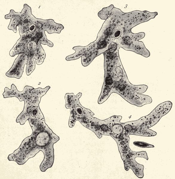
Fig. 5.—Amœba sp.; showing the forms assumed by a single individual in four successive changes. (From life.)
Note that Amœba has no mouth or alimentary canal; no nostrils or lungs, no heart or blood-vessels, no muscles, no glands. It is an animal body not made up of distinct organs and diverse tissues. Its whole body is a simple minute speck of protoplasm, a single animal cell. But it takes in food, it moves, it excretes waste matter from the body, is sensitive to the touch of surrounding objects, and, as we may be able to see, it can reproduce itself, i.e., produce new Amœbæ. Amœba is the simplest living animal.
It is only rarely that we can find an Amœba actually reproducing. The process, in its gross features, is very simple. First the Amœba draws in all of its pseudopodia and remains dormant for a time. Next, certain changes take place in the nucleus, which divides into equal portions, one part withdrawing to one end of the protoplasmic body, the other to the opposite end. Soon the body protoplasm itself begins to divide into two parts, each part collecting about its own half of the nucleus. Finally the two halves pull entirely away from each other and form two new Amœbæ, each like the original, but only half as large. This is the simplest kind of reproduction found among animals.
Amœbæ continue to live and multiply as long as the conditions surrounding them are favorable. But when the pond dries up the Amœbæ in it would be exterminated were it not for a careful provision of nature. When the pond begins to dry up each Amœba contracts its pseudopodia and the protoplasm secretes a horny capsule about itself. It is now protected from dry weather and can be blown by the winds from place to place until the rains begin, when it expands, throws off the capsule and commences active life again in some new pond.
The Slipper Animalcule (Paramœcium sp.)—Technical Note.—Paramœcia can be secured in most pond water where leaves or other vegetation are decaying. However, if specimens are not readily secured place some hay or finely cut dry clover in a glass dish, cover with water and leave in the sun for several days. In this mixture specimens will develop by thousands. Place a drop of water containing Paramœcia on a slide with cover-glass over it. Using a low power, note the many small animals darting hither and thither in the field. Run a thin mixture of cherry gum in water under the cover-glass. In this mixture they can be kept more quiet and be better studied.
How does Paramœcium (fig. 6) differ from Amœba in form and movement? Has the body an anterior and a posterior end? The delicate, short, thread-like processes, on the surface of the body, which beat about very rapidly in the water are called cilia, and they are simply fine prolongations of the body protoplasm. What is their function? Note a fine cuticle covering the body. Note also many minute oval sacs lying side by side in the ectosarc. These are called trichocysts and from each a fine thread can be thrust out.
Note on one side, beginning at the anterior end, the buccal groove leading into the interior through the gullet. Observe also that by the action of the cilia in the buccal groove food-particles are swept into the gullet. Rejected or waste particles are ejected from the body occasionally. Where? Note about midway of the Paramœcium an ovoid body with a smaller oval one attached to its side, the former being the macronucleus, the latter the micronucleus. Note that there are two contractile vacuoles in the Paramœcium; also that the food-vacuoles have a definite course in their movement inside the endosarc.
Make a drawing of a Paramœcium.
In comparing Paramœcium with Amœba it is apparent that the body of the first is less simple than that of the second. The definite opening for the ingress of food, the two nuclei, the fixed cilia, and the definite cell-wall giving[Pg 35] a fixed shape to the body, are all specializations which make Paramœcium more complex than Amœba. But the whole body is still composed of a single cell, and there is, as in Amœba, no differentiation of the body-substance into different tissues, and no arrangement of body-parts as systems of organs.
Paramœcium may occasionally be found reproducing. This process takes place very much as in Amœba. The animal remains dormant for a while, the micronucleus then divides, the macronucleus elongates and finally divides in two, the protoplasm of the body becomes constricted into two parts, each part massing itself about the withdrawn halves of the macro- and micro-nuclei, and lastly the whole breaks into two smaller organisms which grow to be like the original. After multiplication or reproduction has gone on in this way for numerous generations (about one hundred), a fusion of two Paramœcia seems necessary before further divisions take place. This process of fusion, called conjugation, may be noted at some seasons. Two Paramœcia unite with their buccal grooves together, part of the macronucleus and micronucleus of each passes over to the other, and the mixed elements fuse together to form a new macro- and micronucleus in each half. The conjugating Paramœcia now separate, and each divides to form two new individuals.
The single-celled body.—The study of Amœba and Paramœcium has made us acquainted with an animal body very different from that of the toad or the crayfish. These extraordinarily minute animals have a body so simple in its composition, compared with the toad's, that if the toad's body be taken for the type of the animal body, Amœba might readily be thought not to be an animal at all. The body of Amœba is not composed of organs, each with a particular function or work to perform. Whatever an Amœba does is done, we may say, with its whole body. But as we learn the things that this formless viscid speck of matter does, we see that it is truly an animal; that it really does those things which we have learned are the necessary life-processes of an animal. Amœba takes up and digests food composed of organic particles; it has the power of motion; it knows when its body comes in contact with some external object, that is, it can feel or has the power of sensation. Amœba takes in oxygen and gives out carbonic acid gas, and it can produce new individuals like itself, that is, it has the power of reproduction. But for the performance of these various life-processes or functions it has no special parts or organs, no mouth or alimentary canal, no lungs or gills, no legs, no special reproductive organs. We have here to do with one of the "simplest animals." With a minute, organless,[Pg 37] soft speck of viscous matter called protoplasm for a body, the simplest structural condition to be found among living beings, Amœba nevertheless is capable of performing, in the simplest way in which they may be performed, those processes which are essential to animal life.
Paramœcium has a body a little less simple than Amœba. The food-particles are taken into the body always at a certain spot; this might be spoken of as a mouth. And the body has some special locomotory organs, if they may be so called, in the presence of the cilia. The body, too, has a definite shape or form. But, as in Amœba there is no alimentary canal, nor nervous system, nor respiratory system, nor reproductive system. The whole body feels and breathes and takes part in reproduction.
A long jump has been made from the toad and crayfish to Amœba and Paramœcium; from the complex to the simplest animals. But, as will later be seen, the great difference between the bodies of these simplest animals and those of the highly complex ones is only a difference of degree; there are animals of all grades and stages of structural condition connecting the simplest with the most complex. When animals are studied systematically, as it is called, we begin with the simplest and proceed from them to the slightly complex, from these to the more complex, and finally to the most complex. There are hundreds of thousands of different kinds of animals, and they represent all the degrees of complexity which lie between the extremes we have so far studied.
The cell.—The characteristic thing about the body of Amœba and Paramœcium and the other "simplest animals"—for there are many members of the group of "simplest animals," or Protozoa—is that it is composed, for the animal's whole lifetime, of a single cell. A cell is the structural unit of the animal body. As[Pg 38] will be learned in the next exercise, the bodies of all other animals except the Protozoa, the simplest animals, are composed of many cells. These cells are of many kinds, but the simplest kind of animal cell is that shown by the body of an Amœba, a tiny speck of viscous, nearly colorless protoplasm without fixed form. The protoplasm composing the cell is differentiated to form two parts or regions of the cell, an inner denser part, called the nucleus, and an outer clearer part, called the cytoplasm. Sometimes, as in the Paramœcium, the cell is enclosed by a cell-wall which may be simply a denser outer layer of the cytoplasm, or may be a thin membrane secreted by the protoplasm. Thus the cell is not what its name might lead us to expect, typically cellular in character; that is, it is not (or only rarely is) a tiny sac or box of symmetrical shape. While the cell is composed essentially of protoplasm, yet it may contain certain so-called cell-products, small quantities of various substances produced by the life-processes of the protoplasm. These cell-products are held in the protoplasmic body-mass of the cell, and may consist of droplets of water or oil or resin, or tiny particles of starch or pigment, etc. The cell cannot be said to be composed of organs, because the word organ, as it is commonly used in the study of an animal, is understood to mean a part of the animal body which is composed of many cells. But the single cell can be somewhat differentiated into parts or special regions, each part or special region being especially associated with some one of the life-processes. In Paramœcium, for example, the food is always taken in through the so-called mouth-opening; the fine protoplasmic cilia enable the cell to swim freely in the water, the waste products of the body are always cast out through a certain part, and so on. But this is a very simple sort of differentiation, and the whole body is only one of those[Pg 39] structural units, the cells, of which so many are included in the body of any one of the complex animals.
Protoplasm.—The protoplasm, which is the essential substance of the typical animal cell and hence of the whole animal body, is a substance of very complex chemical and physical make-up. No chemist has yet been able to determine its exact chemical constitution, and the microscope has so far been unable to reveal certainly its physical characters. The most important thing known about the chemical constitution of protoplasm is that there are always present in it certain complex albuminous substances which are never found in inorganic bodies. And it is certain that it is on the presence of these substances that the power possessed by protoplasm of performing the fundamental life-processes depends. Protoplasm is the primitive physical basis of life, but it is the presence of the complex albuminous substances in it that makes it so.
The physical constitution of protoplasm seems to be that of a viscous liquid containing many fine globules of a liquid of different density and numerous larger globules of a liquid of still other density. Some naturalists believe the fine globules to be solid grains, while still others believe that numerous fine threads of dense protoplasm lie coiled and tangled in the clearer, viscous protoplasm. But the little we know of the physical structure of protoplasm throws almost no light on the remarkable properties of this fundamental life-substance.
The blood.—Technical Note.—The blood of a frog can be studied as it flows through the small vessels in the membranes between the toes while the animal is alive. Place a frog on a small flat board which has had a hole cut near one end, and with a piece of cloth bind it to the board. Spread the web between two toes over the hole in the board and keep it in place with pins. This done, examine the distended web under the compound microscope first with low then with higher power, and observe the blood-vessels and the blood circulating in them. For a further study of the blood kill a toad or frog and place a drop of the blood on a slide with a cover-glass over it.
Put the prepared slide under the microscope and note that the blood, which as seen with the unaided eye appears to be a red fluid, is made up of a great many yellowish elliptical disks or cells, the blood-corpuscles, floating in a liquid, the blood-plasma. Here and there you may notice amœboid blood-corpuscles. These are irregular-shaped cells which move about by thrusting out pseudopodia. They look like some of the unicellular animals, as the Amœba. Can you distinguish a nucleus and cell-wall in the blood-cells?
Make drawings of these blood-cells.
The skin.—Technical Note.—Keep a live toad or frog in water for some time and note if its skin becomes loose or begins to slip away. If the outer skin, epidermis, comes off, take some of the shed skin and wash it in water, then stain for three or four minutes in a solution of methyl-green and acetic acid (see p. 451). Cut[Pg 41] the pieces of stained skin into small bits and examine one of these under the microscope.
With the low power of the microscope you will note that the skin is made up of a great many flat cells placed edge to edge. Each one has its cell-wall and a central darkly stained nucleus.
Make a drawing of a portion of the toad's skin.
The liver.—Technical Note.—Cut through the fresh liver of a toad, and with a knife-blade scrape from the cut surface some of the liver-cells and place them on a slide with cover-glass.
Examine under the microscope and observe many polygonal cells. Place some of the methyl-green acetic stain under the cover-glass and note, after the cells are stained, that they have definite boundaries and a central nucleus.
Draw some of these scattered liver-cells.
The muscles.—Technical Note.—Take a piece of intestine from a freshly killed toad, wash it thoroughly and place it in a concentrated solution of salicylic acid in 70% alcohol for 24 hours, then gradually heat until about the boiling-point, when the muscles will fall to pieces. Transfer the preparation to a watch-crystal and tease small bits of isolated muscle with dissecting-needles. Place some of the teased muscle-fibres on a slide, cover with cover-glass, and add a drop of the methyl-green acetic acid.
Note the small spindle-shaped muscle-fibres. Each one of these fibres is a cell possessing all of the structures common to cells, namely, cell-wall, nucleus, etc.
Make a drawing of a few isolated fibres of muscle.
From this study of some of the tissues in a toad it will be noted that in the first case we had in the blood separate cells which moved about freely in the plasma. In the second case, that of the epidermis, the cells are fixed edge to edge, thus forming a thin tissue; while in the third and fourth cases, that of the liver and muscle, the cells are not only placed edge to edge, but aggregated[Pg 42] into vast masses or bundles, in one case to form the liver and in the other case a muscle. The entire body of the toad is built up of a colony of simple units (cells) combined in various forms to make all the various tissues and organs.
The many-celled animal body.—In the study of certain of the tissues and organs of the toad we have learned that the body of this animal is composed of many cells, thousands and thousands of these microscopic structural units being combined to form the whole toad. This many-celled or multicellular condition of the body is true of all the animals except the simplest, the unicellular Protozoa. Corals, starfishes, worms, clams, crabs, insects, fishes, frogs, reptiles, birds, and mammals, all the various kinds of animals in which the body is composed of organs and tissues, agree in the multicellular character of the body, and may be grouped together and called the many-celled animals in contrast to the one-celled animals. This division is one which is recognized by many systematic zoologists as being more truly primary or fundamental than the division of animals into Vertebrates and Invertebrates. The one-celled animals are called Protozoa, and the many-celled animals Metazoa.
Differentiation of the cell.—It is apparent at first glance that the cells which compose the body of a many-celled animal are not like the simple primitive cell which makes up the body of the Amœba, nor are they like the more complexly arranged cell of the Paramœcium. Nor are they all like each other. The cells in the toad's blood are of two kinds, the white blood-cells, which are very like[Pg 44] the body of Amœba, and the elliptical disk-like red blood-cells. The cells composing the muscles are, moreover, like neither kind of blood-cells, and the cells of which the liver is composed are not like the cells of the muscles. That is, there are many different kinds of cells in the body of a many-celled animal. While the single cell which composes the whole body of the Amœba is able to do all the things necessary to maintain life, the various cells in the body of a complex animal are differentiated or specialized, certain cells devoting themselves to a certain function or special work, and others to other special functions. For example, the cells which compose the organs of the nervous system, the brain, ganglia, and nerves, devote themselves almost exclusively to the function of sensation, and they are especially modified for this purpose. The highly specialized nerve-cells resemble very little the primitive generalized body-cell of Amœba. The muscle-cells of the complex animal body have developed to a high degree that power of contraction which is possessed, though in but slight degree, by Amœba. These muscle-cells have for their special function this one of contraction, and massed together in great numbers they form the strongly contractile muscular tissue and muscles of the body on which the animal's power of motion depends. The cells which line certain parts of the alimentary canal are the ones on which the function of digestion chiefly rests. And so we might continue our survey of the whole complex body. The point of it all is that the thousands of cells which compose the many-celled animal body are differentiated and specialized; that is, have become changed or modified from the generalized primitive amœboid condition, so that each kind of cell is devoted to some special work or function and has a special structural character fitting it for its special function. In the Protozoan body the single cell can perform and does perform[Pg 45] all the functions or processes necessary to the life of the animal. In the Metazoan body each cell performs, in co-operation with many other similar cells, some one special function or process. The total work of all the cells is the living of the animal.
Technical Note.—Hydra lives in fresh water, attached to stones, sticks, or decayed leaves. It can be found in most open fresh-water ponds not too stagnant, often attached to Chara. There are two species occurring commonly, H. viridis, the green Hydra, and H. fuscus, the brown or flesh-colored Hydra. Both are very small forms and have to be looked for carefully. Specimens should be brought to the laboratory, put into a large dish of water and left in the light. Hydra is best studied alive. Place a living specimen attached to a bit of weed in a watch-crystal filled with water or on a slide with plenty of water and examine with the low power of the microscope.
Note the cylindrical body (fig. 7, A, B) with its flat basal attachment and radial tentacles (varying in number) which crown the upper end and surround the centrally located mouth. Note the movements of Hydra, its powers of contraction, and method of taking in food.
Technical Note.—To feed Hydra, place very small "water-fleas" (Daphnia sp.) in the water with it.
Observe the method by which "water-fleas" are taken into the mouth. Food is caught on stinging cells (to be studied later) and conveyed to the mouth by the tentacles. Note that the cylindrical body encloses a cavity, the digestive cavity. How is this connected with the exterior? If Hydra captures prey too large or is no longer hungry, the prey is released.
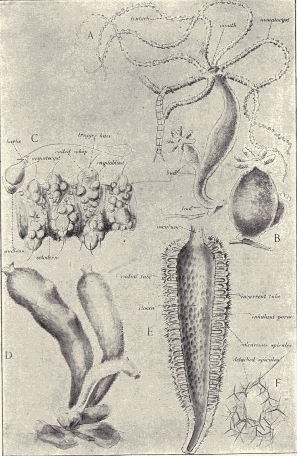
Fig. 7.—A, Hydra fusca, with expanded body and a budding individual; B, H. fusca, contracted; C, H. fusca, part of outer surface of a tentacle, greatly magnified. (A and B drawn from live specimens, C, from a preparation) D, Grantia sp. (a sponge), three individuals; E, Grantia sp., longitudinal section; F, Grantia sp., spicules. (D, E, and F drawn from preserved specimens.)
Technical Note.—Place small slips of paper on the slide near the Hydra, put cover-glass over the whole, and examine with the low power of the microscope.
Note that the whole animal is made up of cells closely joined. Are the cells in the tentacles all alike? Note nodule-like projections above some of the cells; these are stinging cells, or cnidoblasts. In some cases a small hair-like process, the trigger hair or cnidocil, may be seen projecting above the surface of the cell. Note in some of the tentacles dark-colored particles. These are food-particles which have been taken through the mouth into the digestive cavity and have passed thence into the tentacles. The central digestive cavity communicates freely with the cavities in the tentacles, for the tentacles are merely evaginations of the body-wall.
Make drawings of the Hydra expanded and of the same individual contracted.
Technical Note.—From the preparation which you have under the microscope pull out the slips of paper, thus letting the cover-glass drop down on the specimen. With a small pipette put a drop of anilin-acetic stain (see p. 451) on the slide at one side of the cover-glass and with a piece of filter-paper draw the water through from the other side of the cover-glass. When the stain is diffused press down the cover-glass gently and examine the tentacles first under a low power of the microscope, then under a high one.
Note the distortion that the animal has undergone through the action of the reagent. Observe the cnidoblasts of the tentacles and note that many of them have thrown out long whip-like processes (fig. 7, C). On what parts of the body do the cnidoblasts occur? Carefully examine one of the cnidoblasts which has been discharged and note a clear transparent bag-like structure within, the nematocyst, to which is attached the long whip-like process. In another cnidoblast cell which has not been discharged note that the whip-like process is coiled about inside of the bag-like structure. The whole[Pg 49] apparatus is like the inturned finger of a glove which can be blown out by pressure from the inside. The mechanism is simple. The cnidocil or trigger-hair is touched by some animal, an impulse is conveyed to the delicate fibres interspersed among the cells (nerve-cells) which stimulate the cnidoblast cell, whereupon there is a contraction of the contents and, the cnidoblast being compressed, the inverted whip-like process turns wrong side out and impales the animal on its points or barbs.
Technical Note.—The teacher should be provided with microscopical sections, both transverse and longitudinal, of the Hydra stained in some good general stain (hæmatoxylin or borax carmine). If the teacher has no means of making such preparations, they may be procured from dispensers of microscopical supplies.
From the cross-section of the Hydra make out the general structure of the body. Note that it is a hollow cylinder consisting of two well-defined layers of cells, an outside ectoderm layer and an inner endoderm layer. Between these two is yet another thin non-cellular layer called the mesoglœa.
Thus it will be seen that Hydra is made up of two layers of cells, the outer ectoderm or skin, which is specialized to perform the office of capturing prey as well as that of protection, and the inner endoderm, which surrounds the digestive cavity and performs the function of digestion. The endoderm lines the body-cavity, particles taken in as food being digested by certain digestive cells which thrust out amœboid processes and ingest particles of food. Other cells in the endoderm have long flagellate processes which vibrate back and forth in the digestive cavity, thereby creating currents in the water containing food-particles.
Note, in a cross-section, that there are small ovoid or cuboid cells at the bases of the large ectoderm cells. These are the interstitial cells. Some of the interstitial[Pg 50] cells become modified and pushed up between the ectoderm cells to form cnidoblast cells. Many of the endoderm as well as ectoderm cells have muscle-processes which spread out from the base of the cell and which serve to contract and expand the body.
Technical Note.—In the specimens which have been collected perhaps two methods of reproduction will be observed. Place healthy Hydræ in a wide-mouthed jar in the sunlight with plenty of water and food. In a few days active budding will take place.
Observe the method of reproduction in Hydra. Commonly the parent produces small buds, which at first are only evaginations of the body-wall, but which later develop tentacles and a mouth of their own. Subsequently the bud becomes constricted at the base, separates from the parent, and the young Hydra begins a distinct existence.
Another mode of reproduction takes place which, in distinction from the asexual method just mentioned, is called sexual reproduction. This last is the method common to most of the higher organisms. You may note that in some Hydræ there is a swelling or bulging of the ectoderm of the body-wall in the region just below the tentacles. These are the sperm-glands. Within these are produced sperm-cells which break away in great clusters to fertilize the ova, or eggs. Note a larger bulging of the body-wall nearer the lower end of the body which, under high power, has a granular appearance. This is the egg-gland, in which develops a single ovum or egg. The ovum breaks from its covering and is fertilized by sperm-cells from another individual. In forms like Hydra, where both sexes are represented in a single individual, the organism is termed monœcious or hermaphroditic. In connection with reproduction Chapter XIII should be studied.
An instructive experiment can be performed by cutting a Hydra into two or more parts, when (usually) each of the various parts will develop into a complete Hydræ. This process may be called reproduction by fission, but it rarely occurs naturally.
Cell differentiation and body organization in Hydra.—From the examination of Hydra we have learned that there are true many-celled animals which are much less complex in structure than the toad and crayfish. The body of Hydra, like the body of the toad, is composed of many cells, but these cells are of only a few different kinds; that is, show but little differentiation. There is relatively little division of the body into distinct organs. Still, certain parts of the body devote themselves principally to certain particular functions. Thus all the food is taken in through the single "mouth-opening" at the apical free end of the cylindrical body, and there are certain organs, the tentacles, whose special business or function it is to find and seize food and to convey it to the mouth. After the food is taken into the cylindrical body-cavity it is digested by special cells which line the cavity. Some of these cells are unusually large, and each contains one or more contractile vacuoles. From the free ends of these cells, the ends which are next to the body-cavity, project pseudopods or flagella. These protoplasmic processes are constantly changing their form and number. In addition to these large sub-amœboid cells there are, in this inner layer of cells lining the body-cavity, and especially abundant near the base or bottom of the cavity, many long, narrow, granular cells. These are gland-cells which secrete a digestive fluid. The food captured by the tentacles and taken in through the mouth-opening disintegrates in the body-cavity, or digestive cavity as it[Pg 53] may be called. The digestive fluid secreted by the gland-cells acts upon it so that it becomes broken into small parts. These particles are seized by the projecting pseudopods of the sub-amœboid cells and taken into the body-protoplasm of these cells. The cells of the outer layer of the body do not take food directly, but receive nourishment only by means of and through the cells of the inner layer. The body-cavity of Hydra is a very simple special organ of digestion.
In the outer layer of cells there are some specially large cells whose inner ends are extended as narrow pointed prolongations directed at right angles with the rest of the cell. These processes are very contractile and are called muscle-processes. Each one is simply a specially contractile continuation of the protoplasm of the cell-body. There are also in this layer some small cells very irregular in shape and provided with unusually large nuclei. These cells are more irritable or sensitive than the others and are called nerve-cells. We have thus in Hydra the beginnings of muscular organs and of nerve-organs. But how simple and unformed compared with the muscular and nervous systems of the toad and crayfish! There is no circulatory system, nor are there any special organs of respiration.
But Hydra is far in advance of Amœba or Paramœcium. Its body is composed of thousands of distinct cells. Some of these cells devote themselves especially to food-taking, some especially to the digestion of food; some are specially contractile, and on them the movements of the body depend, while others are specially irritable or sensitive, and on them the body depends for knowledge of the contact of prey or enemies. In the cnidoblast cells, those with the stinging threads, there is a very wide departure from the simple primitive type of cells. There is in Hydra a manifest differentiation of the cells into various[Pg 54] kinds of cells. The beginnings of distinct tissues and organs are indicated.
Degrees in cell differentiation and body organization.—In the study of the cellular constitution of the tissues and organs of the toad, we found to what a high degree the differentiation of the cells may attain, and in the study of the anatomy of the toad we found how thoroughly these differentiated cells may be combined and organized into body-parts or organs. The body of the toad is made up of distinct organs, each composed of highly differentiated or specialized cells. The body of Hydra is composed of cells for the most part only slightly differentiated and hardly recognizably grouped or combined into organs. These two conditions are the extremes in the body-structure of the many-celled animals. Between them is a host of intermediate conditions of cell differentiation and body organization. When we come to the study of other members of the great branch of simple many-celled animals to which Hydra belongs (see Chapter XVII), it will be found that some of them show a slight advance in complexity beyond Hydra. Higher in the scale of animal life the forms will be found still more and more complex, with ever-increasing differentiation of the cells, with the combination of the differentiated cells into distinct organs, and the co-ordination of organs into systems of organs up to the extreme shown by the birds and mammals. And hand in hand with this increasing complexity of structure goes ever-increasing complexity or specialization of function. Breathing is a simple function or process with Hydra, where each body-cell takes up oxygen for itself, but it is a complex business with the toad, or with a bird or mammal, where certain complex structures, the lungs and accessory parts, and the heart, blood-vessels and blood all work together to distribute oxygen to all parts of the body.
Technical Note.—As the work of this chapter, or some similar work in getting acquainted with the postembryonic development of a many-celled animal, should be done early in the course, and as most schools open in the fall, it will perhaps be impossible to make this first study of development from live specimens in the field. In such case the examination of a series of prepared specimens, previously obtained by the teacher, must be resorted to. In the spring the development of several kinds of animals, including the toad, can be studied from live specimens in the field or in breeding-cages and aquaria in the laboratory. The eggs of the toad may be found in April and May (the toads are heard trilling at egg-laying time) in ponds. The eggs look like the heads of black pins, and are in single rows in long strings of transparent jelly, which are usually wound around sticks or plant-stems at the bottom of the pond near the shore. Bring some of these strings into the schoolroom and keep them in water in shallow dishes. Keep them in the light, but not in direct sunlight. In the dishes put some small stones and mud from the pond, arranging them in a slope, thus making different depths of water. Stones with green algæ on should be selected, for algæ are the food of the tadpoles. The eggs will hatch in two or three days, and if too many tadpoles are not kept in the dish, and the little aquarium be well cared for, the whole postembryonic development of the toad can be well observed. For the study of the development from prepared specimens the teacher should have a complete series of stages from egg to adult toad in alcohol. The specimens may be examined by the students in connection with a talk from the teacher on the life-history of the toad.
If the study is made from prepared specimens, make drawings of egg-strings, and of a single egg magnified and shaded to indicate its color. Draw each specimen of the series of tadpoles, noting in the youngest the presence of gills and tail and absence of legs and eyes; in the[Pg 56] older the appearance of eyes, the shrivelling of the gills, shrinking of the tail and development of legs; in the still older the characteristic shape, in miniature, of the adult toad.
In observing the course of development of the living specimens there should be made, in addition to the drawings, notes showing the duration of the egg stage, and the time elapsing between all important changes (as seen externally) in the body of the young. Observations and notes on the general behavior of tadpoles should also be made; note the swimming, the feeding, the gradual leaving of the water, etc.
In addition to the easily seen external changes in the body, very important ones in the internal organs take place during development. Perhaps the most important of these concerns the lungs. The young gilled toad breathes as a fish does, but gradually its gills are lost, while at the same time lungs develop and the tadpole comes to the surface to breathe air like any lunged aquatic animal. The toad on leaving the water changes its diet from vegetable to animal food; a tadpole feeds on aquatic algæ; a toad preys on insects. Correlated with the change in habit, the intestine during development undergoes some marked changes, becoming relatively diminished in length.
For an account of the development of the toad see Gage's "Life-history of a Toad" or Hodge's "The Common Toad."
Multiplication.—We know that any living animal has parents; that is, has been produced by other animals which may still be living or be now dead or, as with Amœba, may have changed, by division, into new individuals. Individuals die, but before death, they produce other individuals like themselves. If they did not, their kind or species would die with them. This production of new animals constantly going on is called the reproduction or multiplication of animals. The process is well called multiplication, because each female animal normally produces more than one new individual. She may produce only one at a time, one a year, as many of the sea-birds do or as the elephant does, but she lives many years. Or she may produce hundreds, or thousands, or even millions of young in a very short time. A lobster lays 10,000 eggs at a time. Nearly nine millions of eggs have been taken from the body of a thirty-pound female codfish. As a matter of fact but very, very few of these eggs produce new animals which reach maturity. From the 10,000 eggs produced by the lobster each year an average of but two new mature lobsters is produced. There is always a struggle for food and for place going on among animals, for many more are produced than there are food and room for, and so of all the new or young animals which are born the great[Pg 58] majority are killed before they reach maturity. In a later chapter more attention will be given to this great struggle for life.
In the preceding paragraph it has been stated that "we know that any living animal has parents; that is, has been produced by other animals which may still be living or be now dead." This is a statement, however, which has found complete acceptance only in modern times. It is a familiar fact that a new kitten comes into the world only through being born; that it is the offspring of parents of its kind. But we may not be personally familiar with the fact that a new starfish comes into the world only as the production of parent starfish, or that a new earthworm can be produced only by other earthworms. But naturalists have proved these statements. All life comes from life; all organisms are produced by other organisms. And new individuals are produced by other individuals of the same kind. That these statements are true all modern observations and investigations of the origin of new individuals prove. But in the days of the earlier naturalists the life of the microscopic organisms like Amœba and Paramœcium, and even that of many of the larger but unfamiliar animals, was shrouded in mystery. And various and strange beliefs were held regarding the origin of new individuals.
Spontaneous generation.—The ancients believed that many animals were spontaneously generated. The early naturalists thought that flies arose by spontaneous generation from the decaying matter of dead animals. Frogs and many insects were thought to be generated spontaneously from mud, and horse-hairs in water were thought to change into water-snakes. But such beliefs were easily shown to be based on error, and have been long discarded by zoologists. But the belief that the microscopic organisms, such as bacteria and infusoria, were[Pg 59] spontaneously generated in stagnant water or decaying organic liquids was held by some naturalists until very recent times. And it was not so easy to disprove the assertions of such believers. If some water in which there are apparently no living organisms, however minute, be allowed to stand for a few days, it will come to swarm with microscopic plants and animals. Any organic liquid, as a broth or a vegetable infusion, exposed to the air for a short time becomes foul through the presence of innumerable microscopic organisms. But it has been certainly proved that these organisms are not spontaneously produced in the water or organic fluid. A few of them enter the water from the air, in which there are always greater or less numbers of spores of microscopic organisms. These spores germinate quickly when they fall into water or some organic liquid, and the rapid succession of generations soon gives rise to the hosts of bacteria and one-celled animals which infest all standing water. If all the active organisms and inactive spores in a glass of water are killed by boiling the water, and this sterilized water be put into a sterilized glass, and this glass be so well closed that germs or spores cannot pass from the air without into the sterilized liquid, no living animals will ever appear in it. We know of no instance of the spontaneous generation of animals, and all the animals whose life-history we know are produced by other animals of the same kind.
Simplest multiplication and development.—The simplest method of multiplication and the simplest kind of development shown among animals are exhibited by such simple animals as Amœba and Paramœcium. The production of new individuals is accomplished in Amœba by a simple division or fission of its body (a single cell) into two practically equivalent parts. An Amœba which has grown for some time contracts all of its finger-like[Pg 60] processes, the pseudopodia, and its body becomes constricted. This constriction or fissure increases inwards so that the body is soon divided fairly in two. There are now two Amœbæ, each half the size of the original one; each, indeed, actually one-half of the original one. The original Amœba was the parent; the two halves of it are the young. Each of the young possesses all of the characteristics and powers of the parent; each can move, eat, feel, grow, and reproduce by fission. The only change necessary for the young or new Amœba to become like its parent, is that of simple growth to a size about twice its present size. The development here is reduced to a minimum. Just as the simplest animals perform the other life-processes, such as taking and digesting food, breathing and feeling, in an extremely primitive simple way, so do they perform the necessary life-process of reproduction or multiplication in the simplest way shown among animals.
In the case of Paramœcium the process of multiplication is slightly more complex than that of Amœba in the fact that sometimes before the simple fission of the body takes place the interesting phenomenon of conjugation occurs. Paramœcium may reproduce itself for many generations by simple fission, but a generation finally appears in which conjugation takes place. Two individuals come together and each exchanges with the other a part of its nucleus. Then the two individuals separate and each divides into two. The result of the conjugation, or the coming together, of two individuals with mutual interchange of nuclear substance is to give to the new Paramœcia produced by the conjugating individuals a body which contains part of the body-substance of two distinct individuals. If the two conjugating individuals differ at all—and they always do differ, because no two individual animals, although belonging to the same species, are[Pg 61] exactly alike—the new individual, made up of parts of each of them, will differ slightly from both. Nature seems intent on making every new individual differ slightly from the individual which precedes it. And the method of multiplication which Nature has adopted to produce the result is the method which we have seen exhibited in its simplest form in the case of Paramœcium—the method of having two individuals take part in the production of a new one.
The development of the new Paramœcia is a little more complex than that of Amœba. Not only must the new Paramœcium grow to the size of the original one, but it must develop those slight, but apparent, modifications of the parts of its body which we can recognize in the full-grown, fully developed Paramœcium individual. A new mouth-opening must develop on the new individual formed of the hinder half of the original Paramœcium and new cilia must be developed. Thus there is a slight advance in complexity of development, just as there is in complexity of structure in Paramœcium as compared with Amœba. In the many-celled animals this complexity of development is carried to an extreme.
Birth and hatching.—When a young animal is born alive, it usually resembles in appearance and structure the parent, although of course it is much smaller, and requires always a certain time to complete its development and become mature. A young kangaroo or opossum is carried for some time after its birth in an external pouch on the mother's body and is a very helpless animal. A young kitten is born with eyes not yet opened and must be fed by the mother for several weeks. On the other hand young Rocky Mountain sheep are able to run about swiftly within a few hours after birth.
Most animals appear first as eggs laid by the mother. This is true of the birds, the reptiles, the fishes, the insects, and most of the hosts of invertebrate animals. This egg may be cared for by the parent as with the birds, or simply deposited in a safe place as with most insects, or perhaps dropped without care into the water as with most marine invertebrates. The young animal which issues from the egg may at the time of its hatching resemble the parent in appearance and structural character (although always much smaller) as with the birds, some of the insects, and many of the other animals. Or it may issue in a so-called larval condition, in which it resembles the parent but slightly or not at all, as is the case with the gill-bearing, legless, tailed tadpole of the frog or the crawling, wingless, wormlike caterpillar of the butterfly, or the maggot of the house-fly.
Life-history.—Any animal which hatches from an egg has undergone a longer or shorter period of development within the egg-shell before hatching. The development of an animal from first germ-cell to the time it leaves the egg, for example, the development of the embryo chick from the first cell to time of hatching, is called its embryonic development; and the development from then on, for example, that of the chick to adult hen or rooster, or that of tadpole to frog, is called the post-embryonic development. Beginning students of animals cannot study the embryonic development (embryology) of animals readily, but they can in many cases easily follow the course of the post-embryonic development, and this study will always be interesting and valuable. When the "life-history" of an animal is spoken of in this book, or other elementary text-book of zoology, it is the history of the life of the animal from the time of its birth or hatching to and through adult condition that is meant, not the complete life-history from beginning single egg-cell[Pg 63] to the end. In all of the study of the different kinds of animals to which the rest of this book is devoted, attention will be paid to their life-history.
Basis and significance of classification.—It is the common knowledge of all of us that animals are classified: that is, that the different kinds are arranged in the mind of the zoologist and in the books of natural history, in various groups, and that these various groups are of different rank or degree of comprehensiveness. A group of high rank or great comprehensiveness includes groups of lower rank, and each of these includes groups of still lower rank, and so on, for several degrees. For example, we have already learned that the toad belongs to the great group of back-boned animals, the Vertebrates, as the group is called. So do the fishes and the birds, the reptiles and the mammals or quadrupeds. But each of these constitutes a lesser group, and each may in turn be subdivided into still lesser groups.
In the early days of the study of animals and plants their classification or division into groups was based on the resemblances and the differences which the early naturalists found among the organisms they knew. At first all of the classifying was done by paying attention to external resemblances and differences, but later when naturalists began to dissect animals and to get acquainted[Pg 66] with the structure of the whole body, the differences and likenesses of inner parts, such as the skeleton and the organs of circulation and respiration, were taken into account. At the present time and ever since the theory of descent began to be accepted by naturalists (and there is practically no one who does not now accept it), the classification of animals, while still largely based on resemblances and differences among them, tells more than the simple fact that animals of the same group resemble each other in certain structural characters. It means that the members of a group are related to each other by descent, that is, genealogically. They are all the descendants of a common ancestor; they are all sprung from a common stock. And this added meaning of classification explains the older meaning; it explains why the animals are alike. The members of a group resemble each other in structure because they are actually blood relations. But as their common ancestor lived ages ago, we can learn the history of this descent, and find out these blood relationships among animals only by the study of forms existing now, or through the fragmentary remains of extinct animals preserved in the rocks as fossils. As a matter of fact we usually learn of the existence of this actual blood relationship, or the fact of common ancestry among animals, by studying their structure and finding out the resemblances and differences among them. If much alike we believe them closely related; if less alike we believe them less closely related, and so on. So after all, though the present-day classification means something more, means a great deal more, in fact, than the classification of the earlier naturalists means, it is largely based on and determined by resemblances and differences just as was the old classification. Sometimes the fossil remains of ancient animals tell us much about the ancestry and descent of existing forms. For example, the present-day[Pg 67] one-toed horse has been clearly shown by series of fossils to be descended from a small five-toed horse-like animal which lived in the Tertiary age.
Importance of development in determining classification.—A very important means of determining the relationships among animals is by studying their development. If two kinds of animals undergo very similar development, that is, if in their development and growth from egg-cell to adult they pass through similar stages, they are nearly related. And by the correspondence or lack of correspondence, by the similarity or dissimilarity of the course of development of different animals much regarding their relationship to each other is revealed. Sometimes two kinds of animals which are really nearly related come to differ very much in appearance in their fully developed adult condition because of the widely different life-habits the two may have. But if they are nearly related their developmental stages will be closely similar until the animals are almost fully developed. For example, certain animals belonging to the group which includes the crabs, lobsters, and crayfishes, have adopted a parasitic habit of life, and in their adult condition live attached to the bodies of certain kinds of true crabs. As these parasites have no need of moving about, being carried by their hosts, they have lost their legs by degeneration, and the body has come to be a mere sac-like pulsating mass, attached to the host by slender root-like processes, and not resembling at all the bodies of their relatives the crabs and crayfishes. If we had to trust, in making out our classification, solely to structural resemblances and differences, we should never classify the Sacculina (the parasite) in the group Crustacea, which is the group including the crabs and lobsters and crayfishes. But the young Sacculina is an active free-swimming creature resembling the young crabs and young shrimps. By a[Pg 68] study of the development of Sacculina we find that it is more closely related to the crabs and crayfishes and the other Crustaceans than to any other animals, although in adult condition it does not at all, at least in external appearance, resemble a crab or lobster.
Scientific names.—To classify animals then, is to determine their true relationships and to express these relationships by a scheme of groups. To these groups proper names are given for convenience in referring to them. These proper names are all Latin or Greek, simply because these classic languages are taught in the schools and colleges of almost all the countries in the world, and are thus intelligible to naturalists of all nationalities. In the older days, indeed, all the scientific books, the descriptions and accounts of animals and plants, were written in Latin, and now most of the technical words used in naming the parts of animals and plants are Latin. So that Latin may be called the language of science. For most of the groups of animals we have English names as well as Greek or Latin ones and when talking with an English-speaking person we can use these names. But when scientific men write of animals they use the names which have been agreed on by naturalists of all nationalities and which are understood by all of these naturalists. These Latin and Greek names of animals laughed at by non-scientific persons as "jaw-breakers," are really a great convenience, and save much circumlocution and misunderstanding.
Technical Note.—There should be provided a small set of bird-skins which will serve just as well as freshly killed birds, and which may be used for successive classes, thus doing away with the necessity of shooting birds. The birds suggested for use are among the commonest and most easily recognizable and obtainable. They may be found in any locality at any time of the year. The skins can[Pg 69] be made by some boy interested in birds and acquainted with making skins, or by the teacher, or can be purchased from a naturalists' supply store, or dealer in bird skins. The skins will cost about 25 cents each. This example or lesson in classification can be given just as well of course with other species of birds, or with a set of some other kinds of animals, if the teacher prefers. Insects are especially available, butterflies perhaps offering the most readily appreciated resemblances and differences.
Species.—Examine specimens of two male downy woodpeckers (the males have a scarlet band on the back of the head). (In the western States use Gardiner's downy woodpecker.) Note that the two birds are of the same size, have the same colors and markings, and are in all respects alike. They are of the same kind; simply two individuals of the same kind of animal. There are hosts of other individuals of this kind of bird, all alike. This one kind of animal is called a species. The species is the smallest[4] group recognized among animals. No attempt is made to distinguish among the different individuals of one kind or species of animal as we do in our own case.
Examine a specimen of the female downy woodpecker. It is like the male except that it does not have the scarlet neck-band. But despite this difference we know that it belongs to the same species as the male downy because they mate together and produce young woodpeckers, male and female, like themselves. There are thus two sorts of individuals,[5] male and female, comprised in each species of animal. A species is a group of animals comprising similar individuals which produce new individuals of the same kind usually after the mating together of individuals of two sexes which may differ somewhat in appearance and structure.
Examine a male hairy woodpecker and a female; (in western States substitute a Harris's hairy woodpecker). Note the similarity in markings and structure to the downy. Note the marked difference in size. Make notes of measurements, colors and markings, and drawings of bill and feet, showing the resemblances and the differences between the downy woodpecker and the hairy woodpecker. These two kinds of woodpeckers are very much alike, but the hairy woodpeckers are always much larger (nearly a half) than the downy woodpeckers and the two kinds never mate together. The hairy woodpeckers constitute another species of bird.
Genus.—Examine now a flicker (the yellow-shafted or golden-winged flicker in the East, the red-shafted flicker in the West). Compare it with the downy woodpecker and the hairy woodpecker. Make notes referring to the differences, also the resemblances. The flicker is very differently marked and colored and is also much larger than the downy woodpecker, but its bill and feet and general make-up are similar and it is obviously a "woodpecker." It is, however, evidently another species of woodpecker, and a species which differs from either the downy or the hairy woodpecker much more than these two species differ from each other. There are two other species of flickers in North America which, although different from the yellow-shafted flicker, yet resemble it much more than they do the downy and hairy woodpeckers or any other woodpeckers. We can obviously make two groups of our woodpeckers so far studied, putting the downy and hairy woodpeckers (together with half a dozen other species very much like them) into one group and the three flickers together into another group. Each of these groups is called a genus, and genus is thus the name of the next group above the species. A genus usually includes several, or if there be such, many,[Pg 71] similar species. Sometimes it includes but a single known species. That is, a species may not have any other species resembling it sufficiently to group with it, and so it constitutes a genus by itself. If later naturalists should find other species resembling it they would put these new species into the genus with the solitary species. Each genus of animals is given a Greek or Latin name, of a single word. Thus the genus including the hairy and downy woodpeckers is called Dryobates; and the genus including the flickers is called Colaptes. But it is necessary to distinguish the various species which compose the genus Colaptes, and so each species is given a name which is composed of two words, first the word which is the name of the genus to which it belongs, and, second, a word which may be called the species word. The species word of the Yellow-shafted Flicker is auratus (the Latin word for golden), so that its scientific name is Colaptes auratus. The natural question, Why not have a single word for the name of each species? may be answered thus: There are already known more than 500,000 distinct species of living animals; it is certain that there are no less than several millions of species of living animals; new species are being found, described and named constantly; with all the possible ingenuity of the word-makers it would be an extremely difficult task to find or to build up enough words to give each of these species a separate name. This is not attempted. The same species word is often used for several different species of animals, but never for more than one species belonging to a given genus. And the names of the genera are never duplicated. (There are, of course, much fewer genera than species, and the difficulty of finding words for them is not so serious.) Thus the genus word in the two-word name of a species indicates at once to just what particular genus in the whole animal kingdom the species[Pg 72] belongs, while the second or species word distinguishes it from the few or many other species which are included in the same genus. This manner of naming species of animals and plants (for plants are given their scientific names according to the same plan) was devised by the great Swedish naturalist Linnæus in the middle of the eighteenth century and has been in use ever since.
Family.—Examine a red-headed woodpecker (Melanerpes erythrocephalus) and a sapsucker (Sphyrapicus varius) and any other kinds of woodpeckers which can be got. Find out in what ways the hairy and downy woodpeckers (genus Dryobates), the flickers (genus Colaptes) and the other woodpeckers resemble each other. Examine especially the bill, feet, wings and tail. These birds differ in size, color and markings, but they are obviously all alike in certain important structural respects. We recognize them all as woodpeckers. We can group all the woodpeckers together, including several different genera, to form a group which is called a family. A family is a group of genera which have a considerable number of common structural features. Each family is given a proper name consisting of a single word. The family of woodpeckers is named Picidæ.
We have already learned that resemblances between animals indicate (usually) relationship, and that classifying animals is simply expressing or indicating these relationships. When we group several species together to form a genus we indicate that these species are closely related. And similarly a family is a group of related genera.
Order.—There are other groups[6] higher or more comprehensive[Pg 73] than families, but the principle on which they are constituted is exactly the same as that already explained. Thus a number of related families are grouped together to form an order. All the fowl-like birds, including the families of pheasants, turkeys, grouse and quail, all obviously related, constitute the order of gallinaceous birds called Gallinæ. The families of vultures, hawks and owls constitute the order of birds of prey, the Raptores, and the families of the thrushes, wrens, warblers, sparrows, black-birds, and many others constitute the great order of perching birds (including all the singing birds) called the Passeres.
Class and branch.—But it is evident that all of these orders, together with the other bird orders, ought to be combined into a great group, which shall include all the birds, as distinguished from all other animals, as the fishes, insects, etc. Such a group of related orders is called a class. The class of birds is named Aves. There is a class of fishes, Pisces, and one of frogs and salamanders, Batrachia, one of snakes and lizards called Reptilia, and one of the quadrupeds which give milk to their young called Mammalia. Each of these classes is composed of several orders, each of which includes several families and so on down. But these five classes of Pisces, Batrachia, Reptilia, Aves and Mammals agree in being composed of animals which have a backbone or a backbone-like structure, while there are many other animals which do not have a backbone, such as the insects, the starfishes, etc. Hence these five backboned classes may be brought together into a higher group called a branch or phylum. They compose the branch of backboned animals, the branch Vertebrata; all the animals like the starfishes, sea-urchins and sea-lilies which have the parts of their body arranged in a radiate manner compose the branch Echinodermata; all the animals like the insects and[Pg 74] spiders and centipedes and crabs and crayfishes which have the body composed of a series of segments or rings and have legs or appendages each composed of a series of joints or segments make up the branch Arthropoda. And so might be enumerated all the great branches or principal groups into which the animal kingdom is divided.
In the remainder of this book the classification of animals is always kept in sight, and the student will see the terms species, genus, family, order, etc., practically used. In it all should be kept constantly in mind the significance of classification, that is, the existence of actual relationships among animals through descent.
Of this group the structure and life-history of the Amœba (Amœba sp.) and the Slipper Animalcule (Paramœcium sp.) have already been treated in Chapter VI. Another example is the
Technical Note.—Specimens of Vorticella may usually be found in the same water with Amœba and Paramœcium. The individuals live together in colonies, a single colony appearing to the naked eye as a tiny whitish mould-like tuft or spot on the surface of some leaf or stem or root in the water. Touch such a spot with a needle, and if it is a Vorticellid colony it will contract instantly. Bring bits of leaves, stems, etc., bearing Vorticellid colonies into the laboratory and keep in a small stagnant-water aquarium (a battery-jar of pond-water will do).
Examine a colony of Vorticella in a watch-glass of water or in a drop of water on a glass slide under the microscope. Note the stemmed bell-shaped bodies which compose the colony. Each bell and stem together form an individual Vorticella (fig. 8.) How are the members of the colony fastened together? Tap the slide and note the sudden contraction of the animals; also the details of contraction in the case of an individual. Watch the colony expand; note the details of this movement in the case of an individual.
Make drawings showing the colony expanded and contracted.
With higher power examine a single individual. Note[Pg 76] the thickened, bent-out, upper margin of the bell. This margin is called the peristome. With what is it fringed? The free end of the bell is nearly filled by a central disk, the epistome, with arched upper surface and a circlet of cilia. Between the epistome and peristome is a groove, the mouth or vestibule, which leads into the body. Study the internal structure of the transparent, bell-shaped body. Note the differentiation of the protoplasm comprising the body into an inner transparent colorless endosarc containing various dark-colored granules, vacuoles, oil-drops, etc., and an outer uniformly granular ectosarc not containing vacuoles. Is the stalk formed of ectosarc or endosarc or of both? Note the curved nucleus lying in the endosarc. (This may be difficult to distinguish in some specimens.) Note the numerous large circular granules, the food vacuoles. Note the contractile vesicle, larger and clearer than the food vacuoles. Note the thin cuticle lining the whole body externally. A high magnification will show fine transverse ridges or rows of dots on the cuticle.
Make a drawing showing the internal structure.
Observe a living specimen carefully for some time to determine all of its movements. Note the contraction and extension of the stalk, the movements of the cilia of peristome and epistome, the flowing or streaming of the fluid endosarc (indicated by the movements of the food vacuoles), the behavior of the contractile vesicle.
Make notes and drawings explaining these motions.
Specimens of Vorticella may perhaps be found dividing, or two bell-shaped bodies may be found on a single stem, one of the bodies being sometimes smaller than the other. These two bodies have been produced by the longitudinal division or fission of a single body. In this process a cleft first appears at the distal end of the bell-shaped body, and gradually deepens until the original body is divided quite in two. The stalk divides for a very short distance. One of the new bell-shaped bodies develops a circlet of cilia near the stalked end. After a while it breaks away and swims about by means of this basal circlet of cilia. Later it settles down, becomes attached by its basal end, loses its basal cilia and develops a stalk.
"Conjugation occurs sometimes, but it is unlike the conjugation of Paramœcium in two important points: Firstly, the conjugation is between two dissimilar forms; an ordinary large-stalked form, and a much smaller free-swimming form which has originated by repeated division of a large form. Secondly, the union of the two is a complete and permanent fusion, the smaller being absorbed into the larger. This permanent fusion of a small active cell with a relatively large fixed cell, followed by division of the fused mass, presents a striking analogy to the process of sexual reproduction occurring in higher animals."
Besides the Amœba, Paramœcium, and Vorticella there are thousands of other Protozoa. Most of them live in water, but a few live in damp sand or moss, and some live inside the bodies of other animals as parasites. Of those which live in water some are marine, while others are found only in fresh-water streams and lakes.
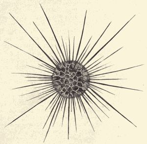
Fig. 9.—Sun animalcule, a fresh-water protozoan with a siliceous skeleton, and long thread-like protoplasmic prolongations. (From life.)
Form of body.—The Protozoa all agree in having the body composed for its whole lifetime of a single cell,[7] but they differ much in shape and appearance. Some of them are of the general shape and character of Amœba, sending out and retracting blunt, finger-like pseudopodia, the body-mass itself having no fixed form or outline but constantly changing. Others have the body of definite form, spherical, elliptical, or flattened, enclosed by a thin cuticle, and having a definite number of fine thread-like or hair-like protoplasmic prolongations called flagella or[Pg 79] cilia. Many of the familiar Protozoa of the fresh-water ponds always have two whiplash-like flagella projecting from one end of the body. By means of the lashing of these flagella in the water the tiny creature swims about. Others have many hundreds of fine short cilia scattered, sometimes in regular rows, over the body-surface. The Protozoan swims by the vibration of these cilia in the water.
Fig. 10.—Stentor sp.; a protozoan
which may be fixed, like Vorticella,
or free-swimming, at will, and
which has the nucleus in the shape
of a string or chain of bead-like
bodies. The figure shows a single
individual as it appeared when fixed,
with elongate, stalked body, and as
it appeared when swimming about
with contracted body. (From life.)
There is no stagnant pool, no water standing exposed in watering-trough or barrel which does not contain thousands of individuals of the one-celled animals. And in any such stagnant water there may always be found several or many different kinds or species. A drop of this water examined with the compound microscope will prove to be a tiny world (all an ocean) with most of its animals and plants one-celled in structure. A few many-celled animals will be found in it preying on the one-celled ones. There are sudden and violent deaths here, and births (by fission of the parent) and active locomotion and food-getting and growth and all of the businesses and functions of life which we are accustomed to see in the more familiar world of larger animals.
Marine Protozoa.—One usually thinks of the ocean as the home of the whales and the seals and the sea-lions, and of the countless fishes, the cod, and the herring, and the mackerel. Those who have been on the seashore will recall the sea-urchins and starfishes and the sea-anemones which live in the tide-pools. On the beach there are the innumerable shells, too, each representing an animal which has lived in the ocean. But more abundant than all of these, and in one way more important than all, are the myriads of the marine Protozoa.
Although the water at the surface of the ocean appears clear and on superficial examination seems to contain no animals, yet in certain parts of the ocean (especially in the southern seas) a microscopical examination of this water shows it to be swarming with Protozoa. And not only is the water just at the surface inhabited by one-celled animals, but they can be found in all the water from the surface to a great depth below it. In a pint of this ocean-water there may be millions of these minute animals. In the oceans of the world the number of them is inconceivable. And it is necessary that these Protozoa exist in such great numbers, for they and the marine one-celled plants (Protophyta) supply directly or indirectly the food for all the other animals of the ocean.
Among all these ocean Protozoa none are more interesting than those belonging to the two orders Foraminifera (fig. 11) and Radiolaria. The many kinds belonging to these orders secrete a tiny shell (of lime in the Foraminifera, of silica in the Radiolaria) which encloses most of the one-celled body. These minute shells present a great variety of shape and pattern, many being of the most exquisite symmetry and beauty. The shells are perforated by many small holes through which project long, delicate, protoplasmic pseudopodia. These fine pseudopodia often interlace and fuse when they touch each[Pg 81] other, thus forming a sort of protoplasmic network outside of the shell. In some cases there is a complete layer of protoplasm—part of the body protoplasm of the Protozoan—surrounding the cell externally.
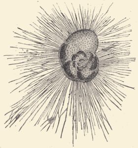
Fig. 11.—Rosalina varians, a marine protozoan (Foraminifera) with calcareous shell. (After Schultze.)
When these tiny animals die their hard shells sink to the bottom of the ocean, and accumulate slowly, in inconceivable numbers, until they form a thick bed on the ocean floor. Large areas of the bottom of the Atlantic Ocean are covered with this slimy ooze, called Foraminifera ooze or Radiolaria ooze, depending on the kinds of animals which have formed it. Nor is it only in present times that there has been a forming of such beds by the marine Protozoa. All over the world there are thick rock strata composed almost exclusively of the fossil shells of these simplest animals. The chalk-beds and cliffs of England, and of France, Greece, Spain, and America, were made by Foraminifera. Where now is land were once oceans the bottoms of which have been gradually[Pg 82] lifted above the water's surface. Similarly the rock called Tripoli found in Sicily and the Barbadoes earth from the island of Barbadoes are composed of the shells of ancient Radiolaria.
It is thus evident that the Protozoa is an ancient group of animals. As a matter of fact zoologists are certain that it is the most ancient of all animal groups. All of the animals of the ocean depend upon the marine Protozoa and the marine Protophyta, one-celled plants, for food. Either they feed on them directly, or prey on animals which in turn prey on these simplest organisms. A well-known zoologist has said: "The food-supply of marine animals consists of a few species of microscopic organisms which are inexhaustible and the only source of food for all the inhabitants of the ocean. The supply is primeval as well as inexhaustible, and all the life of the ocean has gradually taken shape in direct dependence on it." The marine Protozoa are the only animals which live independently; they alone can live or could have lived in earlier ages without depending on other animals. They must therefore be the oldest of marine animals. By oldest is meant that their kind appeared earliest in the history of the world, and as it is certain that ocean life is older than terrestrial life—that is, that the first animals lived in the ocean—it is obvious that the marine Protozoa are the most ancient of all animal groups.
As already learned in the examination of examples of one-celled animals, it is evident that life may be successfully maintained without a complex body composed of many organs performing their functions in a specialized way. The marine Protozoa illustrate this fact admirably. Despite their lack of special organs and their primitive way of performing the life-processes, that they live successfully is shown by their existence in such extraordinary numbers. They outnumber all other animals.[Pg 83] The conditions of life in the surface-waters of the ocean are easy and constant, and a simple structure and simple method of performing the necessary life-processes are wholly adequate for successful life under these conditions.
Technical Note.—Fresh-water sponges may perhaps not be readily found in the neighborhood of the school, but they occur over most of the United States, and careful searching will usually result in the finding of specimens. They are compact, solid-looking masses, sometimes lobed, resting on and attached to rocks, logs, timbers, etc., in clear water in creeks, ponds, or bayous. They are creamy, yellowish-brown or even greenish in color and resemble some cushion-like plant far more than any of the familiar animal forms. They can be distinguished from plants, however, by the fact that there are no leaves in the mass, nor long thread-like fibres such as compose the masses of pond algæ (pond scum). When touched with the fingers a gritty feeling is noticeable, due to the presence of many small stiff spicules. Sponges should be removed entire from the substance they are attached to, and may be taken alive to the laboratory. They die soon, however, and should be put into alcohol before decay begins.
Note the form of the sponge mass. Is it lobed or branched? Examine the surface for openings. These are of two sizes; the larger are osteoles or exhalant openings, while the smaller and more numerous are pores or inhalant openings. The sponge-flesh is called sarcode. Examine a bit of sarcode under the microscope; note the spicules. Have these spicules a regular arrangement? Of what are they composed?
Draw the entire sponge, showing shape and openings; draw some of the spicules.
Embedded in the body-substance, especially near the base, note (if present) numerous small, yellowish, sub-spherical[Pg 85] or disk-like bodies, the gemmules. These are reproductive bodies. Each gemmule is a sort of internal bud. It is composed of an interior group of protoplasmic cells, enclosed by a crust thickly covered with spicules. In winter the sponge dies down and the gemmules are set free in the water. In spring the protoplasmic contents issue through an aperture in the crust, called the micropyle or foraminal opening, and develop and grow into a new sponge.
For a good account of the fresh-water sponge, see Pott's "Fresh-water Sponges."
Technical Note.—For inland schools, specimens preserved in alcohol or formalin must be used. They may be obtained from dealers in naturalists' supplies (see p. 453). Specimens of some species of this genus can be obtained at almost any point on the Atlantic or Pacific coasts of this country.
Examine the external structure of a specimen. Note the elongate, sub-cylindrical form, the attached base, the free end. Note the large exhalant opening, osteole or osculum, at the free end; the numerous small inhalant openings elsewhere on the surface (best seen in dried specimens). Note the spicules covering the surface of the body, and the longer ones surrounding the osculum. Cut the sponge in two longitudinally and note the simple cylindrical body-cavity, the gastric cavity or cloaca. Note the thickness of the body-wall; note the tubes running through the body-wall from cloaca to external surface. Through these tubes water laden with food enters the gastric cavity, where the food is digested, the water and undigested particles passing out through the osculum. Crush a bit of dried sponge, or boil a bit of soft sponge in caustic potash and mount on a glass slide. Examine under a microscope and note the abundance of spicules and the variety in their form. Two kinds may always be found, and[Pg 86] sometimes three. These spicules are composed of carbonate of lime and can be dissolved by pouring on to them a drop of hydrochloric acid.
Some of the sponges may have buds growing out from them near the base. These buds are young sponges developed asexually. If allowed to develop fully the buds would have detached themselves from the parent and each would have become a new sponge.
Make drawings showing the form of a whole sponge; the appearance of the inner face of the sponge bisected longitudinally; the shape of the spicules.
Technical Note.—For the study of the skeleton of an ocean-sponge with more complex body buy several common small bath-sponges without large holes running entirely through them. The teacher should have also a few specimens of small marine sponges preserved in alcohol or formalin. Such specimens should be part of the laboratory equipment (see account of laboratory equipment, p. 450), and can be readily and cheaply obtained from dealers in naturalists' supplies.
The bath-sponge or slate-sponge consists simply of the hard parts or skeleton of a sponge animal. In life all of the skeleton is enclosed or covered by a soft, tough mass composed of layers of cells. Note the many openings on the surface of the sponge. Crush a bit of the skeleton and examine it under the microscope. Note that it is composed of fine fibres of a tough, horny substance called spongin, instead of tiny distinct calcareous spicules.
The sponges are fixed, plant-like aquatic animals. The members of a single family live in fresh water, being found in lakes, rivers, and canals in all parts of the world. All the other sponges, and there are several thousand species known, live in the ocean. They are to be found at all depths, some in shallow water near the shore and[Pg 87] others in deeper water, even to the deepest depths yet explored. They are found in all seas, though especially abundantly in the Atlantic Ocean and Mediterranean Sea.
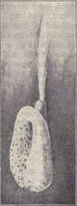
Fig. 12.—The skeleton of a
"glass" sponge (skeleton
composed of siliceous spicules)
from Japan. (From specimen.)
Form and size.—The shape of the simplest sponges is that of a tiny vase or nearly cylindrical cup, hollow and attached at its base. At the free end there is a large opening. But there is a great deal of variety in the form and size of different sponges. There is, indeed, much variation in the shape and general character of different individuals of the same species. Unlike most other animals, sponges are fixed, and the character of the surface to which a sponge is attached has much influence upon its shape. If this surface is rough and uneven the sponge may follow in its growth the sinuosities of the surface and so become uneven and distorted in shape. At best, only a few kinds of sponges have any very even and symmetrical shape. Most of them are very unsymmetrical and grow more like a low compact bushy plant than like the animals we are familiar with. The smallest sponges are only 1 mm. (1/25 in.) high, while the largest may be over a meter (39 in.) in height. In color living sponges may be red, purple, orange, gray, and sometimes blue. Most sponges have the whole body of one color.
Skeleton.—A very few sponges have no skeleton at all. The others have a skeleton or hard parts composed of interwoven fibres of the tough, horny substance called spongin, or of hosts of fine needles or spicules of silica or of carbonate of lime. The siliceous skeletons of some of the so-called glass-sponges (fig. 12) are very beautiful. The lime and siliceous sponge spicules exhibit a great variety of outline, some being anchor-shaped, some cross-shaped, and some resembling tiny spears or javelins.
Structure of body.—The skeleton of a sponge whether composed of interlacing fibres or of short spicules is always invisible from the outside when the sponge is alive. It is embedded in, or clothed by, the soft, fleshy part of the body. The soft part of the sponge is composed simply of two layers of cells, one constituting the external surface of the body, and the other lining the interior cavities and canals of the body. Between these two cell-layers there is a mass of soft gelatinous substance all through which protoplasm ramifies, and in which are embedded numerous scattered cells. There are, as seen in the case of Spongilla and Grantia, no systems of organs such as characterize the higher animals. No heart, lungs, alimentary canal, nervous system, organs of locomotion, eyes, ears, or other organs of special sense; the sponge has none of these. It is simply an aggregate of cells, arranged in two layers, and supported usually by a skeleton of horny fibres or calcareous or siliceous spicules. Its body is usually shapeless, unsymmetrical and without front or back, right or left. It is not to be wondered at that sponges were for a long time believed to be plants.
Feeding habits.—The sponges feed on minute bits of animal or plant substance and on the microscopic unicellular plants or animals which float in the water which bathes their bodies. The water entering the sponge-body through the various openings of the surface is moved[Pg 89] along by the waving or lashing of the flagella of the cells which line the canals, and these currents of water bear with them the tiny organisms which are taken up by these same cells and digested. The incoming currents of water meet in the central cavity or cavities of the body and pass out through the large opening called the osculum at the free end of the vase-like body, or if the body is branched, through the large openings at the tips of these branches.
The same currents of water bring also oxygen for the sponge's breathing and carry away the carbonic acid gas given out by the body-cells.
As a German naturalist has said, the one necessary condition for the life of a sponge is the streaming of water through its body. All sponges have a system of canals for this water-current and all have means, in the waving flagella or cilia with which these canals are lined, for producing these currents. When a live sponge is put into a vessel of water, currents are immediately set up, and they always flow into the body through the many fine openings and out of the body through the osculum.
Development and life-history.—Although the sponge in its adult condition is permanently attached by its base to the sea-bottom or to some rock or shell, when it is first born it is an active free-swimming creature. The sponges reproduce in two ways, asexually and sexually. The asexual mode of reproduction of the fresh-water sponge by gemmules has already been described. The ocean sponges also reproduce asexually either by forming interior gemmules or external buds. In this latter method a bud forms on the outer surface of the body which increases in size and finally grows into a new sponge individual. In some species this new sponge does not become separated from the body of the mother, but remains attached to it like a branch to a tree-trunk. By the continued production of such non-separating individuals,[Pg 90] a colony of sponges is formed which has the general appearance of a branching plant. In other species the new sponge formed by the development and growth of a bud falls off and becomes a distinct separate individual.
In the sexual mode of reproduction, male or sperm-cells and female or egg-cells are developed in the same individual. The sperm-cells are motile and swim about in the cavities and canals of the sponge-body until they find egg-cells, which they fertilize. The fertilized eggs begin to develop and pass through their first stages in the sponge-body. Finally the embryo sponge, which is usually a tiny oval or egg-shaped mass of cells, escapes from the body of the parent into the water. The young sponge has some of its outer cells provided with cilia, and by means of these it swims about. After a while it comes to rest on the ocean-floor or on some rock or shell, attaches itself, and begins to take on the form and character of the parent. It leads hereafter a fixed sedentary life.
The sponges of commerce.—The sponge-skeletons which are the "sponges" that we use all belong to a few species, not more than half a dozen. Most of the commercial sponges come from the Mediterranean Sea, though some come from the Bahama Islands, some from the Red Sea, and a few from the coasts of Greece, Asia Minor, and Africa. The commercial sponges do not live in very deep water; they are usually found not deeper than 200 feet. The living sponges are collected by divers, or are dragged up by men in boats using long-poled hooks, or dredges. "When secured they are exposed to the air for a limited time, either in the boats or on shore, and then thrown in heaps into the water again in pens or tanks built for the purpose. Decay thus takes place with great rapidity, and when fully decayed they are fished up[Pg 91] again, and the animal matter beaten, squeezed, or washed out, leaving the cleaned skeleton ready for the market. In this condition after being dried and sorted, they are sold to the dealers, who have them trimmed, re-sorted and put up in bales or on strings ready for exportation. There are many modifications of these processes in different places, but in a general way these are the essential-steps through which the sponge passes before it is considered suitable for domestic purposes. Bleaching-powders or acids are sometimes used to lighten the color, but these unless very delicately handled injure the durability of the fibres."
Classification.—The sponges are classified according to the character of the skeleton. In one group are put all those sponges which have a skeleton of calcareous spicules, and this group is called the Calcarea. All other sponges are grouped as Non-Calcarea, the members of this group either having no skeleton at all, or having a skeleton composed of siliceous spicules or of spongin fibres. According to the absence or presence of a skeleton and the character of the skeleton when it exists the Non-Calcarea are subdivided into smaller groups.
The structure and life-history of an example of the polyps (the Fresh-water Hydra, Hydra sp.) has been studied in Chapters X and XI.
Technical Note.—The teacher should have, if possible, several pieces of coral and a few specimens of Cœlenterates in alcohol or formalin, which will show the external character, at least, of these animals (see account of laboratory equipment, p. 450). If the school is on the coast, the pupils should be shown the sea-anemones of the tide-pools.
The animals which are included in the branch Cœlenterata are, at least in living condition, unfamiliar to most of us. Like the sponges, they are almost all inhabitants of the ocean; a few, like Hydra, live in fresh water. Like the sponges, too, most of the members of this branch are fixed, and in their general appearance suggest a plant rather than an animal. The name zoophytes, or plant-animals, which is often applied to these animals is based on this superficial resemblance. But many of the Cœlenterates lead an active free-swimming life. This is true of the jellyfishes which float or swim about on or near the surface of the ocean. Many of the zoophytes spend part of their life in an active free-swimming condition before settling down, becoming attached and thereafter[Pg 93] remaining fixed. In localities near the seashore many animals belonging to this great group can be readily found and observed. The beautiful sea-anemones with their slowly-waving tentacles, the fine many-branched truly plant-like hydroids with their hosts of little buds, and the soft colorless masses of jelly, the jellyfishes, which are cast up on to the beaches by the waves are all animals belonging to the branch Cœlenterata.
General form and organization of body.—The general or typical plan of body-structure for the Cœlenterata, these animals which come next to the sponges in degree of complexity, can best be understood by imagining the typical cylindrical or vase-like body of the simple sponges to be modified in the following way: The middle one of the three layers of the body-wall not to be composed of scattered cells in a gelatinous matrix, but to be simply a thin non-cellular membrane; the body-wall not to be pierced by fine openings or pores, but connected with the outside only by the single large opening at the free end, and this opening to be surrounded by a circlet of arm-like processes or tentacles, which are continuations of the body-wall and similarly composed. Such a body-structure, which we saw well shown by Hydra, is the fundamental one for all polyps, sea-anemones, corals, and jellyfishes. The variety in shape of the body and the superficial modifications of this type-plan are many and striking, but after all the type-plan is recognizable throughout the whole of this great group of animals.
The two chief body-shapes represented in the branch are those of the polyps on the one hand, and the jellyfishes or medusæ on the other. The polyp-shape is that of a tube with a basal end blind or closed, attached to some firm object in the water and with the free end with an opening, the mouth-opening. At this mouth-end there is a circlet of movable, very contractile tentacles.[Pg 94] The mouth may open directly into the interior of the body, which interior may be called the digestive cavity, or it may lead into a simple short tube produced by the invagination or bending in of the body-wall, which may be looked on as the simplest kind of œsophagus. This œsophageal tube opens into the body-cavity or digestive cavity. This cavity may be incompletely divided by longitudinal partitions which project from the sides into the cavity.
The jellyfish or medusoid body-form corresponds in general to an umbrella or bell. Around the edge of this umbrella are disposed numerous threads or tentacles (corresponding to the circlet of tentacles in the polyp). The mouth-opening is at the end of a longer or shorter projection which hangs down from the middle of the under side of the umbrella. The interior body-cavity or digestive cavity extends out into the umbrella-shaped part of the body, usually in the condition of canals radiating from the centre and a connecting canal running around the margin of the umbrella.
Structure.—Although the Cœlenterata show little indication of the complex composition of the body out of organs, as it exists among the higher animals, yet they do show an unmistakable advance on the simple, almost organless body of the sponges. This is chiefly shown by the differentiation among the cells which compose the body. In the polyps and jellyfishes some of the cells are specialized to be unmistakable muscle-cells, some to be nerve-cells and fibres, and so on. A very simple nervous system consisting of small groups of nerve-cells connected by nerve-fibres exists. Some very simple special sense-organs may occur. The digestive system, although in the simpler Cœlenterates consisting merely of the cylindrical body-cavity enclosed by the body-wall and opening by the single hole at the free end of the body, in some is[Pg 95] rather complex and is composed of different parts. Those Cœlenterates which are not fixed but lead an active, free-swimming life, viz., the jellyfishes or medusæ, are the most highly organized.
The tentacles which surround the mouth-opening and serve to grasp food and carry it into the mouth, and the stinging or lasso threads with which these tentacles are provided are special organs possessed by most of these animals.
Skeleton.—Like the sponges, some of the Cœlenterata possess a hard skeleton. This skeleton is always composed of calcium carbonate and is called coral. Those polyps which form such a skeleton are called the corals. Coral will be described in connection with the account of the coral-polyps.
Development and life-history.—The polyps and jellyfishes reproduce both asexually and sexually. The asexual mode is usually that of budding. On a polyp a bud is formed by a hollow outgrowth of the body-wall. The bud grows, an opening appears at its distal end, a circlet of tentacles arises about this mouth-opening and a new polyp individual is formed. This individual may separate from the parent or it may remain attached to it. By the development of numerous buds, and the remaining attached of all of the individuals developing from these buds, a colony of polyp individuals may be formed, plant-like in appearance. The various polyp individuals of a colony may differ somewhat among themselves, and these differences are correlated with a division of labor. Thus some of the individuals may devote themselves to getting food for the colony, and these have mouth and tentacles. Others may be devoted to the production of new individuals by budding or by producing germ-cells, and may not have any mouth-opening or any food-grasping tentacles.
In case of many polyps all or some of the new individuals which arise by budding do not become polyps, but develop into medusæ or jellyfish, which separate from the fixed polyp and swim off through the water. These medusæ or jellyfish produce sperm-cells and egg-cells. The sperm-cells fertilize the egg-cells and a new individual develops from each fertilized egg. This new individual is at first an active free-swimming larva called a planula, which does not resemble either a medusa or polyp. After a while it settles down, becomes fixed and develops into a polyp. Thus a polyp may produce a medusa or jellyfish which, however, produces not a new jellyfish, but a polyp. This is called an alternation of generations, and is not an uncommon phenomenon among the lower animals. It results from such an alternation of generations that a single species of animal may have two distinct forms. This having two different forms is called dimorphism. Sometimes, indeed, a species may appear in more than two different forms; such a condition is called polymorphism.
Not all medusæ or jellyfish are produced by polyp individuals, nor do jellyfish always produce polyps and not jellyfishes. There are some jellyfishes (we might call them the true jellyfishes) which always have the jellyfish form, producing new jellyfishes either by budding or by eggs, and there are some polyps which always have the true polyp form, producing new individuals, either by budding or by eggs, always of polyp form and never of jellyfish form. That is, some species of Cœlenterata exist only in polyp form, some species exist only in jellyfish form, while some species (those having an alternation of generations) exist in both polyp and jellyfish form, these two forms appearing as alternate generations.
Classification.—The branch Cœlenterata is divided into four classes: (1) the Hydrozoa, including the fresh-water[Pg 97] polyps, numerous marine polyps, many small jellyfishes and a few corals; (2) the Scyphozoa, including most of the large jellyfishes; (3) the Actinozoa, including the sea-anemones and most of the stony corals; (4) the Ctenophora, including certain peculiar jellyfishes.
Fig. 13.—The Portuguese Man-of-War (Physalia sp.). (From specimen from Atlantic Coast.)
The polyps, colonial jellyfishes, etc. (Hydrozoa).—To the class Hydrozoa belongs the Hydra already studied. There are a few other fresh-water polyps and they all belong to this class. The most interesting members of the class are the "colonial jellyfishes," constituting the order Siphonophora. These[Pg 98] colonial jellyfishes are floating or swimming colonies of polypoid and medusoid individuals in which there is a marked division of labor among the individuals, accompanied by marked differences in structural character. The individuals are accordingly polymorphic, that is, appear in various forms, although all belong to the same species. Because these various individuals forming a colony have given up very largely their individuality, combining together and acting together like the organs of a complex animal, they are usually not called individuals, nor on the other hand organs, but zooids, or animal-like structures. The beautiful "Portuguese man-of-war" (fig. 13) is one of these colonial jellyfishes. It appears as a delicate bladder-like float, brilliant blue or orange in color, usually about six inches long, and bearing on its upper surface which projects above the water a raised parti-colored crest, and on its under surface a tangle of various appendages, thread-like with grape-like clusters of little bell- or pear-shaped bodies. Each of these parts is a peculiarly modified polyp- or medusa-zooid produced by budding from an original central zooid. The Portuguese man-of-war is very common in tropical oceans, and sometimes vast numbers swimming together make the surface of the ocean look like a splendid flower-garden.
Usually the central zooid in a Siphonophore to which the other zooids are attached is not a bladder-like float, but is an upright tube of greater or less length. In the Siphonophore shown in figure 14, the compound body is composed of a long central hollow stem with hundreds or thousands of variously shaped parts, each of which is reducible to either a polyp or medusazooid, attached around it. The upper end is enlarged to form an air-filled chamber, a sac-like boat, by means of which the whole colony is kept afloat. Around the upper end of the[Pg 99] central stem are many medusoid structures, the swimming-bells, by means of whose opening and closing the whole colony is made to swim through the water. Each swimming-bell is a modified medusa-zooid, without tentacles, without digestive or reproductive organs, but exercising the power of swimming by contracting and forcing the water out of the hollow bell just as is done by the free medusæ. Below the swimming-bells, at the lower end of the central stem, are grouped many structures presenting at first sight a confusion of variety and complexity, but on careful examination revealing themselves to be polyp- and medusa-zooids modified to form at least five kinds of particularly functioning structures. There are many flattened scale-like parts whose function is simply that of affording a passive protection,[Pg 100] in times of danger, to the other structures. These protecting-scales are greatly modified medusa-zooids, each consisting of a simple cartilage-like gelatinous mass penetrated by a food-carrying canal. Under the broad leaves of these protecting-zooids are a number of pear-shaped bodies which have a wide octagonal mouth-opening at their free end, and possess in their interior certain digestive glands. Each one is provided with a very long flexible tentacle which bears many fine stinging-threads. The tentacle waves back and forth in the water, and on coming in contact with an enemy or with prey its poisonous stinging-threads shoot out and paralyze or wound the unfortunate animal. These pear-shaped bodies are the feeding structures, each being a modified polyp-zooid. Scattered among these dangerous structures are many somewhat similarly shaped but wholly harmless structures, the sense-structures. Each of these has a pear-shaped body but without mouth-opening, and also a long, very sensitive, tentacle-like process. The sense of feeling is highly developed in these tentacles, and they discover for the colony the presence of any strange body. These sense-structures are modified polyp-zooids. Finally there are two other kinds of structures, usually arranged in groups like bunches of grapes, which are the reproductive structures, male and female. They are modified medusa-zooids grown together and without tentacles. This whole colony, or this compound animal, floats or swims about at the surface of the ocean, and performs all of the necessary functions of life as a single animal composed of organs might. Yet the Siphonophore is more truly to be regarded as a community in which the hundreds or thousands of animals, representing five or six kinds of individuals, all of one species, are fastened together. Each individual performs the particular duties devolving upon its kind or class. Thus there are food-gathering individuals,[Pg 101] locomotor individuals, sense individuals, and reproductive individuals. The modifications of the various kinds of individuals are more extreme than in the case of the various kinds of individuals composing a bee-community, for example, but the holding together or fusing of all into one body or corporation is a condition which makes this greater modification necessary and not unexpected. And there is no difficulty in seeing that each of these parts is really, structurally considered, a modified polyp or medusa.
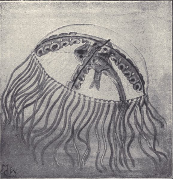
Fig. 15.—A jellyfish or medusa, Gonionema vertens, eating two small fishes. (From specimen from Atlantic Coast.)
The large jellyfishes, etc. (Scyphozoa).—To the class Scyphozoa belong most of the common large jellyfishes. When one walks along the sea-beach soon after a storm one may find many shapeless masses of a clear jelly-like[Pg 102] substance scattered here and there on the sand. These are the bodies or parts of bodies of jellyfishes which have been cast up by the waves. Exposed to the sun and wind the jelly-like mass soon dries or evaporates away to a small shrivelled mass. The body-substance of a jellyfish contains a very large proportion of water; in fact there is hardly more than 1 per cent of solid matter in it.
The jellyfishes occur in great numbers on the surface of the ocean and are familiar to sailors under the name of "sea-bulbs." Some live in the deeper waters; a few specimens have been dredged up from depths of a mile below the surface. They range in size from "umbrellas" or disks a few millimeters in diameter to disks of a diameter of two meters (2-1/6 yards). They are all carnivorous, preying on other small ocean animals which they catch by means of their tentacles provided with stinging-threads. The tentacles of some of the largest jellyfishes "reach the astonishing length of 40 meters, or about 130 feet." Many of the jellyfishes are beautifully colored, although all are nearly transparent. Almost all of them are phosphorescent, and when irritated some emit a very strong light.
The sea-anemones and corals (Actinozoa).—Almost everywhere along the seashore where there are rocks and tide-pools a host of various kinds of sea-anemones can be found. When the tide is out, exposing the dripping seaweed-covered rocks, and the little sand- or stone-floored basins are left filled with clear sea-water, the brown and green and purple "sea-flowers" may be found fixed to the rocks by the base with the mouth-opening and circlet of slowly-moving tentacles hungrily ready for food (fig. 16). Touch the fringe of tentacles with your fingertip and feel how they cling to it and see how they close in so as to carry what they feel into the mouth-opening. A host of individuals there are, and scores of different kinds;[Pg 103] some small, some large, some with the body covered outside with tiny bits of stone and shell so that they are hardly to be distinguished from the rock to which they cling; some of bright and showy colors. These are the most familiar members of the class Actinozoa.
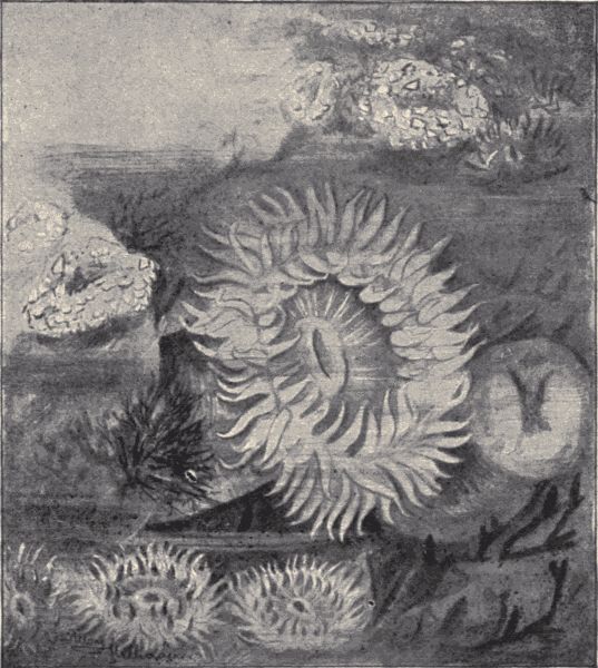
Fig. 16.—Sea anemones, Bunodes californica, open and closed individuals. The closed individuals in upper right-hand corner show the external covering of small bits of rock and shell, characteristic of most individuals of this species. (From living specimens in a tide-pool on the Bay of Monterey, California.)
But in other oceans, along the coasts of other lands, especially those of the tropics and sub-tropics, there are[Pg 104] some other members of the class which are of unusual interest. They are the corals, or coral polyps. We know these animals chiefly by their skeletons (fig. 17). The specimens of corals which one sees in collections, or made into ornaments, are the calcareous skeletons of various kinds of the coral polyps. Some of the corals live together in enormous numbers, forming branching colonies fixed as closely together as possible, and secrete while living a stony skeleton of carbonate of lime. These skeletons persist after the death of the animals, and because of their abundance and close massing form great reefs or banks and islands. These coral reefs and islands occur only in the warmer oceans. In the Atlantic they are found along the coasts of Southern Florida, Brazil and the West Indies; in the Pacific and Indian Oceans there are great coral reefs on the coast of Australia, Madagascar and elsewhere, and certain large groups of inhabited islands like the Fiji, Society, and Friendly Islands are exclusively of coral formation. Coral islands have a great variety of form, although the elongated, circular, ring-shaped and crescent forms predominate. How such islands are first formed is described as follows by a well-known student of corals:
"A growing coral plantation, with its multitudinous life, oftentimes arises from great depths of the ocean, and the sea-bed upon which it rests is probably a submarine bank or mountain, upon which have lodged and slowly aggregated the hard skeletons of pelagic forms of life. When, through various sources of increase, this submarine bank approaches the depth of from one hundred to one hundred and fifty feet from the surface of the water, there begins on its top a most wonderful vital activity. It is then within the bathymetric zone of the reef-building corals. Of the many groups of marine life which then take possession of the bank, corals are not the only[Pg 105] animals, but they are the most important, as far as its subsequent history goes. As the bank slowly rises by their growth, it at last approaches the surface of the water, and at low tide the tips of the growing branches of coral are exposed to the air. This, however, only takes place in sheltered localities, for long before it has reached this elevation it has begun to be more or less changed and broken by the force of the waves. As the submarine bank approaches the tide level, the delicate branching forms have to meet a terrific wave-action. Fragments of the branching corals are broken off from the bank by force of the waves, and falling down into the midst of the growing coral below fill up the interstices,[Pg 106] and thus render the whole mass more compact. At the same time larger fragments are broken and rolled about by the waves and are eventually washed up into banks upon the coral plantation, so that the island now appears slightly elevated above the tides. This may be called a first stage in the development of a coral island. It is, however, little more than a low ridge of worn fragments of coral washed by the high tides and swept by the larger waves—a low, narrow island resting on a large submarine bank."
When the coral island rises thus a little above the surface of the water, the waves break up some of the coral into fine sand, which fills in the interstices, and offers a sort of soil in which may germinate seeds brought in the dried mud on the feet of ocean birds or carried by the ocean currents. With the beginning of vegetable growth the soil is more firmly held, is fertilized and ready for the seeds of plants which need a better soil than lime sand. Flying insects find their way to the island, especially if it be near the mainland, birds begin to nest on it, and soon it may be the seat of a luxuriant plant and animal life.
For an account of coral islands see Darwin's "The Structure and Distribution of Coral Reefs."
There are over 2000 kinds of coral polyp known, and their skeletons vary much in appearance. Because of the appearance of the skeleton certain corals have received common names, as the organ-pipe coral, brain coral, etc. The red coral, of which jewelry is made, grows chiefly in the Mediterranean. It is gathered especially on the western coast of Italy, and on the coasts of Sicily and Sardinia. Most of this coral is sent to Naples, where it is cut into ornaments.
There are other interesting members of the class Actinozoa like the beautiful sea-pens, sea-feathers and[Pg 107] sea-fans, delicate, branching, tree-like forms found all over the world.
Ctenophora.—The members of this class are mostly small, peculiar jellyfishes which do not form colonies, and are extremely delicate, being usually perfectly transparent. They swim by means of cilia. They never appear in a polyp condition, but are always medusoid in shape.
Technical Note.—The species of Asterias are widely distributed on both coasts of the United States and may be procured on almost any rocky shore at low tide. Teachers in inland schools can obtain preserved material from the dealers mentioned on p. 453. Most of the specimens should be placed in alcohol or 4% formalin. If fresh material can be had it is well to place at least one specimen for each student in a 20% solution of nitric acid in water for two or three hours, when all of the calcareous parts will have been dissolved, and after a thorough washing the specimen will be ready for use.
External structure (figs. 18 and 19.)—In a fresh specimen or one which has been preserved in alcohol or formalin note the raying out of parts of the body from a common centre. This is characteristic of the body organization of all Echinoderms, and is known as radial symmetry. The lower surface of the body is called the oral (because the mouth is on this surface), while the upper is called the aboral surface. The central part of the body is called the disk. Note on the aboral surface of the disk a small striated calcareous plate, the madreporite or madreporic plate. In the middle (or very nearly in the middle) of this surface of the disk there is a small pore, the anal opening. The entire aboral surface as well as a greater part of the oral side is thickly studded with the calcareous ossicles of the body-wall. These ossicles support numerous short stout spines arranged in irregular rows. Note that some of the ossicles[Pg 109] support certain very small pincer-like processes, the pedicellariæ. In the interspaces between the calcareous plates are soft fringe-like projections of the inner body-lining, the respiratory cæca. Note at the tip of each arm or ray a cluster of small calcareous ossicles and within each cluster a small speck of red pigment, the eye-spot or ocellus.
Make a drawing of the aboral surface showing all these parts.
On the oral surface note the centrally-located mouth, the ambulacral grooves, one running longitudinally along each ray, and in each groove two double rows of soft tubular bodies with sucker-like tips. These are called the tube-feet and are organs of locomotion. Make a drawing of the oral surface.
Internal structure (figs. 18 and 19).—Technical Note.—Take a specimen which has been immersed for some time in the nitric acid solution, and with a strong pair of scissors, or better, bone-cutters, cut away all the aboral wall of the disk except that immediately around the madreporite and the anus. Now begin at the tip of each ray and cut away the aboral wall of each, leaving, however, a single arm intact. When the roof of each arm has been carefully dissected away the specimen should appear as in fig. 18.
Note the large alimentary canal, which is divided into several regions. Note the short œsophagus leading from the mouth on the oral surface directly into a large membranous pouch, the cardiac portion of the stomach. By a short constriction the cardiac portion is separated from the part which lies just above, i.e., the pyloric portion of the stomach. From the pyloric portion large, pointed, paired glandular appendages extend into each ray. These are the pyloric cæca. Their function is digestive, and oftentimes they are spoken of as the digestive glands or "livers." The pyloric cæca, as well as the cardiac portion of the stomach, are held in place by paired muscles which extend into each arm. Note two sets of these muscles, one set for thrusting the cardiac portion of the stomach out through the mouth and another for pulling it back, the protractor muscles and retractor muscles, respectively. The starfish obtains its food by enclosing it in its everted stomach and then withdrawing stomach and food into the body. Note that the pyloric portion of the stomach opens above into a short intestine terminating[Pg 111] in the anus, and observe that there is attached to the intestine a convoluted many-branched tube, the intestinal cæcum.
Carefully remove a pair of pyloric cæca from one of the rays and note the short duct which connects them with the pyloric chamber of the stomach. Note in the angle of each two adjoining rays paired glandular masses which empty by a common duct on the aboral surface. These glands are the reproductive organs. Note the small bulb-like bladders extending in two double rows on the floor of each ray. These are the water-sacs or ampullæ, and each one is connected directly with one of the locomotor organs, the tube-feet.
Make a drawing of the organs in the dissection which have so far been studied.
Technical Note.—For a careful study of the locomotor organs a fresh starfish should be injected. This can usually be accomplished by cutting one ray off squarely, and inserting the needle of a hypodermic syringe (which has been previously filled with a watery solution of carmine or Berlin blue), into the end of the radial water-tube which runs along the floor of the ray. By injecting here, the whole system of vessels, tube-feet, and ampullæ are filled.
Note a ring-shaped canal which passes around the alimentary canal near the mouth from which radial vessels run out beneath the floor of each ray and from which a hard tube extends to the madreporite. This hard tube is the stone canal, so called because its walls contain a series of calcareous rings, while the circular tube is the ring canal or circum-oral water-ring from which radiate the radial canals. In some species of starfish there are bladder-like reservoirs, Polian vesicles, which extend interradially from the ring canal.
Note that the ampullæ and tube-feet are all connected with the radial canals. By a contraction of the delicate[Pg 112] muscles in the walls of the ampullæ the fluid in the cavity is compressed, thereby forcing the tube-feet out. By the contraction of muscles in the tube-feet they are again shortened while the small disk-like terminal sucker clings to some firm object. In this way the animal pulls itself along by successive "steps." This entire system, called the water-vascular system, is characteristic of the branch Echinodermata. In addition to the fluid in the water-vascular system there is yet another body-fluid, the perivisceral fluid, which bathes all of the tissues and fills the body-cavity.
Technical Note.—Take a drop of the perivisceral fluid from a living starfish and examine under high power of microscope, noting the amœboid cells it contains.
The perivisceral fluid is aerated through outpocketings of the thin body-wall which extend outward between the calcareous plates of the body. These outpocketings have[Pg 113] already been mentioned as the respiratory cæca (see p. 109). Surrounding the stone canal is a thin membranous tube, and within it and by the side of the stone canal is a soft tubular sac. The function of these organs is not certainly known.
Work out the nervous system; note, as its principal parts, a nerve-ring about the mouth, and nerves running from this ring beneath the radial canals along each arm.
Life-history and habits.—The starfishes are all marine forms. They hatch from eggs, and in their early stages are very different in appearance from the adults. At first they are bilaterally symmetrical, their radial symmetry being acquired later. Thousands of eggs and sperm-cells are extruded into the sea-water, where fertilization and development take place. The young swim freely in the open sea, feeding on microscopic organisms, and then undergo very radical changes in the course of their development. The adults are for the most part carnivorous, feeding on crabs, snails, and the like. The live prey is surrounded by the extruded stomach which secretes fluids that kill it, after which the soft parts are digested. (See general account of the life-history of Echinoderms on p. 119.)
External structure.—Technical Note.—If fresh or alcoholic specimens or even the dry "tests" of the sea-urchin (fig. 20) are to be had, the general characteristics of the external structure can be made out.
How does the external surface of the sea-urchin differ from that of the starfish? Can you find the very long tube-feet? Where is the mouth-opening? With what is it surrounded? Each tooth is enclosed in a calcareous framework. The whole structure is known as "Aristotle's lantern."
Technical Note.—Remove the spines from the underlying shell or test (fig. 21) and wash the test until perfectly clean, or place in a solution of lye for a short time and then wash.
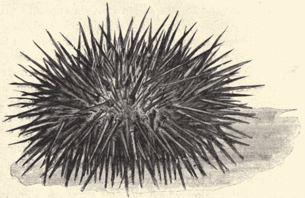
Fig. 20.—A sea-urchin, Strongylocentrotus franciscanus. (From specimen from Bay of Monterey, Calif.)
Note the characteristic radial symmetry of the shell or test. Note on the aboral aspect, diverging from the medial anal aperture, five double rows of pores. What are these for? Each of the five divisions set with pores is called an ambulacral area, while the intervening segments which support the long spines are called the interambulacral areas. Note on the aboral surface, surrounding the median-placed anal aperture, a series of small plates. Those which are located in the interambulacral areas are the genital plates. Through these plates the ducts from the reproductive organs open by small pores. Note a very much enlarged plate with a striated appearance. This is the madreporite, which, as in the starfish, is the external opening of the stone canal and water-vascular system. Note the small ocular plate at the tip of each ambulacral area. The ocular plates contain[Pg 115] small pigment-cells and communicate with the nervous system.
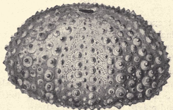
Fig. 21.—"Test" of sea-urchin, Strongylocentrotus franciscanus, with spines removed. (From specimen.)
From a general inspection of the sea-urchin's shell the Echinoderm characteristics, namely, radial symmetry and the presence of the water-vascular system, are readily seen. While at first glance there is apparent little similarity between the starfish and sea-urchin, nevertheless careful examination shows that the two animals are alike in their fundamental structure. Both are radially symmetrical. The position of the anal opening makes both starfish and sea-urchin slightly asymmetrical. In both the madreporite and anus are on the aboral side, while the mouth is centrally located on the oral side. In the starfish we noted five ambulacral areas, one on the under side of each arm; similarly we find five in the sea-urchin. In both cases also we find the ocular spots at the tips of the ambulacral areas. The genital apertures are situated interradially in the starfish. In the sea-urchin they are similarly placed. The dissimilarity between the two forms is largely due to the very much developed outer spines and the dorso-ventral thickening of the disk in the sea-urchin. The starfish is carnivorous, while the sea-urchin lives on vegetable matter consisting[Pg 116] for the most part of green algæ and the red sea-weeds. Correlated with this difference in food-habits there are certain differences in the structure of the internal organs. For example, the alimentary canal in the sea-urchin winds in about two and one-half turns within the body-cavity before it reaches the anus.
Without exception all the Echinoderms, under which term are included the starfishes, sea-urchins, brittle-stars, feather-stars, and sea-cucumbers, live in the ocean. Some of them, the starfishes and sea-urchins, are among the most common and familiar animals of the seashore. Most of them are not fixed, but can move about freely, though slowly. Some of the feather-stars are fixed, as the sponges and polyps are.
Shape and organization of body.—The body-shape of the Echinoderm varies from the flat, rayed body of the starfish to the thick, flattened egg-shape of the sea-urchin, the melon-like sac of the sea-cucumber and the delicate many-branched head of the sea-lily sometimes borne on a slender stalk. But in all these shapes can be seen more or less plainly a symmetrical, radiate arrangement of the parts of the body. The Echinoderm body has a central portion from which radiate separate arm or branch-like parts, as in the starfishes and sea-lilies, or about which are arranged radiately the internal body-parts, although the external appearance may at first sight give no plain indication of the radiate arrangement. This is the case with the sea-urchins and sea-cucumbers, yet, as has been seen in the sea-urchin, the radiate arrangement can be readily perceived by closer examination of the surface of the egg- or sac-like body. The radiating parts of the[Pg 117] body are usually five. In the body of an Echinoderm can be usually recognized an upper or dorsal surface and a lower or ventral surface. The mouth is usually situated on the ventral side and the anal opening on the dorsal. Echinoderms agree also in having a calcareous outer skeleton or body-wall usually in the condition of definitely-shaped plates or spicules fitted either movably or rigidly together. This outer body-wall or exoskeleton may bear many tubercles or spines. These spines are sometimes movable. The body-wall of the sea-urchin shows very well the exoskeleton composed of plates on which are borne movable strong spines.
Structure and organs.—As has been learned from the dissection of the starfish, the Echinoderms have well-developed systems of organs. The body-structure in its complex organization presents a marked advance beyond the structural condition of the polyps and jellyfishes. There is a well-organized digestive system with mouth, alimentary canal, and anal opening. The alimentary canal is either a simple spiral or coiled tube, or it is a tube in which can be recognized different parts, namely, œsophagus, stomach, intestine, cæca, and special glands secreting digestive fluids. This alimentary canal is not, as in the polyps, simply the body-cavity, but it is an inclosed tubular cavity lying within the general body-cavity. At the mouth-opening there is in some Echinoderms, notably the sea-urchins, a strong masticating apparatus consisting of five pointed teeth which are arranged in a circle about the opening. The nervous system consists of a central ring around the œsophagus or mouth, from which branches extend into the radiately arranged arms or regions of the body. There is no brain as in the higher animals, but the central nerve-ring is composed of both nerve-cells and nerve-fibres as in the nerve-centres of higher forms. Of organs of special sense there are[Pg 118] special tactile or touch organs in all the Echinoderms, and the starfishes have very simply composed eyes or eye-like organs at the tips of the rays.
While some of the Echinoderms breathe simply through the outer body-wall, taking up by osmosis the air mixed with the water, some of them have special, though very simple, gill-like respiratory organs. These organs consist of small membranous sacs which are either pushed out from the body into the water, or lie in cavities in the body to which the water has access. There is also a distinct circulatory system, but the "blood" which is carried by these organs and which fills the body-cavity consists mainly of sea-water, although containing a number of amœboid corpuscles containing a brown pigment. There is no organ really corresponding to the heart of the higher animals. There are distinct organs for the production of the germ or reproductive cells. The sexes are distinct (except in a few species), each individual producing only sperm-cells or egg-cells, but the organs or glands which produce the germ-cells are very much alike in both sexes. There is no apparent difference between male and female Echinoderms except in the character or rather in the product of the germ-cell producing organs. A few species are exceptions, certain starfishes showing a difference in color between males and females.
As all of the Echinoderms except some of the feather-stars can move about, they have organs of locomotion, and well-defined muscles for the movement of the locomotory organs. The external organs of locomotion, the tube-feet (in the sea-urchins the dermal spines aid also in locomotion), are parts of a peculiar system of organs characteristic of the Echinoderms, called the ambulacral or the water-vascular system. This system is composed of a series of radial tubular vessels which rise from a central[Pg 119] circular or ring vessel and which give off branches to each of the tube-feet. The water from the outside enters the ambulacral system through a special opening, the madreporic opening, and flowing to the tube-feet helps extend them. The tube-feet usually have a tiny sucking disk at the tip, and by means of them the Echinoderm can cling very firmly to rocks.
Development and life-history.—Differing from the sponges and the polyps and jellyfishes, the reproduction of the Echinoderms is always sexual; young or new individuals are never produced by budding, or in any other asexual way. The new individual is always developed from an egg produced by a female and fertilized by the sperm of a male. The eggs are usually red or yellow, are very small (about 1/50 in. in diameter in certain starfishes), and are fertilized by the sperm-cells of the males after leaving the body of the female. That is, both sperm-cells and unfertilized egg-cells are poured out into the water by the adults, and the motile sperm-cells in some way find and fertilize the egg-cells.
From the egg there hatches a tiny larva which does not at all resemble the parent starfish or sea-urchin. It is an active free-swimming creature, more or less ellipsoidal in shape and provided with cilia for swimming. Soon its body changes form and assumes a very curious shape with prominent projections. The larvæ of the various kinds of Echinoderms, as the starfishes, sea-urchins, sea-cucumbers, etc., are of different characteristic shapes. The naturalists who first discovered these odd little animals did not associate them in their minds with the very differently shaped starfishes and sea-urchins, but believed them new kinds of fully developed marine animals, and gave them names. Thus the larvæ of the starfishes were called Bipinnaria, the larvæ of the sea-urchins Pluteus, and so on. These names are still used[Pg 120] to designate the larvæ, but with the knowledge that Bipinnaria are simply young starfishes, and that a Pluteus is simply a young sea-urchin. From these larval stages the adult or fully developed starfish or sea-urchin develops by very great changes or metamorphoses. The Echinoderms have in their life-history a metamorphosis as striking as the butterflies and moths, which are crawling worm-like caterpillars in their young or larval condition.
Most of the Echinoderms have the power of regenerating lost parts. That is, if a starfish loses an arm (ray) through accident, a new ray will grow out to replace the old. And this power of regeneration extends so far in the case of some starfishes that if very badly mutilated they can practically regenerate the whole body. This amounts to a kind of asexual reproduction. Some species, too, have the peculiar habit of self-mutilation. "Many brittle stars and some starfishes when removed from the water, or when molested in any way, break off portions of their arms piece by piece, until, it may be, the whole of them are thrown off to the very bases, leaving the central disc entirely bereft of arms. A central disc thus partly or completely deprived of its arms is capable in many cases of developing a new set; and a separated arm is capable in many cases of developing a new disc and a completed series of arms." In some of the sea-cucumbers "it is the internal organs, or rather portions of them, that are capable of being thrown off and replaced, the œsophagus ... or the entire alimentary canal, being ejected from the body by strong contractions of the muscular fibres of the body-wall, and in some cases, at least, afterwards becoming completely renewed."
Classification.—The Echinodermata are divided into five classes, viz., the Asteroidea or starfishes, "free Echinoderms with star-shaped or pentagonal body, in[Pg 121] which a central disc and usually five arms are more or less readily distinguishable, the arms being hollow and each containing a prolongation of the body-cavity and contained organs"; the Ophiuroidea, or brittle-stars, "star-shaped free Echinoderms, with a central disc and five arms, which are more sharply marked off from the disc than in the Asteroidea and which contain no spacious prolongations of the body-cavity"; the Echinoidea, or sea-urchins, "free Echinoderms with globular, heart-shaped, or disc-shaped body enclosed in a shell or corona of close-fitting, firmly united calcareous plates"; the Holothuroidea, or sea-cucumbers, "free Echinoderms with elongated cylindrical or five-sided body, ... with a circlet of large oral tentacles"; and the Crinoidea, or feather-stars, "temporarily or permanently stalked Echinoderms with star-shaped body, consisting of a central disc, and a series of five bifurcate or more completely branched arms, bordered with pinnules."
Starfishes (Asteroidea).—The starfishes feed on other marine animals, especially shell-fish and crabs. They are also reputed to destroy young fish. By means of their sucking-tubes, or tube-feet with sucker tips, they can seize and hold their prey firmly. They do much injury to oyster-beds by attacking and devouring the oysters. When attacking prey too large to be taken into the mouth the starfish everts its stomach over the prey and devours it. The stomach is afterward drawn back into the body-cavity by special muscles.
Starfishes vary much in size, color and general appearance, although all are readily recognizable as starfishes (fig. 22). The number of arms or rays varies from five to thirty or more in different species; some have the interradial spaces filled out nearly to the tips of the rays, making the animal simply a pentagonal disc. In size starfishes vary from a fraction of an inch in diameter to[Pg 122] three feet; in color they are yellow or red or brown or purple.
Fig. 22.—A group of Echinoderms; the upper one, a starfish, Asterina mineata, the one at the right a starfish, Asterias ocracia, at the left a brittle-star, species unknown, and at bottom two sea-urchins, Strongylocentrotus franciscanus. (From living specimens in a tide-pool on the Bay of Monterey, California.)
Brittle-stars (Ophiuroidea).—The brittle-stars, or serpent-stars (fig. 22) as they are also called, resemble the starfishes in external appearance, that is, they are flat and composed of a central disc with radiating arms (always five in number, although each arm may be several times branched). The central disc is always sharply distinguished from the arms, and the arms are usually slender and more or less cylindrical.[Pg 123] The distinguishing difference between the brittle-stars and the starfishes is that the body-cavity and the stomach which extend out into the arms in the starfishes are in the brittle-stars limited to the central disc, or to the disc and bases of the arms. The tube-feet also have no suckers at the tips. More than 700 species of brittle-stars are known. They feed on marine shell-fish, crabs and worms.
Sea-urchins (Echinoidea).—The sea-urchins (figs. 20, 21 and 22) of which more than 300 species are known, have no arms or rays, and they are usually not flat like the starfishes but globular, with poles more or less flattened. As has been noted in the examination of the body-wall or "shell," the radiate character of the body is shown by the five radiating zones of tube-feet. The mouth, with its five strong "teeth," is on the ventral surface, and the anal opening and madreporic opening are on the dorsal surface. The calcareous plates (seen distinctly in a specimen from which the spines have been removed) which constitute the firm part of the body-wall, are more or less pentagonal in shape and are usually firmly united at the edges. The spines which are so characteristic of the sea-urchins vary much in size and number and firmness, but are present in some form on all of them.
While most of the sea-urchins live near the shore, being very common in tide-pools, some live only on the bottom of the ocean at great depths. Their food consists of small marine animals and of bits of organic matter which they collect from the sand and débris of the ocean floor. Many of the sea-urchins are gregarious, living together in great numbers. Some have the habit of boring into the rocks of the shore between tide-lines. I have seen thousands of small beautifully colored purple sea-urchins lying each in a spherical pit or hole in hard conglomerate rock on the California coast. How they[Pg 124] are enabled to bore these holes is not yet known. There is great variety in size and color among the sea-urchins. The colors are brown, olive, purple red, greenish blue, etc.
A few kinds of sea-urchins have a flexible shell or test. The Challenger expedition dredged up from sea-bottom some sea-urchins, and when placed on the ship's deck "the test moved and shrank from touch when handled, and felt like a starfish." The cake-urchins or sand-dollars are sea-urchins having a very flat body with short spines. They lie buried in the sand, and are often very brightly colored. Their hollow bleached tests with the spines all rubbed off are common on the sands of both the Atlantic and Pacific coasts.
Sea-cucumbers (Holothuroidea).—The sea-cucumbers (fig. 23) show at first glance little resemblance to the other radiate animals. The body is an elongate, sub-cylindrical sac, resembling a thick worm or sausage or cucumber in shape. At one end it bears a group of branched tentacles which are set in a ring around the mouth-opening. The body-wall is muscular and leathery, but contains many small separated calcareous spicules. There are usually five longitudinal rows of tube-feet. In some species, however, tube feet are wholly wanting; in others they are scattered over the surface.
Although there are known about five hundred species of sea-cucumbers many of which live along the shores, they are much less familiar to us than the starfishes and sea-urchins. They usually rest buried in the sand by day, feeding at night. Some of them attain a large size. A great orange-red species of the genus Cucumaria, which is found in the Bay of Monterey, California, is three feet long.
The people of some nations use sea-cucumbers as food. They are called "trepang" in the orient. The trade of[Pg 125] preparing the trepang is almost entirely in the hands of the Malays, and every year large fleets set sail from Macassar and the Philippines to the south seas to catch sea-cucumbers.
Feather-stars (Crinoidea).—The feather-stars or sea-lilies or crinoids (fig. 24), as they are variously called, differ from the other Echinoderms in having the mouth on the upper side of the central disc, and in the fact that all of the species are fixed, either permanently or for a part of their life, being attached to rocks on the sea-bottom by a longer or shorter stalk which is composed of a series of rings or segments. The central disc is small and the[Pg 126] radiating arms are long, slender, sometimes repeatedly branched, and all the branches bear fine lateral projections called pinnulæ. Most of the feather-stars live in deep water and are thus only seen after being dredged up. They feed on small crab-like animals, and on the marine unicellular animals and plants.
Technical Note.—Obtain live earthworms of large size, killing some in 30% alcohol and hardening and preserving them in 80% alcohol, and bringing others alive to the laboratory. The worms may be found during the daytime by digging, or at night by searching with a lantern. They often come above ground in the daytime after a heavy rain. Live specimens may be kept in the laboratory in flower-pots filled with soil. "They may be fed on bits of raw meat, preferably fat, bits of onion, celery, cabbage, etc., thrown on the soil."
External structure (fig. 25).—Examine the external
structure of live and dead specimens. Which is the ventral
and which the dorsal surface? Which the anterior and
which the posterior end? Note the segmented condition
of the body; the number of segments or somites, and their
relative size and shape. Note absence of appendages such
as limbs and the presence of locomotor setæ (short bristles).
How many setæ are there on each segment and what is
their disposition? The mouth is covered by a dorsal
projection called the prostomium. The anal opening is
situated in the posterior segment of the body. The broad
thickened ring or girdle including several segments near[Pg 128]
[Pg 129]
the anterior end of the body is the clitellum, a glandular
structure which secretes the cases in which the eggs are
laid. On the ventral surface of the fourteenth and
fifteenth segments (in most species) are two pairs of small
pores; two other pairs of small openings (usually difficult
to find), one between segments 9 and 10, and one between
segments 10 and 11, are present. All these are the
external openings of the reproductive organs.
Make drawings showing the external structure of the earthworm.
Examine a live specimen placed on moist paper or wood. Note the characteristics of its locomotion, and the movements of its body-parts. How do the setæ aid in locomotion?
Internal structure (figs. 25, 26 and 28).—Technical Note.—With a fine-pointed pair of scissors make a dorsal median incision, not too deep, behind the clitellum and cut forward as far as the first segment. Put the specimen into dissecting-dish, carefully pin back the edges of the cut and cover with clear water or, better, 50% alcohol.
Note the long body-cavity divided by the thin septa which have been torn away for the most part by the pinning process. Note the thin transparent covering of the body, the cuticle. Just beneath this note a less transparent layer, the epidermis, and underneath this a layer of muscles. The muscular layer is made up of two clearly recognizable sets, an outer circular layer and an inner longitudinal layer the fibres of which are continuous with the septa.
Note, as the most conspicuous internal organ, the long alimentary canal, of which a number of distinct parts may be recognized. Most anteriorly is a muscular pharynx, which is followed by a narrow œsophagus, leading directly into the thin-walled crop; next comes the muscular[Pg 130] gizzard, and next the intestine which opens externally in the terminal segment through the anus. The anterior end of the alimentary canal is more or less protrusible, while the posterior portion is held more firmly in place by the septa which act as mesenteries. Surrounding the narrow œsophagus are the reproductive organs, three pairs of large white bodies and two pairs of smaller sacs.
Note the dorsal blood-vessel lying along the dorsal surface of the alimentary canal, from the anterior portion of which arise several circumœsophageal rings or "hearts." These hearts are contractile and serve to keep the blood in motion through the blood-vessels (see later). In the most anterior of the body segments note the pear-shaped brain or cerebral ganglion.
Technical Note.—Lift carefully to right and left the reproductive organs, thus exposing the œsophagus.
Note three pairs of bag-like structures projecting from the œsophagus. The front pair is the œsophageal pouches; the next two pairs are the œsophageal or calciferous glands. They communicate with the alimentary canal, and their secretion is a milky calcareous fluid.
Make a drawing that will show all the parts so far studied.
Technical Note.—Cut transversely through the alimentary canal in the region of the clitellum and carefully dissect the anterior portion of the canal away from the surrounding organs.
Note the dorsal fold of the intestine, typhlosole, extending into the lumen. This fold gives a greater surface for digestion, and in it are a great many hepatic or special digestive cells. The entire alimentary canal is lined with epithelium. Observe just beneath the alimentary canal the ventral blood-vessel, and still beneath this blood-vessel the ventral nerve-cord. There is a slight swelling on the nerve-cord in each segment of the body. These[Pg 131] swellings are the ganglia. How many pairs of nerves are given off from each ganglion? Observe in each segment, posterior to the first three or four, the successive pairs of convoluted tubes, the nephridia, or organs of excretion. Each nephridium opens internally through a ciliated funnel, the nephrostome, within the body-cavity, while it opens externally by a small excretory pore between the setæ on the ventral surface of the segment behind that in which the nephridium chiefly lies. The function of the nephridia is to carry off waste matter from the fluid which fills the body-cavity.
Trace the ventral nerve-cord forward to its connection with the cerebral ganglion. Note the throat nerve-ring or circumœsophageal collar connecting the ventral cord with the brain.
Make a drawing of the nervous system showing its relation to other organs.
Life-history and habits.—The earthworm lives in soft moist soil which is rich in organic matter. Its food is[Pg 132] taken into the mouth mixed with dirt and sand. As this mixture passes through the long alimentary canal the organic particles are taken up and digested. As we have already seen, there are in each worm two sets of reproductive glands, namely, male and female organs. Each earthworm produces both egg-cells and sperm-cells, but the sperm-cells of one worm are not used to fertilize the eggs of the individual producing them. When the eggs are ready to be discharged from the body, the clitellum becomes very much swollen and its glands begin an active secretion which hardens and forms a collar-like structure about the body of the worm. As this collar moves forward toward the anterior end of the body it collects the eggs and also the sperm-cells previously received from[Pg 133] another worm, and finally slips off the head end of the animal. The entire structure with the contained eggs and sperm-cells as it passes off from the body becomes closed at both ends, thus forming a horny capsule which lies in the earth until the young worms emerge. Only a part of the eggs develop in each capsule, the rest being used as food for the growing young. The young earthworms, though of very small size, are fully formed before they leave the egg-capsule. Earthworms are more or less gregarious, large numbers often being found together.
For an interesting account of the habits of earthworms see Darwin's "The Formation of Vegetable Mold."
The branch Vermes comprises so large a number of kinds of animals presenting such great differences in structure and habit that it is impossible to give a brief statement in general or summary terms of their external body-characters, of the structural and functional condition of their various organs and systems of organs, and of the course of their development and life-history as has been done for the preceding branches. Many zoologists, indeed, do not include all the worms or worm-like animals in one branch, but consider them to form several distinct branches.
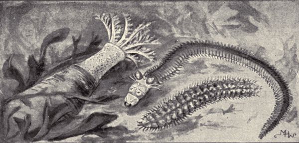
Fig. 29.—A group of marine worms: at the left a gephyrean, Dendrostomum cronjhelmi, the upper right-hand one a nereid, Nereis sp., the lower right-hand one, Polynoe brevisetosa. (From living specimens in a tide-pool on the Bay of Monterey, California.)
In certain very general characters all of the animals which compose the branch Vermes do agree. All, or nearly all, have an elongate body which is bilaterally symmetrical, that is, which could be cut by a median longitudinal cutting in two similar halves. In most of them also the body is composed of a number of successive segments or somites which are more or less alike. This kind of segmented or articulated body is also possessed by[Pg 134] the insects and crabs. Almost all of the worms have the power of locomotion; usually that of crawling. For this crawling they do not have legs composed of separate segments or joints as do the higher articulated animals, the crabs and insects, but either have fleshy unjointed legs, or various kinds of bristles or spines, or suckers, or even no external organs of locomotion at all. As regards their internal structure they have well-organized systems of organs, which show great variety in character and degree of complexity. The special sense-organs are usually of simple character and low degree of functional development. Reproduction occurs both sexually and asexually; in some species the sexes are distinct, while in others both sperm-cells and egg-cells are produced by the same individual. Asexual reproduction is by budding or by a kind of simple division or fission. The worms live either in salt or fresh water, or in moist, muddy or slimy places or as parasites in the bodies of other animals or in plants. While most worms feed on animal substance[Pg 135] either living or dead, some feed on living or decaying plant matter.
Classification.—There is great lack of agreement among zoologists in the matter of the classification of the worms. Not only are the various groups which by some are called classes held by others to be distinct branches, co-ordinate in rank with the Echinodermata, Cœlenterata, etc., but the limits of these groups are also constantly called in question. It will require a great deal better knowledge of the structure and life-history of these diverse animals before the matter of their classification is satisfactorily settled. We shall consider briefly four of the various groups (which we may consider as classes) which include worms either specially familiar to us or of special interest or importance. One or two examples of each group (the groups being selected primarily because of the examples) will be described in some detail. By this means we may get an idea of the extremely diverse character of the animals which are included in the heterogeneous branch Vermes.
Earthworms and leeches (Oligochætæ).—The various species of earthworms, an example of which has been studied are found in all parts of the world; they occur in Siberia and south to the Kerguelen Islands. They are absent from desert or arid regions, and some can live indifferently either in soil or in water. Some near allies of the earthworms are aquatic, living in fresh or brackish water, some in salt water near the shore. In size earthworms vary from 1 mm. (1/25 in.) to 2 metres (2-1/6 yds.) in length. All show the distinct segmentation of the body noticeable in the common earthworm already studied.
The leeches, some of which are familiar animals, are closely related to the earthworms, although at first glance the similarity in structure is not very noticeable.
Technical Note.—Some common water-leeches, alive or preserved in alcohol, should be examined by the class. The animals are not unfamiliar to boys who "go in swimming" in the small streams of the country. The body of a leech should be examined carefully, and drawings of it showing the external structural characters should be made.
The body of a leech is flattened dorso-ventrally, instead of being cylindrical as in the earthworm, and tapers at both ends. In the live animal the body can be greatly elongated and narrowed or much shortened and broadened. It is composed of many segments (not as many as there are cross-lines however; each segment is transversely annulated), and bears at each end on the ventral surface a sucker, the one at the posterior end being the larger. These suckers enable the leech to cling firmly to other animals. The mouth is at the front end of the body on the ventral surface and is provided with sharp jaws. Leeches live mostly on the blood of other animals which they suck from the body. The common leech "fastens itself upon its victim by means of its suckers, then cuts the skin, fastens its oral sucker over the wound and pumps away until it has completely gorged itself with blood, distending enormously its elastic body, when it loosens its hold and drops off." Its biting and sucking cause very little pain, and in olden days physicians used the leeches when they wanted to "bleed" a person. A common European species of leech much used for this purpose is known as the "medicinal leech." All leeches are hermaphroditic, that is, the sexes are not distinct, but each individual produces both sperm-cells and egg-cells. Most of the leeches lay their eggs in small packets or cocoons. This cocoon is dropped in soil on the banks of a pond or stream so that the young may have a moist but not too wet environment. The young issue from the eggs in four or five weeks, but they grow very[Pg 137] slowly and it is several years before they attain their full size. Leeches are long-lived animals, some being said to live for twenty years.
Flatworms (Platyhelminthes).—Technical Note.—Collect some live fresh-water planarians (see fig. 30), which are to be found on the muddy bottom of most fresh-water ponds, and examine them while alive in watch-glasses of water. Make drawings showing the external appearance, and as much of the internal anatomy as can be seen. The branching alimentary canal can be seen in more or less detail, and with higher power of the microscope parts of the nervous system can be seen also. Have also a tapeworm preserved in alcohol or formalin to show the very flat and many-segmented body.
The flatworms include a large number of forms which vary much in shape and habits. They are all, however, characteristically flat; in some this condition is very marked. Some are active free-living animals, as the planarians (figs. 30 and 31), while many live as parasites in the alimentary canal of other animals, as do the sheep-fluke and the tapeworms.
The fresh-water planarians (fig. 30), which live commonly in the mud of the bottom of ponds, are small, being less than half an inch long. They are very thin and rather broad, tapering from in front backwards. On the upper surface near the front they have a pair of eyes; the mouth is on the under surface a little behind the middle of the body. The alimentary canal is composed of three main branches, each with numerous small side branches. One main branch runs forward from the mouth, and the other two run backwards, one on each side of the body. There is no anal opening, and the[Pg 138] alimentary canal thus forms a system of fine branches closed at the tips, and extending all through the body. The nervous system is composed of a ganglion or brain in the front end of the body from which two main branches extend back throughout its whole length. From these main longitudinal branches arise many fine lateral branches.
Of the parasitic flatworms the tapeworms are the best known. There are numerous species of them, all of which live in the bodies of vertebrate animals. In the adult or fully developed stage the tapeworms live in the alimentary canal, holding on to its inner surface by hook-like clinging organs and being nourished by the already digested food by which they are bathed. In the young or larval stage tapeworms live in other parts of the body of the host, and usually, indeed, in other hosts not of the same species as the host of the adult worm.
The common tapeworm of man, Tænia solium (there are several other species of Tænia which infest man, but solium is the common one), may serve as an example of the group. In the adult condition its body, which is found attached to the inner wall of the intestine, is like a long narrow ribbon: it may be two or three metres long. It is attached by one end, the head, which is very small and provided with a score of fine hooks. Behind the head the thin ribbon-like body grows wider. The body is composed of many (about 850) joints called proglottids. There is no mouth or alimentary canal, the liquid food being simply taken in through the skin. Each proglottid produces both sperm-cells and egg-cells; one by one these proglottids or joints with their supply of fertilized eggs break off and pass from the alimentary canal with the excreta. If now one of these escaped proglottids or the eggs from it are eaten by a pig, the embryos issue from the eggs in the alimentary canal of the pig, bore through the walls of the canal and lodge in the muscles. Here they increase greatly in size and develop into a sort of rounded sac filled with liquid. If the flesh of the pig be eaten by a man, without its being first cooked sufficiently to kill the larval sac-like tapeworms, these young tapeworms lodge in the alimentary canal of the man and develop and grow into the long ribbon-like many-jointed adult stage.
The life-history of the other tapeworms which infest the various vertebrate animals is of this general type. There is almost always an alternation of hosts, the larval tapeworm living in a so-called intermediate host, and the adult in a final host. Of the domestic animals the dog is the most frequently attacked. At least ten different species of tapeworms have been found in the dog. The intermediate hosts of these dog tapeworms include rabbits, sheep, mice, etc. Some of the domestic fowl,[Pg 140] ducks, geese and chickens, for instance, are also infested by tapeworms, and the intermediate hosts in these cases are usually insects or small aquatic crustaceans like the familiar Cyclops.
Roundworms (Nemathelminthes).—Technical Note.—Vinegar-eels from mouldy vinegar, and hair-worms from fresh-water pools, can usually be readily obtained. They should be examined, and drawings should be made of them, showing their shape and simple external structural character. If a specimen of trichinosed pork be obtained, the encysted stage of the Trichina, described in the following account, can be shown.
The roundworms are slender, smooth, cylindrical worms pointed at both ends. They are all very long in proportion to their diameter, although their actual length may be short. Some species are of microscopic size; as the Trichina worm, which is about 1/20 in. long; while the guinea-worm, one of the worst parasites of man, may reach a length of six feet. Many of the roundworms are parasites living in the various organs of other animals. Some, however, lead an independent free life in water or in damp earth.
Familiar examples of roundworms are the so-called vinegar-eels (Anguillula) (fig. 32) to be found in weak vinegar, and other species of this same genus which live in water or moist ground or in the tissues of plants, doing much injury. The hair-worms (Gordius) or horse-hair snakes, which are believed by some people, to be horse-hairs dropped into water and turned into these animals, are also familiar examples of roundworms. They are often found abundantly in little pools after a rain, and[Pg 141] it is sometimes said that these worms come down with the rain. They have in reality come from the bodies of insects in which they pass their young or larval stages as parasites. The hair-worms all live as parasites during their larval stage, and as free independent animals in their adult stage. Some of them require two distinct hosts for the completion of their larval life, living for a while in the body of one, and later in the body of another. The first host is usually a kind of insect which is eaten by the second host. The eggs are deposited by the free adult female in slender strings twisted around the stems of water-plants. The young hair-worm on hatching sinks to the bottom of the pond, where it moves about hunting for a host in which to take up its abode.
The terrible Trichina spiralis (fig. 33), which produces the disease called trichinosis, is another roundworm of which much is heard. This is a very small worm which in its adult condition lives in the intestine of man as well as in the pig and other mammals. The young, which are borne alive, burrow through the walls of the intestine, and are either carried by the blood, or force their way, all over the body, lodging usually in muscles. Here they form for themselves little cells or cysts in which they lie. The forming of these thousands of tiny cysts injures the muscles and causes great pain, sometimes[Pg 142] death, to the host. Such infested muscle or flesh is said to be "trichinosed," and the flesh of a trichinosed human subject has been estimated to contain 100,000,000 encysted worms. To complete the development of the encysted and sexless Trichinæ the infested flesh of the host must be eaten by another animal in which the worm can live, e.g. the flesh of man by a pig or rat, and that of a pig by man. In such a case the cysts are dissolved by the digestive juices, the worms escape, develop reproductive organs and produce young, which then migrate into the muscles and induce trichinosis as before. But however badly trichinosed a piece of pork may be, thorough cooking of it will kill the encysted Trichinæ, so that it may then be eaten with impunity. Some people, however, are accustomed to eat ham, which is simply smoked pork, without cooking it, and in such cases there is always great danger of trichinosis.
Wheel animalcules (Rotifera).—Technical Note.—Live specimens of Rotifers can be found in almost any stagnant water. Examine a drop of such water with the compound microscope, and find in it a few small, active, transparent creatures, larger than the Paramœcium and other Protozoa in the water and which have the appearance shown in fig. 34. They may be known by the constant whirling, or rather vibrating, circlet or wheel of cilia at the larger or head end of the body. These wheel animalcules may be studied alive by the class. Although usually darting about, the animalcules occasionally cease to move, when, because of their transparency, almost the whole of their anatomy can be made out. Their feeding habits can also be readily observed, and the food itself watched as it moves through the body. Make drawings showing as much of the anatomy as can be worked out. Note especially the "mastax" or gizzard-like masticating apparatus in the alimentary canal.
The wheel animalcules (fig. 34) or Rotifers look little like the other worms we have studied. But they are nevertheless more nearly related to the worms than to any other branch of animals. They are all small, about 1/3 mm. long, and have a compact body. They are aquatic and feed on smaller animals and plants or on bits of organic[Pg 143] matter which they capture by means of the currents produced by the vibrating cilia of the "wheel." Small as they are they have a complex body-structure, with well-organized systems of organs. For a long time, however, they were classed by naturalists with the Protozoa on account of their size. They are found all over the world, mostly in fresh water; a few are marine. More than 700 species of them are known.
An interesting thing about the Rotifers is their remarkable power to withstand drying-up. When the water in a pond or ditch evaporates some of the Rotifers do not die, but simply dry up and lie in the dust, shrivelled and apparently lifeless, yet really in a state of suspended animation. On being put into water they will gradually fill out to their full size and shape, and finally resume all their normal activities. In this dried-up condition Rotifers may persist for a long time, several years even, although otherwise their natural life is short, being probably of not over two weeks' duration. Certain other of the lower animals have this same power of withstanding desiccation.
The great branch Arthropoda includes a host of familiar animals. It contains more species than any other branch of the animal kingdom. To it belong the crayfishes, shrimps, crabs, lobsters, water-fleas, and other animals which compose the class Crustacea; the centipeds and thousand-legged worms which compose the class Myriapoda; the true or six-footed insects forming the class Insecta, which includes nearly two-thirds of all the known species of animals; and the scorpions, mites, ticks, and spiders which constitute the class Arachnida. There is also a fifth class in the branch Arthropoda which includes a few species of animals unfamiliar to us but of great interest to zoologists.
All these varied kinds of animals have a body on the annulate or segmented type-plan, like that shown by most worms, but they differ from the worms in possessing jointed appendages, used for locomotion or food taking. There is typically or racially one pair of these jointed or segmented appendages on each segment of the body, but in all of the Arthropoda some of the segments have lost their appendages. The body is covered by a firm cuticle or outer body-wall called the exoskeleton. This exoskeleton serves not only to enclose and protect the soft parts of the body but also for the attachment of the body[Pg 145] muscles. It may be flexible as in the sutures between the body-segments in most insects, or hard and rigid as in the sclerites of the segments. The firmness is due primarily, and in the insects usually solely, to a deposit in the cuticle of chitin, a substance probably secreted by the underlying cells of the true skin, or it may be due chiefly, as in the crabs, to a calcareous deposit. In such cases it becomes a veritable armor. The internal organs of the Arthropods show a more or less obvious segmentation corresponding with the segmentation of the body-wall. The alimentary canal runs longitudinally through the center of the body from mouth to anal opening. The nervous system consists of a brain lying above the œsophagus and a double nerve-chain running backward from beneath the œsophagus, along the median line of the ventral wall, to the posterior extremity of the body. This ventral nerve-chain consists of a pair of longitudinal commissures or cords and a series of pairs of ganglia, arranged segmentally. The two ganglia of each pair are fused more or less nearly completely to form a single ganglion, and the nerve-cords are partially fused, or at least lie close together. In addition there is a smaller sympathetic system composed of a few small ganglia and certain nerves running from them to the viscera, this system being connected with the main or central nervous system. In this group the organs of special sense reach for the first time a high stage of development. Compound eyes are peculiar to Arthropoda. The heart lies above the alimentary canal. Respiration is carried on by gills in the aquatic forms, and by a remarkable system of air-tubes or tracheæ in the land forms (insects). The sexes are usually distinct, and reproduction is almost universally sexual. Most of the species lay eggs.
The Arthropods are animals of a high degree of organization. The extremely diverse life-habits of the various[Pg 146] kinds among them have led to much modification and to great specialization of structure. The course of development, too, is made very complicated by the elaborate metamorphosis undergone by many of the members of the branch.
We shall study the Arthropoda by getting acquainted with a few examples of each class and thus learning the special class characteristics.
Structure.—The structure of the crayfish has been already studied (see Chapter IV and figs. 3 and 4).
Life-history and habits.—Crayfish frequent fresh-water lakes, rivers, and springs in most parts of the United States. Many of them perish whenever the small prairie ponds dry up. But some burrow into the earth when the dry season comes. There may be noticed in meadows where water stands for certain seasons of the year many scattered holes with slight elevations of mud about them. These are mostly the burrows of crayfish. During the dry season the crayfish digs down until it reaches water, or at least a damp place, where it rests until wet weather brings it to the surface once more. One of these burrows, followed in digging a mining shaft, extended vertically down to a distance of twenty-six feet, where the crayfish was found tucked snugly away.
The eggs are carried by the female on her abdominal appendages. Previous to the laying of the eggs the female rubs off all foreign matter from the appendages, thus preparing them for the reception of the eggs. This cleaning is done with the fifth pair of legs. When the[Pg 147] eggs are ready to be laid, which is during the last of March or in April in the Central States, a sticky secretion passes out of the openings at the base of the walking legs and smears the pleopods of the abdomen. The eggs as they pass out are fertilized and caught on the pleopods, where they remain attached in clusters. After some weeks the young crayfishes issue from the eggs. In general appearance they are not very unlike the adults. They grow very rapidly at this stage. As the animal is enclosed in a hard shell, growth can only take place during the period just following the molt, for the crayfish casts its skin periodically, and it is while the new shell is forming that the animal does its growing. The crayfish when it molts casts not only the exoskeleton, but also the lining of part of the alimentary canal. After the females have hatched their young many die in the shallow pools, in which places the dried-up skeletons are noticeable during the summer months.
Most of the crustaceans live in water, a few being found in damp soil or in other moist places. Some are fresh-water animals and some marine. They vary in size from the tiny water-fleas, a millimeter long, to crabs two feet across the shell or sixteen feet from tip to tip of legs. They present great differences in form and general appearance of body, being adapted for various conditions of life. Some crustaceans live as parasites on other animals, in some cases on other crustaceans. Such parasitic species have the body much modified and are hardly to be recognized as members of the class.
Body form and structure.—In structural character and body organization the Crustaceans show, of course, the general characteristics already attributed to the Arthropoda, the branch to which they belong. The characteristics[Pg 148] which distinguish them from other Arthropods are the possession of gills for respiration (some insects have gills, but of a very different kind as will be seen later), and the bi-ramose condition of the body appendages, each appendage (excepting the antennules) consisting of a single basal segment from which arise two branches made up of one or more segments. Of the form of the crustacean body few generalizations can be made.
"There is no [other] class in the animal kingdom which presents so wide a range of organization as the Crustacea, or in which the deviations in structure from the 'type form' are so striking and so interesting from their obvious adaptation to the mode of life." For this reason no attempt will be made to discuss in general terms the form of the crustacean body, but brief accounts will be given of a few of the more familiar kinds of Crustacea which will serve to illustrate this remarkable diversity of body form.
Similarly impossible is it also to give a general account of the development of the crustaceans. The sexes are distinct in most Crustacea, and there is often great difference in form between the male and female. A certain amount of metamorphosis takes place in the development of all crustaceans; that is, the young when hatched from the egg differs, often decidedly, in appearance and structure from the parent, and in the course of its post-embryonic development undergoes more or less striking change or metamorphosis. This metamorphosis is often very marked.
Water-fleas (Cyclops).—Technical Note.—The water-fleas are common in the water of ponds or of slow streams; they may often be found in the school aquarium. They are, though small (about 1 mm. long), readily seen with the unaided eye; they are white, rather elongate, and have a rapid jerky movement. Examine specimens alive in water in a watch glass. Note the "split pear" shape, broadest near the front, tapering posteriorly, flat beneath,[Pg 149] convex above; note the forked stylets at tip of abdomen; also the two pairs of antennæ, the single median eye, the mandibles, two pairs of maxillæ, and five pairs of legs (last pair very small). There are no gills. Some of the specimens, females, may have attached to the first abdominal segment on either side an egg sac. Make drawings showing all these structural details. Watch the Cyclops capturing and feeding on Paramœcium or other small animals.
The water-fleas (Cyclops) (fig. 35) are among the smallest of the Crustacea. They are extremely abundant, having great power of multiplication. "An old Cyclops may produce forty or fifty eggs at once, and may give birth to eight or ten broods of children living five to six[Pg 150] months. As the young begin to reproduce at an early age, the rate of multiplication is astonishing. The descendants of one Cyclops may number in one year nearly 4,500,000,000, or more than three times the total population of the earth, provided that all the young reach maturity and produce the full number of offspring." The Cyclops feed on smaller aquatic animals such as Protozoa, Rotifera, etc. They in turn serve as food for fishes; and because of their immense numbers and occurrence in all except the swiftest fresh waters "they form the main food of most of our fresh-water fishes while young." Many aquatic insect larvæ feed almost exclusively on them.
Related to the Cyclops are a host of other kinds of minute Crustaceans. Among these the so-called fish-lice are specially interesting because of their parasitic habits and greatly modified and degenerate structure. There are many kinds of these parasitic crustaceans infesting fishes, whales, molluscs, and worms. "As on land almost every species of bird or mammal has its own parasitic insects, so in the water almost every species of fish or larger invertebrate has its parasitic crustaceans." Some of the most common of these parasites attach themselves to the gills of fishes. Here they cling, sucking the blood or animal juices from the host. In form of body they do not at all resemble other Crustaceans, but are strangely misshapen. They are often worm-like, or sac-like, without legs or other locomotory appendages. As with other parasites (see Chapter XXX) an inactive dependent life results in the atrophy and loss by degeneration of the body-parts concerned with locomotion and orientation.
Wood lice (Isopoda).—Technical Note.—Specimens of wood lice, pill bugs, or damp bugs, as they are variously called, may be readily found in concealed moist places, as under stones or boards on damp soil. They are often common in houses, near drains or in dark, damp places. Examine some live wood lice, and some dead specimens (killed by chloroform or in an insect-killing bottle).[Pg 151] Note the division of the body into the head, thorax, and abdomen; find the eyes, the antennæ and the mouthparts (mandibles and maxillæ are usually pressed closely together). All the locomotory appendages are adapted for walking or running, not swimming. Note the number of pairs of legs; the structure of a leg; find gills and gill-covers. Some females may be found with eggs on the under side of the thorax near the bases of the legs, the eggs being covered by thin membranous plates. Make drawings showing the general form and character of body and details of legs, gills, etc. Compare with the crayfish and Cyclops.
The wood-lice (fig. 36) are among the few Crustacea which have a wholly terrestrial life. They run about quickly and feed chiefly on decaying vegetable matter. They are night scavengers. They have the body oval and convex above, rather purplish or grayish brown, and smooth. Although they do not live in the water they breathe partly at least by means of gills (though they may breathe partly through the skin). It is therefore necessary for them to live in a damp atmosphere so that the gill membranes may be kept damp. If not kept moist they could not serve as osmotic membranes.
Lobsters, Shrimps and Crabs (Decapoda).—Technical Note.—Teachers living near the sea-shore can get specimens of live and dead lobsters, shrimps, and crabs in the markets. Schools in the interior should have a few preserved specimens for examination. These specimens should be compared with the crayfish; although differences in shape of body are evident, the character and arrangement of body parts will be found to be very similar.
The largest and most familiar Crustaceans, as the crayfishes, lobsters, shrimps, prawns and crabs, all belong to the order Decapoda, or ten-legged Crustacea. The members of this order have, including the large claws, ten walking feet; they all have eyes on movable stalks,[Pg 152] and the front portion of the body is covered by a horny fold of the body-wall called the carapace.
The lobsters are large ocean-inhabiting crustaceans which are very like the fresh-water crayfish in all structural characters. They live on the rocky or sandy ocean-bottom at shallow depths. They feed largely on decaying animal matter. They are caught in great numbers in so-called "lobster pots," a kind of wooden trap baited with refuse. "The number thus taken upon the shores of New England and Canada amounts to between twenty and thirty million annually." Live lobsters are brownish or greenish with bluish mottling; they turn red when boiled. A single female will lay several thousand eggs. The eggs are greenish and are carried about by the mother until the young hatch. The young are free-swimming larvæ, until they reach a length of half an inch.
The shrimps and prawns are mostly marine, though some species live in fresh water. They are, like the lobsters, used for food. Some of the species are gregarious in habit, occurring in great "schools" of individuals. Like the lobsters they crawl about on the sea-bottom feeding on decaying animal matter. Shrimps are very abundant near San Francisco, where extensive "shrimp fishing" is done by the Chinese.
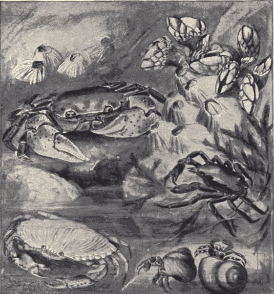
Fig. 37.—Some crabs and barnacles of the Pacific coast; the short sessile acorn barnacles in the upper left-hand corner belong to the genus Balanus; the stalked barnacles in the upper right-hand corner are of the species Pollicipes polymenus; the largest crab (upper left-hand) is Brachynotus nudus; the one in left-hand lower corner is a young rock-crab, Cancer productus; the crab in the sea-weed at the right is a kelp-crab, Epialtus productus, while the two in snail-shells in lower corner are hermit-crabs, Pagurus samuelis. (From living specimens in a tide-pool on the Bay of Monterey, California.)
The crabs (fig. 37) differ from the lobsters and crayfishes and shrimps in having the body short and broad, instead of elongate. This is due to the special widening of the carapace and the marked shortening of the abdomen. The abdomen, moreover, is permanently bent underneath the body, so that but little of it is visible from the dorsal aspect. The number of abdominal legs or appendages is reduced. When the tide is out the rocks and tide-pools of the ocean shore are alive with crabs. They "scuttle" about noisily over the rocks, withdrawing into crevices or sinking to the bottom of the pools[Pg 153] when disturbed. They move as readily backward or sidewise, "crab-fashion," as forward. They are of various colors and markings, often so patterned as to harmonize very perfectly with the general color and appearance of the rocks and sea-weeds among which they[Pg 154] live. The spider-crabs are especially strange-looking creatures with unusually long and slender legs and a comparatively small body-trunk. They include the Macrocheira of Japan, the largest of the crustaceans. Specimens of this crab are known measuring twelve to sixteen feet from tip to tip of extended legs; the carapace is only as many inches in width or length. The soft-shelled crab is a species common along our Atlantic coast. It is "soft-shelled" only at the time of molting, and has to be caught in the few days intervening between the shedding of the old hard shell and the hardening of the new body-wall. The little oyster-crabs (Pinnotheres) which live with the live oyster in the cavity enclosed by the oyster shell are well-known and interesting crabs. They are not parasites preying on the body of the oyster, but are simply messmates feeding on particles of food brought into the shell by the currents of water created by the oysters.
Among the most interesting crabs are the hermit crabs (fig. 37), familiar to all who know the seashore. There are numerous species of these crabs, all of which have the habit of carrying about with them, as a protective covering into which to withdraw, the spiral shell of some gastropod mollusc. The abdomen of the crab remains always in the cavity of the shell; the head and thorax and legs project from the opening of the shell, to be withdrawn into it when the animal is alarmed or at rest. The abdomen being always in the shell and thus protected loses the hard body-wall, and is soft, often curiously shaped and twisted to correspond to the cavity of the shell. It has on it no legs or appendages except a pair for the hindmost segment which are modified into hooks for holding fast to the interior of the shell. As the hermit crab grows it takes up its abode in larger and larger shells, sometimes killing and removing piece-meal[Pg 155] the original inhabitant. Some hermit crabs always have attached to the shell certain kinds of sea-anemones. It is believed that both crab and sea-anemone derive advantage from this arrangement. The sea-anemone, which otherwise cannot move, is carried from place to place by the crab and so may get a larger supply of food, while the crab is protected from its enemies, the predaceous fishes, by the stinging threads of the sea-anemone, and also perhaps by the concealment of the shell its presence affords. This living together by two kinds of animals to their mutual advantage is called commensalism or symbiosis (see Chapter XXX). The hermit crabs are not true crabs, but are more nearly related to the crayfishes and shrimps than to the true broad-bodied, short-tailed crabs.
Barnacles.—Technical Note.—Specimens of barnacles may be got readily from the tide rocks or from piles in a harbor. Interior schools should have, if possible, specimens preserved in alcohol or formalin for examination. The "shells" of acorn (sessile) barnacles may often be found on oyster shells (get at restaurants).
Crustaceans which at first glance are hardly recognizable as such are the stalked or sessile barnacles (fig. 37) which live fixed in great numbers on the rocks between the tide lines, or on the piles supporting wharves, or on the bottom of ships or even on the body-wall of whales and other ocean animals. In the stalked forms the stalk is a flexible stem or peduncle covered with a blackish finely-wrinkled skin bearing at its free end the greatly modified body of the barnacle. This body is enclosed in a sort of bivalved shell or carapace formed by a fold of the skin and stiffened by five calcareous plates. Within this curious shell is the compact, rather worm-like body-mass, showing little or no indication of segmentation. The legs, of which there are usually six pairs, are much modified, being long, feathery, and divided nearly to the[Pg 156] base. These feathery feet project from the opened shell when the animal is undisturbed, and waving about in the water catch small animals which serve as the barnacle's food. When disturbed the barnacle withdraws its feet and closes tightly its strong protecting shell. The acorn-barnacles have no stalk, but look like a low bluntly-pointed pyramid, this appearance being due to the converging arrangement of six calcareous plates in its body-wall.
The barnacles present several unusual conditions with regard to the internal organs. They have no heart nor any blood-vessels; most of the species are hermaphroditic; and there are other indications of a degenerate condition. This degeneration of the barnacles is due to their fixed life, the results of which are like those of a parasitic life. The young barnacles when hatched from the egg are free-swimming larvæ as with the other Crustacea. They finally attach themselves and undergo the changes, some of them of degenerative nature, which produce the body-structure of the adult. It was long a belief among many people that the barnacle produced the barnacle goose. Pictures in ancient books show the young barnacle geese issuing from the opened shell of the barnacle. The early naturalists believed barnacles, on account of the shell, to be a kind of shell-fish or mollusc, but when their development was thoroughly worked out, it became evident that they belong to the Crustacea.
Technical Note.—Locusts or grasshoppers are common and familiar insects all over the country. The genus Melanoplus includes numerous species, one or more of which are to be found in almost any locality. The common red-legged locust (M. femur-rubrum) of the East, the Rocky Mountain migratory locust (M. spretus), of the West, the large differential (M. differentialis) and two-striped (M. bivittatus) locusts of the Southwest, are especially common species. All the members of the genus have their hind wings uncolored, and the front wings marked with a longitudinal series of small dots more or less distinct, or with a longitudinal line. There is a small blunt spine or process projecting from the ventral aspect of the prothorax. If a species of Melanoplus cannot be found, any other locust may be used, although there are some slight variations in the external structure of the various species. Fresh specimens killed in a cyanide bottle (for preparing see p. 463) are preferable in the study of the external structure, but specimens preserved in alcohol will do.
External structure (fig. 38).—Note that the body of the grass-hopper is composed of successive rings or segments grouped into three regions, the head (anterior), thorax (median), and abdomen (posterior). In which region of the body are the segments most readily distinguished? Of how many segments does the head appear to be composed? The thorax is composed of three segments of which the most anterior, to which is attached the front pair of legs, differs from the succeeding two, being freely movable and bearing a large hood- or saddle-shaped piece on its dorsal aspect. To the other two thoracic segments the second and third pair of legs are[Pg 158] attached, as are also the two pairs of wings. The remaining segments of the body compose the abdomen.
Note the smooth, rather firm and horny character of the body. This is due to the fact that the skin is everywhere covered with a cuticle in which is deposited a horny substance called chitin. The cuticle is not uniformly firm over the body. At the junction of the body segments in the abdomen, in the neck and between the segments of the legs, in fact, wherever motion is desirable, the cuticle is flexible, thus making bending of the body-wall possible. Elsewhere, however, it is hard and stiff, serving not only as a protective coat or armor over the body, but also affording firm places for the attachment of muscles.
Insects (and all other Arthropods) have no[9] internal skeleton, but, in this firm cuticle, an exoskeleton.
Although the head is apparently a single segment, it[Pg 159] is really composed of six or seven body segments greatly modified and firmly fused together. Note that it bears a pair of large compound eyes and three much smaller simple eyes or ocelli.
Technical Note.—Strip off a bit of the outer covering of a compound eye, mount on a glass slide and examine under the microscope.
Note that, as in the crayfish, each compound eye is composed externally of many small hexagonal facets, the outer covering, the cornea, being simply the cuticular covering of the body, in this place transparent and divided into small facets. Besides the eyes, the head bears also several movable appendages, namely the antennæ, and the mouth-parts. Note the number, place of insertion, and segmented character of the antennæ. These antennæ are sense-organs and are used for feeling, smelling, and, in some insects, for hearing. Note that the mouth-parts consist of an upper, broad, flap-like piece, the[10]labrum; of a pair of brown, strongly chitinized, toothed jaws or mandibles; of a second pair of jaw-like structures, the maxillæ, each of which is composed of several parts; and of an under, freely-movable flap, the labium, also composed of several pieces. Each maxilla bears a slender feeler or palpus composed of five segments. The labium bears a pair of similar palpi, which are, however, only three-segmented. The mandibles and maxillæ, which are the insect jaws, move laterally, not vertically as with most animals.
Make drawings of the lateral aspect of the head; of a bit of the cornea; of the dissected out mouth-parts.
Of the three segments of the thoracic region of the body, the most anterior one is called the prothorax. It is freely movable and has a large hood or saddle-shaped[Pg 160] piece, the pronotum, on its dorsal aspect, and a blunt-pointed tubercle on the ventral aspect. The foremost pair of legs is attached to the prothorax. The next segment is the mesothorax, which is immovably fused to the next thoracic segment. What appendages does it bear? The third segment is the metathorax, which besides being fused with the mesothorax in front, is similarly fused with the foremost abdominal segment behind. What appendages does the metathorax bear?
Examine one of the fore legs and note that it is composed of a series of unequal parts or segments. The segment nearest the body is sub-globular and is called the coxa; the second segment is smaller than the coxa and is called the trochanter; the third, known as the femur, is the largest of all; the fourth, tibia, is long and slender; and the next three, the last of which is the terminal one and bears a pair of claws and between them a little pad, the pulvillus, are called the tarsal segments. Most insects have five tarsal segments. Note the great size of the hindmost or leaping legs. Determine the segments of the middle and hindmost legs. Make a drawing of a fore leg.
Examine the wings. In what ways do the front wings differ from the hind wings? The front wings are known as the wing covers or tegmina. Note how the hind wings fold up like a fan, and are covered and protected by the wing covers. Draw the wings.
The abdomen is composed of a number of segments most of which resemble each other. The first segment (immediately behind the metathorax) has its dorsal and ventral parts widely separated by the cavities for the insertion of the hindmost legs. The ventral part of this segment is dovetailed into the ventral part of the metathorax and appears to be part of it. In the dorsal part of this segment there is on each side a spot where the[Pg 161] cuticle is only a thin membrane. At these places are the auditory organs or ears of the locust. The thin membranes are the tympana. Only the various kinds of locusts and those insects closely related to them have ears of this kind. Most other insects are believed to have the sense of hearing situated in the antennæ.
The abdominal segments from second to eighth are ring-like in form and are without appendages. There is on the side of each of these segments near its front margin a tiny opening or pore called a spiracle. These spiracles are the breathing pores of the locust, which does not take in air through its mouth or any other opening in the head. There is a spiracle near each ear in the first abdominal segment, and one on each side of the mesothorax near the insertion of the middle legs.
The terminal segments of the abdomen are provided with certain processes which are different in male and female. The female has at the tip of its abdomen two pairs of strong, curved pointed pieces which compose the ovipositor, or egg-laying organ. The opening of the oviduct lies between the pieces. The male has a swollen rounded abdominal tip, with three short inconspicuous pieces on the dorsal surface.
Make a drawing of the lateral aspect of the abdomen of a female locust; also, of a male.
For a more detailed account of the external anatomy of a locust see Comstock and Kellogg's "Elements of Insect Anatomy," chap. II.
The external structure of the grasshopper should be carefully compared with that of the crayfish; pay special attention to the mouth-parts and legs.
The teacher should point out the homologies and modifications.
Life-history and habits.—The eggs of the locust are laid in the autumn in the ground in bare dry places,[Pg 162] as roadsides, closely-grazed pastures, etc. The female thrusts her strong ovipositor into the soil, and by opening and shutting it, thus boring, pushes in the abdomen for about two thirds its length. The eggs, about one hundred, are then deposited in a capsule or pod. The young locusts hatch in the following spring. When just hatched they resemble the parent locust in general appearance and structure except that they lack wings, and are of course very small. The young locusts are gregarious, congregating in warm and sunny places. They feed on green plants and travel about by walking and hopping. At night they try to find shelter under rubbish in the fields. They feed voraciously and grow rapidly, reaching maturity in about two months. During this post-embryonic development and growth they molt (shed the chitinous exoskeleton) five times. After the first molt indications of the wings appear in the shape of small backward and downward prolongations of the posterior margins of the dorsum of the mesothorax and metathorax. With each succeeding molt these wing-pads, or developing wings, are larger and more wing-like, until after the last molting they appear fully developed. With each molting, too, there is a marked increase in size of the locust, the average length of the body just before the first moult being 4.3 mm., before the second 6.8 mm., before the third 9 mm., before the fourth 14 mm., before the fifth 17 mm., and after the fifth (the full-grown stage) about 26 mm.
The molting is an interesting process, and can be readily observed. The young locust ready for its last molt crawls up some post, weed, grass stalk, or other object, and clutches this object securely with the hind feet. The head is generally downward. The locust remains motionless in this position for several hours, when the skin suddenly splits along the back from the middle[Pg 163] of the head to the base of the abdomen. By steady swelling and contracting and slight wriggling, lasting for half an hour to three-fourths of an hour, the old skin is completely shed, and the wings spread out. In an hour the wings are dry and the new chitinized exoskeleton firm enough for flying, or crawling about, and in another hour the locust begins to eat.
The red-legged locust does considerable damage to cultivated crops, but its injuries are insignificant compared with the tremendous losses occasioned by a near relative, the Rocky Mountain Locust (Melanoplus spretus). This locust has its breeding-grounds on the high plateaus of the Rocky Mountain region, but it sometimes migrates in countless numbers southeast over the plains and into the great grain-fields of the Mississippi valley. Such migrations occurred in 1866, 1867, 1874 (in this year eighteen hundred and forty two families in Kansas were reduced to destitution by the utter wiping out of their crops by the locusts) and 1876. With the settling-up of the regions in which the Rocky Mountain locust breeds, there seems to have come a change of conditions, so that no great migrations have occurred since 1876.
Technical Note.—The great water-scavenger beetles are large, black, elliptical insects common in quiet pools where they may be found swimming through the water, or crawling among the plants growing on the bottom. They are an inch and a half long and are readily distinguishable from all other water insects except the predaceous diving beetles (Dyticus). The antennæ of Hydrophilus, however, are thickened (clavate) at the tip, while those of Dyticus are thread-like for their whole length. The beetles may be readily collected with a water-net, and kept alive in glass jars or aquaria in water containing decaying vegetation.
External structure (fig. 39).—Is the body of the water-beetle
composed of segments? Can you make out three
body-regions, head, thorax and abdomen? As in the
locust the metathorax is fused with the first abdominal[Pg 164]
[Pg 165]
segment and with the mesothorax, while the prothorax
is freely movable, and is covered above by a strong shield.
The chitin armor of the whole body is specially heavy
and strong, affording a great protection to the insect.
On the flattened head note the compound eyes and the peculiarly-shaped nine-segmented antennæ. Are there any ocelli? Dissect out the mouth-parts. The beetle's mouth is fitted for biting, the mouth-parts being in general character like those of the locust, with distinct flap-like labrum, dentate mandibles, jaw-like maxillæ with long, slender, four-segmented palpi and lip-like labium with three-segmented palpi. Make drawings of the antennæ and mouth-parts.
Note the character of the thoracic segments. Examine the wings and legs. The fore wings are modified into strong horny sheaths, or elytra, which completely cover and protect the folded hind wings. The hind wings are large and membranous. How are they folded? Note the adaptation of the middle and hind legs for swimming. Determine the various segments of the legs, i.e. coxa, trochanter, femur, tibia and tarsus. Note the long longitudinal median keel on the ventral aspect of the thorax.
The abdomen articulates with the metathorax by the full width of the broad first abdominal segment. It is composed of a series of segments without appendages, of about equal length but decreasing in width from in front backwards. Of how many segments does the abdomen seem to be composed when viewed from the ventral aspect? From the dorsal?
Make a drawing of the ventral aspect of the whole body.
Technical Note.—After examining the abdomen thus far, remove it from the rest of the body, and boil it in dilute potassium hydrate (KOH) in a test-tube. This will soften and partially bleach the body wall.
Examine the softened specimen, and note that at least two additional segments are to be found retracted or telescoped into the apparently last segment. The character of these terminal abdominal segments differs in male and female individuals, and specimens of both sexes should be examined. (The males can be distinguished from the females by the peculiar pad-like expansion of the last tarsal segment of the fore legs.) Pull out the retracted segments, and note that they are unevenly chitinized, parts of their surface being simply membranous. Projecting backwards are several long-pointed processes. The female has but one retracted segment. Though the females of many insects possess more or less elaborately developed egg-laying organs, this is not the case with the beetles. Look for spiracles near the lateral margins of the dorsal surface of the abdomen. How many pairs are present?
Internal structure (fig. 40).—Technical Note.—If fresh specimens are to be had, kill by dropping into the cyanide bottle (see p. 463). Specimens preserved in a 5% solution of chloral hydrate may be used if necessary. When putting specimens into this solution a small slit should be cut through the body wall to allow the preservative to enter the body cavity. When ready to dissect a specimen cut off the elytra and wings close to the base, and carefully remove all of the dorsal wall of the abdomen and thorax and the median portion of the dorsal wall of the head. Pin out, ventral side down, under water in a dissecting-dish.
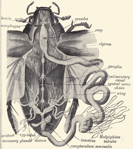
Fig. 40.—Dissection of female great water-scavenger beetle, Hydrophilus sp., the heart and tracheæ being cut away.
Note in the median dorsal line of the abdomen a pale transparent longitudinal vessel, the heart or dorsal vessel. Note on each side of it six prominent triangles or "Vs" with apex of each directed laterally, the posterior three smaller than the anterior three of each side. These triangles are formed by respiratory tubes or tracheæ. From each spiracle or breathing-pore there extends into the body a respiratory tube or trachea. These lateral tracheæ join a main longitudinal trachea on each side, from which[Pg 167] are given off branches, which in turn repeatedly subdivide, until all parts of the body are ramified by tracheæ, large and small, bringing air to all the tissues. The oxygen is taken up from this air, and carbonic-acid gas is given up to it, when it passes out of the body again through the spiracles. Thus in the insects oxygen and carbonic-acid[Pg 168] gas are not carried by the blood but by special air-tubes. The respiratory system of insects is very different from that of other animals.
Mount a bit of trachea in glycerine on a glass slide and examine under the microscope. Note the fine spiral line (looking like transverse annular striations) which is a thickening of the chitinous inner wall of the tube and which by its elasticity keeps the tracheal tubes open.
The heart, already noted, is composed of a longitudinal series of very thin-walled chambers, each with a pair of lateral openings into the body-cavity and with terminal openings into the adjacent chambers. The blood, which is colorless or greenish or yellowish, is sent forward through the successive heart chambers by regular contractions until it finally pours from the most anterior chamber freely into the body-cavity. Here it bathes the body-tissues, flowing perhaps in regular paths, giving up food to the tissues and taking up food from the alimentary canal, until it finds its way through the lateral openings into the heart chamber again. There are no arteries or veins.
Note the large mass of muscles in the metathorax. Note, by attempting to remove it, that the anterior part of the muscle mass is attached to a chitinous partition-wall between the meso- and meta-thorax. Remove this partition-wall (and one between the metathorax and abdomen) and note that certain muscles run deeply down into the body. By pulling on the bits of chitin to which the muscles are attached, the muscles (if they have not been cut) can be stretched to the length of three-quarters of an inch. When released they will contract. (This stretching and contracting takes place only in fresh specimens.) What are these large and numerous muscles of the thorax for?
Remove the thin membrane stretching over the abdomen[Pg 169] and in which the heart and tracheal "Vs" lie, and note immediately underneath it the large coiled intestine with a knot of greenish yellow threads in the centre. Carefully uncoil and pin out the intestine, cutting away the tying tracheæ, but being careful not to cut other structures. Work out the full length of the alimentary canal, noting the œsophagus, the widened crop behind it, and the long intestine. From the intestine arise several greenish yellow threads, the Malpighian tubules. These are the excretory organs of the insect. What is the total length of the alimentary canal?
The reproductive organs, consisting of a pair of glands (egg-glands or sperm-glands) with a pair of tubes which unite before reaching the body-wall and have a common external opening, may now be seen. These should be removed, thus exposing the ventral nerve-chain in the abdomen. To expose the chain in the thorax it will be necessary to pick away carefully the muscles. As in the crayfish, the central nervous system in the beetle consists of a ventral nerve-chain, a brain or supra-œsophageal ganglion and a pair of circum-œsophageal commissures connecting the brain and the foremost ganglion (infra-œsophageal) in the ventral chain. There are, in the ventral chain, four ganglia in the thorax and four in the abdomen. The large nerves running from the brain to the compound eyes and to the antennæ can be traced.
Make a drawing showing the nervous system.
Life-history and habits.—The eggs, usually about one hundred, are deposited in a silken sac or case which is spun by the female, and either floats freely or is attached to the under sides of the leaves of aquatic plants. This egg-case is not wholly filled with eggs but has a considerable air-chamber in it, causing it to float. It is oval in shape, and has a peculiar curved horn-like projection at the upper end. In sixteen or eighteen days the young[Pg 170] water-scavenger beetles hatch as elongate, wingless, active larvæ, provided with three pairs of legs and strong jaws. They remain for a short time after hatching in the egg-case, feeding on each other! After they issue from the case they feed on flies or other insects which fall into the water, and on snails. They breathe through a pair of spiracles situated at the posterior tip of the abdomen, coming to the surface and thrusting this tip up so that the spiracles are out of water. They grow rapidly, molting three times before becoming full grown. They attain a length of nearly three inches. When full grown they leave the water, crawling out on the damp shore of the pond or stream, and burrow into the soil for a few inches. Here they molt again, or pupate as it is called, changing to a non-feeding, quiescent stage called the pupal stage. The pupa is the stage in which the great changes from wingless, crawling and swimming, short-legged, long, slender-bodied larva to winged, swimming and flying, long-legged, compact, broad-bodied adult are completed. Late in the summer or in the fall the pupal skin breaks and the adult issues. It works its way to the surface of the ground, and betakes itself to the nearest water.
The water-scavenger beetle shows in its post-embryonal development a "complete metamorphosis" as contrasted with the "incomplete metamorphosis" of the locust. Wherever among insects similar changes occur, the young issuing from eggs as larvæ only remotely resembling the parent, and these active feeding larvæ changing finally into more or less quiescent, strictly non-feeding pupæ, which finally change into the active adults, a complete metamorphosis is said to exist. All the beetles, the butterflies and moths, the two-winged flies, the ants, bees and wasps, and certain other groups of insects undergo in their post-embryonic development a complete metamorphosis. The crickets, katydids, the sucking bugs, the[Pg 171] May-flies, the white ants and numerous other insects have, like the locust, an incomplete metamorphosis, that is, the young when hatched resemble in most respects, except in the absence of wings, their parents.
The adult water-scavenger beetle feeds chiefly on decaying vegetation in the water, but instances of the taking of other insects and of snails have been noted. Although an aquatic insect the beetle, like its larva, has no gills for breathing the air which is mixed with the water, but has to come to the surface occasionally to obtain air. This it does in an interesting way, which should be carefully observed by the pupils. The air is received and held by a covering of fine hairs on the ventral surface of the body, so that a considerable supply may be carried about by the beetle while underneath the surface. The beetles often leave the water by night, flying abroad to other ponds or streams. In winter the beetles hibernate, burying themselves in the banks of the ponds which they inhabit.
For a good account, with illustrations, of the water-scavenger beetle's life-history see Miall's "Natural History of Aquatic Insects," pp. 61-87.
Technical Note.—The Monarch or Milkweed butterfly is distributed all over the country. It is large, and red-brown in color, and lays its eggs on milk weeds where the greenish yellow and black-banded larvæ (caterpillars) may be found feeding. The covering of scales conceals the outlines of the various external parts, but these scales may be easily removed with dissecting needle and a small brush. In brushing the scales from the head care must be taken not to break off the mouth-parts.
External structure (fig. 41).—Note the three body-regions, head, thorax and abdomen. Is the body segmented? Note the dark color and firm character of the chitinized cuticle.
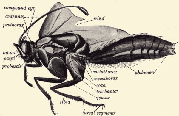
Fig. 41.—Body of the monarch butterfly, Anosia plexippus, with scales removed to show the external parts.
Note on the head the large compound eyes. Note the tumid convex clypeus which composes most of the anterior aspect of the head. Are ocelli present? Compare the antennæ with those of the locust and water-beetle. Compare also the mouth-parts and note that they differ radically from those of the locust and beetle. They are not fitted for biting, but for sucking up liquid food (the nectar of flowers). Note the absence of a movable flap-like labrum (a minute narrow stiff piece, bearing at each latera end a small group of fine brown hairs, represents the labrum), the entire absence of mandibles, and the absence of a movable flap-like labium. The labium is a fixed chitinized triangular piece forming part of the floor of the head. Note the long slender proboscis coiled up like a watch-spring. (In fresh specimens this proboscis can be uncoiled and will be found flexible. If dried or alcoholic specimens are being studied, the head of the butterfly[Pg 173] should be removed and softened in warm water before the mouth-parts are examined.) On either side of this proboscis is a peculiar pointed process which rises from the under side of the head. These processes are the labial palpi and serve to protect the sucking proboscis. The proboscis itself is composed of the two greatly modified maxillæ. Instead of being short, jaw-like and composed of several pieces as in the locust, in the butterfly each maxilla is a slender, flexible half tube applied against its mate on the opposite side in such a way as to form a perfect tube long enough to reach into the nectaries of flowers when in use and capable of being compactly coiled up at other times. Cut across the proboscis and note the canal in the centre. Try to separate the two maxillæ which compose it.
Make a drawing of the frontal aspect of the head with the eyes and appendages.
Compare the thorax with that of the beetle and that of the locust. The prothorax is a freely movable narrow ring or collar. The mesothorax and metathorax are fused to form a large convex mass, of which fully five-sixths is mesothorax and only one-sixth metathorax. Try to distinguish the boundaries of the two segments. Note the three pairs of legs; the differences in size among them, and the differences between them and the legs of the locust and water-beetle. In one of the legs determine the coxa, trochanter, femur, tibia and tarsal segments. Note the differences between the wings of the butterfly and those of the locust and beetle. Note that the wings are membranous, but are covered with many fine scales (fig. 42), as is, indeed, the whole body. Rub off some of these scales on a glass slide and examine; note shape, little stem or pedicel of insertion, and longitudinal striations. Examine under microscope a bit of wing from which some of the scales have been rubbed. How are the scales[Pg 174] attached to the wing membranes? How are the scales arranged? Note that the wing is colorless where the scales have been removed. All the colors and patterns of the wings of butterflies are produced by the scales.
Make drawings of scales; of parts of denuded wings, and of bit of wing covered with scales.
Remove all or nearly all the scales from a wing and note the arrangement of the veins (venation). Compare with venation in wings of locust.
Make drawing showing venation in the butterfly's wings.
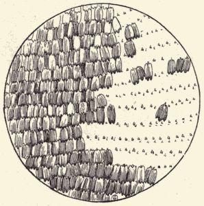
Fig. 42.—Bit of wing of Monarch butterfly,
Anosia plexippus, magnified to show the
scales; some scales removed to show the
insertion-pits and their regular arrangement.
(From specimen.)
The venation of insects' wings is much used in insect classification, and the various veins have been given names. The names of the veins in the butterfly's wings are given in fig. 43. When the veins in the wings of all the various groups of insects are studied, it is evident that the principal ones are the same in all insects, so that the costa, sub-costa, radius, media, cubitus and anal veins of the butterfly's wings can be compared with the corresponding veins in the wings of a beetle or wasp or fly. Noting the differences in the number and character of branching of these principal veins, and the number and disposition of the cross-veins which connect the longitudinal veins, the various kinds of insects can be to a large extent properly grouped or classified. A detailed account of the wing-veins of insects is given[Pg 175] in Comstock and Kellogg's "Elements of Insect Anatomy," chap. VII.
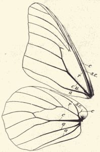
Fig. 43.—Wings of monarch butterfly,
Anosia plexippus, to show venation; c,
costal vein; sc, sub-costal vein; r, radial
vein; cu, cubital vein; a, anal veins.
In addition most insects have a vein
lying between the sub-costal and radial
veins called the median vein.
Of how many segments is the abdomen composed? The first or basal segment is depressed, while the others are more or less compressed. The spiracles are, as in the locust, situated on the lateral aspects of the abdominal segments. What segments bear spiracles? The terminal segments of the abdomen differ in the two species. In the female the dorsal part of the (apparently) last segment is longer than the ventral part and is bent down over it forming a sort of hood over a space enclosed partly by this hood, partly by a bluntly-pointed projection from the ventral surface, and partly by the lateral margins of the segment. In this chamber lies the opening from which the eggs issue. In the male there are several backward-projecting, horny, thin processes.
Make a drawing of the lateral aspect of the whole body.
Life-history and habits.—The tiny, conical, yellowish-green eggs of the monarch butterfly are deposited on the under side of the leaves of milkweeds (Asclepias) and when examined under the microscope are seen to be very beautiful little objects finely ribbed with longitudinal and transverse striæ. The eggs are laid in April and May (depending on the latitude and season) by females which[Pg 176] have hibernated in the adult condition. From the eggs the minute, cylindrical, pale-green, black-headed larvæ hatch in four or five days. As soon as hatched the larva devours the eggshell from which it has escaped and then feeds voraciously on the milkweed leaves. It grows rapidly, and in three or four days a blackish band or ring appears on each segment, and for the rest of its life it is very conspicuously colored with its black rings on a yellowish-green background. It molts three times, and in from twelve to twenty days is ready to pupate, or change to a chrysalis.
When ready to pupate the larva usually leaves the milkweed plant, and seeks some such protected place as the under side of a fence-rail or jutting rock. Here it attaches its posterior extremity by a small silken web to the rail or rock, and casting its larval skin appears as a beautiful pale-green chrysalis with ivory black and golden spots. It hangs motionless, and of course without taking food, for from a week to two weeks (according to season and temperature), when the pupal cuticle breaks and the great red-brown butterfly (fig. 165) issues.
The butterfly feeds (as is indicated by the structure of its mouth-parts) very differently from the larva; it sucks up by means of its long tubular proboscis the nectar of flowers, nor does it confine itself at all to the flowers of milkweeds. It is a fine flyer and a great traveller. Many thousands of these butterflies often make long flights or migrations together. At other times tens of thousands of these butterflies congregate in a certain limited area, clinging sometimes to the branches of a few trees in such numbers and so closely together as to give the tree a brown color. Such a "sembling" of monarch butterflies occurs every year near the Point Pinos lighthouse on the Bay of Monterey, California. The object of this assembling together is not understood. Both the larvæ and adults of the monarch butterfly are distasteful to birds,[Pg 177] by their possession of an acrid body-fluid. The species is thus protected against the most dangerous enemies of butterflies, a fact which chiefly accounts for the great abundance and wide distribution of the monarch (see p. 137). For a full account of the life-history of the monarch butterfly, see "Scudder's Life of a Butterfly."
Technical Note.—For directions for finding and identifying the larvæ of the monarch butterfly see p. 171. If larvæ (caterpillars) of Anosia cannot be found, those of any other butterfly or moth will do. Use naked, smooth kinds like cutworms, cabbage worms and the like, rather than hairy or spiny ones. Use large specimens. Kill the caterpillar with ether or in a cyanide bottle.
Structure (fig. 44).—As we have learned from the study of the life-history of the locust, water-beetle and butterfly, some insects are hatched from the egg in a condition resembling that of the parents in most structural characters. This is true of the locust. Other insects, as the beetle and butterfly, are hatched in a form and condition apparently very different from that of the parents. The external appearance of a beetle or butterfly larva differs much from that of the adult or imago of the same individual. It will be of interest to examine more particularly the structural condition of one of these larvæ and to compare it with the structure of the adult.
Is the body segmented? Is the body composed of
head, thorax and abdomen? Note the soft, flexible,
weakly-chitinized condition of the body-wall. How many
pairs of legs are there? Where are they situated? Is
there any difference in the various legs? If so, what is
the difference? Which of the legs of the larva correspond
with the legs of the butterfly? Why? The prothoracic
segment and the abdominal segments 1 to 8 each bear a
pair of spiracles (small blackish spots on the sides). Are
both compound and simple eyes present? How many eyes[Pg 178]
[Pg 179]
are there? Are there antennæ? Dissect out the mouth-parts.
How do they differ from those of the butterfly?
Are they more like the mouth-parts of the butterfly or
more like those of the locust?
With fine sharp-pointed scissors make a shallow longitudinal incision along the whole length of the dorsal wall. In a freshly-killed specimen a drop of pale greenish blood will issue as the scissors' point is first thrust through the skin. Put a droplet of this blood on a glass slide, cover with cover glass and examine with high power of the microscope. Note that the blood is a fluid containing numerous sub-circular or elliptical bodies, the blood-corpuscles. Note at least two kinds of corpuscles: most abundant a granular, circular kind, the true blood-corpuscles; and rarer, a larger, clear, usually elliptical or oval, but sometimes irregular and amœbiform kind, generally spoken of as fat-cells.
Make a drawing of the corpuscles in the field of the microscope.
After making the dorsal longitudinal incision pin out the caterpillar in the dissecting-dish with dorsal aspect uppermost. When the edges of the skin are pinned back, the organs most conspicuous in the body-cavity will be the flocculent masses of adipose tissue, the large, simple, tubular alimentary canal usually dark or greenish because of the color of its contents, and the numerous silvery tracheal tubes. In those caterpillars which spin a silken cocoon, the silk or spinning-glands are usually long and prominent. They lie on either side of the anterior part of the alimentary canal, and open by a common duct on the labium. Rising from behind the middle of the alimentary canal may be found the long, whitish, folded and twisted Malpighian tubules. By picking away the fat masses, expose the full length of the alimentary canal. Note its great size (large diameter). Is it divided into[Pg 180] distinct regions such as crop, proventriculus, stomach, intestine, etc.? How is it held in place? Trace the principal longitudinal tracheal trunks. Find, if you can, a pair of small compact bodies usually somewhat elongate, one lying on each side of the posterior part of the alimentary canal. These are the rudimentary reproductive organs.
Remove the alimentary canal by cutting it off at its posterior tip and also in the prothoracic segment. Work out now the ventral nerve-cord and ganglia, and the supra-œsophageal (brain) and infra-œsophageal ganglia and the commissures in the head.
In the body of the caterpillar we have found the same general disposition of organs as in the body of an adult insect, but several differences are nevertheless noticeable, viz., the presence of a large quantity of fatty tissue, the great size and simple character of the alimentary canal, and the undeveloped condition of the reproductive organs.
The class Insecta includes those Arthropods which have one pair of antennæ (sense appendages), three pairs of mouth-parts (oral appendages), and three pairs of legs (locomotory appendages). The insects, in further contradistinction to the crustaceans, are mostly land animals and breathe by means of tracheæ or tracheal gills. They are the most familiar of land invertebrates, and, as already mentioned, include more species than are comprised in all the other groups of animals taken together. Beetles, moths and butterflies, flies, wasps and bees, dragonflies and grasshoppers are familiar members of the class of insects, but spiders, mites, scorpions, centipeds and thousand-legged worms are not true insects and should[Pg 181] not be so miscalled. These last belong to the branch Arthropoda but to other classes than the class Insecta. While insects are found living under most diverse conditions on land, that is, on the ground, in the leaves, fruits and stems of plants, in the trunks of trees or in dead wood, in the soil, in decaying animal or plant matter, as parasites on or in other animals, and in all fresh-water ponds and streams, they do not live in ocean water. A few species live habitually on the surface of the ocean, and a few other forms are found habitually on the water-drenched rocks and seaweeds between tide lines. The varied habits of insects, their economic relations with man, the beauty and grace of many of them, and the readiness with which they may be collected, reared and studied, renders them unusually fit animals for the special attention of beginning students of zoology.
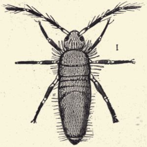
Fig. 45.—A wingless insect; the American
spring-tail. Lepidocyrtus americanus,
common in dwelling-houses. The short
line at the right indicates the natural
size. (From Marlatt.)
Body form and structure.—The segments composing the body of an insect are grouped to form three body-regions, the head, thorax, and abdomen. The head of an adult insect appears to be a single segment or body-ring, but in reality it is composed of several segments, probably seven, completely fused. The head bears the eyes, antennæ and the mouth-parts. The thorax is made up of three segments, each segment bearing a pair of legs. From the dorsal side of the hinder two thoracic segments[Pg 182] arise the two pairs of wings which are the most striking structural features of insects. Not all insects are winged, (fig. 45), and of those which are a few have only one pair of wings, but the great majority of them have two pairs of well-developed wings (fig. 46), which give them, as compared with the other animals we have studied, a new and most effective means of locomotion. The great numbers of insects and their preponderance among living animals is undoubtedly largely due to the advantage derived from their power of flight. The hindmost part of the body, the abdomen, is composed of from seven to eleven segments, only the last one or two of which are ever provided with appendages. When such posterior abdominal appendages are present they form egg-laying or stinging or clasping organs.
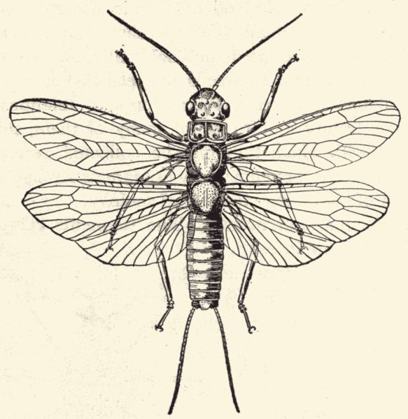
Fig. 46.—A four-winged insect; a stone fly, Perla sp., common about brooks. (From Jenkins and Kellogg.)
The body-wall is usually firm and rigid, with thinner flexible places between the segments and body-parts for the sake of motion. The body-wall is composed of a cellular skin or hypoderm, and an outer non-cellular cuticle in which is deposited a horny substance called chitin. This chitinous cuticle or exoskeleton serves as an armor or protective covering for the soft body within, and also as a point of attachment for the many muscles of the body.
Fig. 47.—Piece of trachea (air-tube) from the larva of the giant-cranefly. (Photo-micrograph by Geo. O. Mitchell.)
Insects vary a great deal in regard to shape and appearance of the body, and certain of the external organs are greatly modified in different insects to adapt them to the varied conditions under which they live. Especially interesting and important are the variations in the character of the mouth-parts and wings, the organs of food-getting and locomotion. In our consideration later of some of the more important groups of insects the modification of these parts will be specially referred to. Despite the great number of insects, however, and their varied habits of life, a strong uniformity of body-structure is noticeable, all of them holding pretty closely to the typical body-plan.
The most interesting feature of the internal anatomy of the insect body is the respiratory system. Insects breathe through tiny paired openings, called spiracles, in the sides of the abdominal (and sometimes the thoracic) segments (the number and disposition of the pairs of spiracles varying much in different insects). These spiracles are the external openings of an elaborate system of air-tubes or tracheæ (fig. 47) which ramify throughout the whole body and carry air to all the organs and tissues. The blood has apparently nothing to do with respiration as it has in the vertebrate animals, where it carries oxygen to all the body tissues.
The other systems of organs are well developed and in many respects more complex and elaborate than those of[Pg 184] any of the other invertebrates. The muscular system comprises a large number of distinct muscles, usually small and short, which are disposed so as to make very effective the various complex motions of antennæ, mouth-parts, legs, wings, and egg-laying organs. The muscles appear to be very delicate, being almost colorless when fresh, but they have a high contractile power. The alimentary canal is divided into various special regions, as pharynx, œsophagus, crop, fore stomach or gizzard, digesting stomach, and small and large intestine. From the canal just at the point of union of the digesting stomach (ventriculus) and the small intestine rise the so-called Malpighian tubules, which are excretory organs. They are long slender diverticula of the alimentary canal, and are typically six (three pairs) in number. The circulatory system is composed of a tubular vessel running longitudinally through the body in the median line just under the dorsal wall. It is composed of a series of chambers or segmental parts, which by a rhythmic contraction and expansion propel the blood anteriorly and into a short, narrow, unsegmented anterior portion of the vessel[Pg 185] which may be called the aorta. There are no other arteries or veins, the blood simply pouring out of the anterior end of the dorsal vessel into the body-cavity. It bathes the body tissues, flowing usually in regular channels without walls. It re-enters the dorsal vessel through paired lateral openings in the chambers.
Fig. 48.—The antenna of a carrion beetle, with the terminal three segments enlarged and flattened, and bearing many "smelling-pits", the antenna thus serving as an olfactory organ. (Photo-micrograph by Geo. O. Mitchell.)
The main or central nervous system consists of a large ganglion, the "brain," situated in the head above the œsophagus, which sends nerves to the antennæ and eyes, a ganglion in the head below the œsophagus connected with the brain by a short commissure on each side of the œsophagus, and sending nerves to the mouth-parts; and a ventral nerve-chain composed of a pair of longitudinal[Pg 186] commissures lying close together and running from the head to the next to the last abdominal segment, which bears a series of segmentally disposed ganglia, each ganglion being composed of two ganglia more or less nearly completely fused. There is, in addition, a lesser system called the sympathetic system, which comprises a few small ganglia and certain nerves which run from them to the viscera. The function of the nervous system of insects reaches a very high development among the so-called "intelligent insects" and certain extraordinarily complex and interesting instincts are possessed by many forms. The social or communal habits of the ants, bees, and wasps and the habits connected with the deposition of the eggs and the care of the young exhibited by the digger wasps and other insects are of extreme specialization. The organs of special sense are highly specialized, the sense of smell (fig. 48) reaching in particular a high degree of perfection. One of the compound eyes (figs. 49 and 50) may contain as many as 30,000 distinct eye-elements or ommatidia, but the sight is probably in no insect very sharp or clear. Among insects there are organs of hearing of two principal kinds. In one kind the organ for taking up the sound-waves is a group of vibratile hairs usually situated on the antennæ, as is the case with the mosquito; in the other kind, it is a stretched membrane or tympanum such as is found in the fore leg of a cricket or katydid or on the first abdominal segment of the locust (fig. 51).
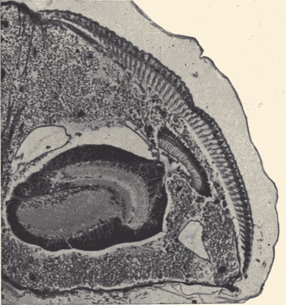
Fig. 49.—A section through the compound eye (in late pupal stage) of the blow-fly, Calliphora romitoria. In the centre is the brain, with optic lobe, and on the right-hand margin are the many ommatidia in longitudinal section. (Photo-micrograph by Geo. O. Mitchell.).
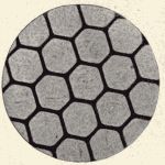
Fig. 50.—Part of cornea, showing
facets, of the compound
eye of a horse-fly (Therioplectes
sp.). (Photo-micrograph
by Geo. O. Mitchell.)
The sexes are distinct in insects, and there is often a[Pg 187] marked sex dimorphism; in numerous species the males are winged while the females are wingless, and in a few cases this condition is reversed. Where there is a difference in size between male and female, the females are usually the larger. Fertilization of the egg takes place in the body of the female and, strangely, this fertilization is effected after the eggshell has been formed. In all insect eggs there is a minute opening in one pole of the eggshell called the micropyle through which the sperm-cells enter. In a few cases the young are born alive, but such a viviparous condition is exceptional. In[Pg 188] a few species, too, young are produced parthenogenetically, that is, are produced from unfertilized eggs. And in the case of a few insect species male individuals are not known.
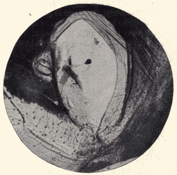
Fig. 51.—The auditory organ of a locust (Melanoplus sp.). The large clear part in centre of the figure is the thin tympanum, with the auditory vesicle (small black pear-shaped spot) and auditory ganglion (at left of vesicle and connected with it by a nerve) on its inner surface. (Photo-micrograph by Geo. O. Mitchell.)
Development and life-history.—The young insect when just hatched from the egg either resembles, except for the absence of wings, its parent in general appearance as in the case of the locust, or it may, as in the butterfly, emerge in a form very unlike the parent. In the first case the young has simply to grow, that is, to increase in size, to develop wings, and to make some other not very obvious developmental changes in order to become fully grown. But in the case of the butterfly, and similarly in the case of all other insects as the flies, beetles, bees et al., whose young hatch in a larval condition differing markedly from the adult, some radical and striking developmental changes occur before maturity is reached. Such insects are said to undergo complete metamorphosis in their development, while those insects like the locusts, the sucking-bugs, white ants, and others,[Pg 189] the just hatched young of which resemble their parents, are said to have an incomplete metamorphosis (fig. 52).
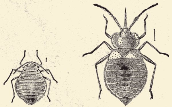
Fig. 52.—The young (at left) and adult (at right) of the bed-bug, Acanthia lectularia, a wingless insect with incomplete metamorphosis. (After Riley.)
In the case of insects with complete metamorphosis, the young hatches as an active grub or worm-like feeding larva which increases in size, casting its skin or molting several times in its growth. Finally after the last larval molt (fig. 53) called pupation the insect appears in a quiescent non-feeding stage called the pupa (fig. 54), and encased in an extra thick and firm chitinous exoskeleton. The immovable pupa is sometimes concealed underground, sometimes enclosed in a silken cocoon spun by the larva just before pupation, or is in some other way specially protected. It is in this pupal condition that the great changes from wingless, often legless, worm-like larva to[Pg 190] winged, six-legged, graceful imago of adult stage are completed, and with the molting of the chitinous pupal cuticle the metamorphosis or development of the insect is completed. As a matter of fact many of the special organs of the adult, the legs and wings, for example, begin to develop as little buds or groups of cells in the body of the larva, and when the larva is ready to pupate these imaginal wings and legs are drawn out to the external surface of the body, and may be readily recognized as they lie on the ventral surface of the pupa folded and closely pressed to the body surface. In recent years the study of the post-embryonic development of insects with complete metamorphosis has revealed some remarkable changes of the internal organs which result in a nearly complete disintegration or breaking down of[Pg 191] most of the internal organs of the larva (fig. 55) and a rebuilding of the organs of the adult from primitive beginnings.
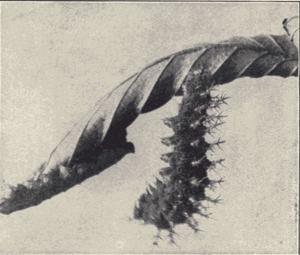
Fig. 53.—The larva of the violet tip butterfly, Polygonia interragationis, making its last molt, i.e. pupating. (Photograph from life.)
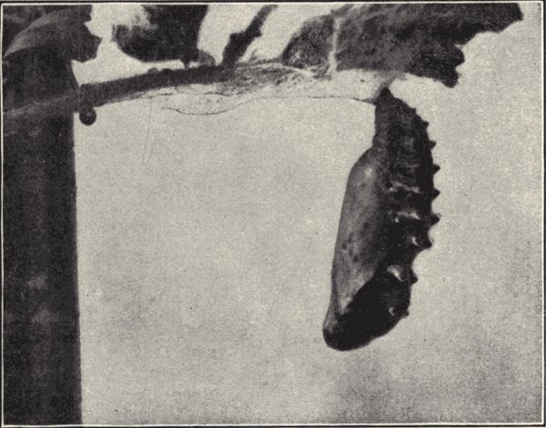
Fig. 54.—Chrysalid (pupa) of the violet tip butterfly, Polygonia interragationis. From this chrysalid issues the full fledged butterfly. (Photograph from life.)
The habits of the larvæ of insects with complete metamorphosis and of the young of some insects with incomplete metamorphosis often differ markedly from the habits of the adults, and as the habits and instincts of insects are remarkably specialized, the study of their behavior and of the structural and physiological modification which their varied habits of life have brought about is of much interest and significance. In later paragraphs this phase of insect study will be again referred to.
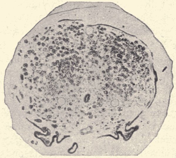
Fig. 55.—A cross-section of the body of the pupa of a honey-bee, showing the body cavity filled with disintegrated tissues, and (at the bottom) a budding pair of legs of the adult, the larva being wholly legless. (Photo-micrograph by Geo. O. Mitchell.)
Classification.—Much attention has been paid to the classification of insects and the 300,000 (approximately) known species have been variously grouped together into orders by different entomologists. A subdivision of the class Insecta into five orders was proposed by Linnæus about 1750 and was used until comparatively recently. Since then, however, numerous other arrangements have been proposed, all of them agreeing in increasing the[Pg 192] number of orders by breaking up some of the old ones into two or more new ones. The classification adopted in the text-book[11] of zoology which we have made our reference in classification is an 8-order system. The latest English[12] text-book in entomology adopts a 9-order system, while the principal American[13] text-book on this subject divides the insects into nineteen orders.
The classification depends chiefly on the character of the post-embryonic development, that is, on whether the metamorphosis is complete or incomplete, and on the structural character of the mouth-parts and wings. In the following paragraphs a few of the larger insect orders, with some special representatives of each, will be briefly considered.
The best American text-book of the classification and habits of insects is Comstocks' "Manual of Insects." For an account of the structure of the wings and mouth-parts of various insects see Comstock and Kellogg's "Elements of Insect Anatomy."
Orthoptera: the locusts, cockroaches, crickets, katydids, etc.—Technical Note.—Obtain specimens of crickets or katydids, and cockroaches, and compare the external body structure with that of the grasshopper; examine especially the wings, mouth-parts, legs, and egg-laying organs. Note that the hindmost legs of the cockroach are not fitted for leaping but for running. Note the sound-making (stridulating) organs on the bases of the fore wings of the male katydids and crickets. Note the auditory organs (tympana) in the fore tibiæ of the katydids and crickets. Crickets can be easily kept alive in breeding-cages in the laboratory and their feeding habits and much of their life-history observed. The growth of the young and the development of the wings can be noted, and will be found to be essentially similar to the conditions already found in the case of the locust.
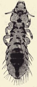
Fig. 57.—A bird louse, Nirmus
præstans, from a tern,
Sterna maxima. Most birds
are infested with small,
wingless, biting insects,
called bird-lice, which are
external parasites feeding
on the feathers of the bird
host. The bird louse
figured is about 1/12 in. long.
(Photo-micrograph by Geo.
O. Mitchell.)
The locust studied as one of the examples of the class Insecta belongs to the order Orthoptera, which also includes[Pg 193] the cockroaches, crickets (fig. 56), katydids and green grasshoppers, the walking-stick or twig insects, the praying mantis and others. The members of this order all have an incomplete metamorphosis, and in all the mouth-parts are fitted for biting and the fore wings are more or less thickened and modified to serve as covers or protecting organs for the broad, plaited, membranous hind wings, which are the true flight organs. The hind legs of locusts, grasshoppers, crickets, and katydids are very large, and enable the insects to leap; the legs of the cockroaches are fitted for swift running; the fore legs of the praying mantis are fitted for grasping other insects which serve as their food, and the legs of the walking-stick (fig. 162) are long and slender and fitted for slow walking. The shrill singing of the crickets and katydids and the loud "clacking" of the locusts are all made by stridulation, that is, by rubbing two roughened parts of the body together. The sounds of insects are not made by vocal cords in the throat. The male crickets and katydids (for only the males sing) have the veins of the fore wings modified so that when the bases of the wings are rubbed together (and when the cricket or katydid is at rest the base of one fore wing overlaps the base of the other) a part of one wing called the "scraper" rubs against a part of the other called the "file" and the shrilling is produced. The sounds of locusts are produced by the rubbing of the inside of the hind leg against the outside of the fore wing when the insect is at rest, or by striking the front[Pg 194] margin of each hind wing against the hind margin of each fore wing when the locust is flying. For hearing the Orthoptera are provided with auditory organs having the character of tympana or vibrating membranes. In the locusts these ears (fig. 51) are situated on the dorsal surface of the first abdominal segment; in the katydids and crickets they are in the tibiæ of the fore legs. The food of locusts, crickets, and katydids is vegetable, being usually green leaves; the cockroaches eat either plant or animal substances fresh or dry, while the praying mantis is predaceous, feeding on other insects which it catches in its strong grasping fore legs. The walking-stick or twig insect is an excellent example of what is called "protective resemblance" among animals. Indeed most of the Orthoptera are so colored and patterned as to be almost indistinguishable when on their usual resting- or feeding-grounds. Some of the tropical Orthoptera carry to a marvelous degree this modification for the sake of protection. (In this connection read Chapter XXXI referring to "Protective Resemblances".)
Odonata and Ephemerida: the dragon-flies and May-flies.—Technical Note.—Obtain specimens of adult and immature dragon-flies. The young dragon-flies (fig. 59) may be got by raking out some of the slime and aquatic vegetation from the bottom of a small pond. Compare the external structure of the adult dragonflies with that of the grasshopper; note the large eyes, the narrow nerve-veined wings, the biting mouth-parts, and the short antennæ.[Pg 195] Compare the young dragon-flies with the adults; note the developing wings and the peculiar modification of the lower lip into a protrusible, grasping organ which when at rest is folded like a mask over the face. Examine the interior of the posterior part of the alimentary canal to find the rectal gills. Obtain specimens of adult and young May-flies. The young may be found on the under side of stones in a "riffle" in almost any stream. They live also in ponds. They may be recognized by reference to fig. 61. Compare adult May-flies with the dragon-flies; note the weakly chitinized, delicate body-wall, and the difference in size between fore and hind wings; note the biting mouth-parts of the young and their absence or presence in vestigial condition only in the adults.
The young of both dragon-flies and May-flies may easily be kept alive in the laboratory aquarium (fruit-jars or battery-jars with pond water in), and their feeding habits, their swimming, their respiration, and much of their development observed. The young May-flies should be got from ponds, not running streams. Put one of these semi-transparent May-fly nymphs into a watch-glass of water, and examine under the microscope. The movements of the gills, heart, and alimentary canal, and much of the anatomy can be readily made out. The emergence of the adult from the nymphal skin can be seen if close watch is kept. The young dragon-flies may be seen to capture and devour their prey. They may also transform into adults, but for this it will be necessary to obtain nymphs nearly ready for transformation.
Among the most familiar and interesting insects are the dragon-flies (fig. 58), sometimes called "devil's darning-needles." They are commonly seen flying swiftly about over ponds or streams catching other flying insects. The dragon-flies are the insect-hawks; they are predaceous and very voracious, and are probably the most expert flyers of all insects. There are many species, and their bright iridescent colors and striking wing-patterns make them very beautiful. The young dragon-flies (fig. 59) are aquatic, living in streams and ponds, where they feed on the other aquatic insects in their neighborhood. They catch their prey by lying in wait until an insect comes close enough to be reached by the extraordinarily developed protrusible grasping lower lip (fig. 60). When at rest this lower lip lies folded on the face so as to conceal the great jaws. The young dragon-flies breathe[Pg 196] by means of gills which do not project from the outside of the body, as do the gills of other aquatic insects, but line the inner wall of the posterior or rectal part of the alimentary canal. Water enters the canal through the anal opening and bathes these gills, bringing oxygen to them and taking away carbonic acid gas. The aquatic immature life of the dragon-flies lasts from a few months to two years. When ready to change to adult, the young crawls out of the water and clinging to a rock or plant makes its last molt.
Other abundant and interesting pond and brook insects are the May-flies. The young May-flies (fig. 61) are aquatic, living in streams and ponds and feeding on minute organisms such as diatoms and other algæ. The immature life lasts a year, or even two or three in some species, and then the May-fly crawls out of the water upon a plant-stem or projecting rock and, molting, appears as the winged adult. The adult May-fly, having its mouth-parts atrophied (a few May-flies have functional mouth-parts), takes no food, and lives only a few hours or at most perhaps a few days. It has the shortest life (in adult stage) of all insects. The female drops her eggs into the water.
Hemiptera: the sucking-bugs.—Technical Note.—Obtain specimens of water-striders (narrow elongate-bodied insects with long spider-like legs which run quickly about on the surface of ponds or quiet pools in streams), water-boatmen (mottled grayish insects about half an inch long which swim and dive about in ponds and stream-pools), back-swimmers (which are usually in company with the water-boatmen, but which swim with back downwards and are marked with purplish-black and creamy white patches), cicadas (the dog-day locusts), and plant-lice (the "green fly" of rose-bushes and other cultivated plants). Compare the external structure of some of these Hemiptera with the other insects already examined; note especially the sucking beak, composed of the elongate tube-like labium in which lie the greatly modified flexible needle-like maxillæ and mandibles, the whole forming an equipment for piercing and sucking. Obtain immature specimens of some of these insects (distinguished by their smaller size and the wing-pads); note that the metamorphosis is incomplete, the young resembling the parents in general appearance. Both immature and adult specimens of water-boatmen (Corisa), back-swimmers (Notonecta), and water-striders[Pg 198] (Hygrotrechus) can be easily kept in the laboratory aquaria- and their swimming, breathing, and feeding habits observed. Note especially the carrying of air down beneath the water.
The Hemiptera are characterized particularly by their highly specialized sucking mouth-parts, no other of the sucking insects having the proboscis composed in the same manner. The palpi of both maxillæ and labium are wholly wanting in Hemiptera and the flexible needle-like maxillæ and mandibles are enclosed in the tubular labium. This order is a large one and includes many well-known injurious species, as the chinch-bug (Blissus leucopterus), which occurs in immense numbers in the grain-fields of the Mississippi valley, sucking the juices from the leaves of corn and wheat, the grape Phylloxera (Phylloxera vastatrix), so destructive to the vines of Europe and California, the scale insects (Coccidæ) (figs.[Pg 199] 62 and 63), the worst insect pests of oranges, the squash-bugs and cabbage-bug and a host of others. Some of the Hemiptera, for example, the lice and bed-bugs, are predaceous, sucking the blood of other animals.
The water-striders (fig. 64) catch other insects, both those that live in the water and those which fall on to its surface, and holding the prey with their seizing fore legs they pierce its body with their sharp beak and suck its blood. They lay their eggs in the spring glued fast to water-plants. The young water-striders are shorter and stouter in shape than the adults.
The water-boatmen (fig. 65) and back-swimmers swim and dive about in the water, coming more or less frequently to the surface to get a supply of air. This air they hold under the wings, or on the sides and under part of the body entangled in the fine hairs on the surface. The insects appear to have silvery spots on the body, due to the presence of this air. The "rowing" legs of the water-boatmen (Corisa) are the hindmost pair; in the back-swimmers (Notonecta) they are the middle legs.
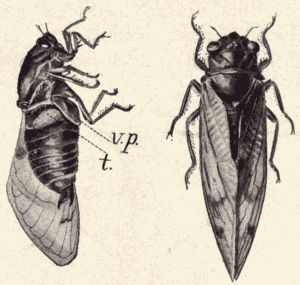
Fig. 66.—The seventeen-year cicada, Cicada
septendecim; the specimen at left
showing sound-making organ, v. p., ventral
plate; t, tympanum. (From specimen.)
The cicadas (fig. 66) are the familiar insects of summer[Pg 200] which sing so shrilly from the trees, the seventeen-year cicada (Cicada septendecim) (oftentimes called locust) being the best known of this family. Its eggs are laid in slits cut by the female in live twigs. The young, which hatch in about six weeks, do not feed on the green foliage, but fall to the ground, burrow down to the roots of the tree and there live, sucking the juices from the roots, for sixteen years and ten or eleven months. When about to become adult, the young cicada crawls up out of the ground and clinging to the tree-trunk molts for the last time, and flies to the tree-tops.
The plant-lice (Aphididæ) are small soft-bodied Hemiptera which have both winged and wingless individuals. In the early spring a wingless female hatches from an egg which, laid in the preceding fall, has passed the winter in slow development. This wingless female, called the stem-mother, lays unfertilized eggs or more often perhaps gives birth to live young, all of which are similarly wingless females which reproduce parthenogenetically. This reproduction goes on so rapidly that the plant-lice become overcrowded on the food-plant and then a generation of winged[14] individuals is produced from[Pg 201] time to time. These winged plant-lice fly away to new plants. In the autumn a generation of males and females is produced; these individuals mate and each female lays a single large egg which goes over the winter, and produces in the spring the wingless agamic stem-mother. Plant-lice produce honey-dew, a sweetish substance much liked by ants, and the lice are often visited, and sometimes specially cared for, by the ants for the sake of this honey-dew. Small as they are, plant-lice occur in such numbers as to do great damage to the plants on which they feed. The apple-aphis, cherry-aphis, pear-aphis, cabbage-aphis and others are well-known pests. The most notoriously destructive plant-louse is the grape Phylloxera, which lives on the roots and leaves of the grape-vine. Immense losses have been caused by this pest, especially in the wine-producing countries of southern Europe.
Diptera: the flies.—Technical Note.—Obtain specimens of the adult and young stages of the blowfly and the mosquito. All the young stages of the blowfly may be obtained, and its life-history studied, by exposing a piece of meat to decay in an open glass jar. The larvæ of the mosquito are the familiar wrigglers of puddles and ponds, and by collecting some of them and keeping them in a glass jar of water covered with a bit of mosquito-netting, the life-history of the mosquito is easily studied. If the eggs can be obtained from the pond so much the better; they are in little black masses floating on the surface of the water, and resemble at first glance nothing so much as a floating bit of soot. The external structure of the adult flies should be compared with that of the other insects studied, noting especially the condition of mouth-parts and wings, and the substitution of balancers for the hind wings. The mouth-parts of the mosquito are in the form of a long proboscis composed of six slender needle-like stylets lying in a tube narrowly open along its dorsal surface. The tube is the labium, and the stylets are the two maxillæ, two mandibles, and two other parts known as the epipharynx and the hypopharynx. Two additional thicker elongate segmented processes lying outside of and parallel with the tube are the maxillary palpi. The male mosquito (distinguished from the female by the more hairy or bushier antennæ) lacks[Pg 202] the pair of needle-like mandibles. The mouth-parts of the blowfly are composed almost exclusively of the thick fleshy proboscis-like labium, which is expanded at the tip to form a rasping organ.
The Diptera or true flies are readily distinguishable from other insects by their having a single pair of wings instead of two pairs, the hind wings being transformed into small knob-headed pedicels called balancers or halteres. The flies undergo complete metamorphosis, and their mouth-parts are fitted for piercing and sucking (as in the mosquito) or for rasping and lapping (as in the blowfly). Nearly 50,000 species of flies are known, more than 4,000 being known in North America alone.
The blowfly (Calliphora vomitoria) is common in houses, but can be distinguished from the house-fly by its larger size and its steel-blue abdomen. It lays its eggs on decaying meat (or other organic matter) and the white footless larvæ (maggots) hatch in about twenty-four hours. They feed voraciously and become full grown in a few days. They then change into pupæ which are brown and seed-like, being completely enclosed in a uniform chitinized case which wholly conceals the form of the developing fly. The house-fly has a life-history and immature stages like the blowfly, but its eggs are deposited on manure.
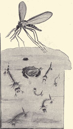
Fig. 67.—The mosquito, Culex sp.; showing eggs (on surface of water), larvæ (long and slender, in water), pupa (large headed, at surface), and adult (in air). (From living specimens.)
The mosquito (Culex sp.) (fig. 67) lays its eggs in a sooty-black little boat-shaped mass which floats lightly on the surface of the water. In a few days the larvæ, or "wrigglers," issue and swim about vigorously by bending the body. The head end of the body is much broader than the other, the thoracic segments being markedly larger than the abdominal ones. The head bears a pair of vibrating tufts of hairs, which set up currents of air that bring microscopic organic particles in the water into the wriggler's mouth. At the posterior tip of the body are two projections, one the breathing-tube (the wriggler[Pg 203] coming often to the surface to breathe), and the other the real tip of the abdomen. The wriggler, although heavier than water, can hang suspended from the surface film by the tip of its breathing-tube. It changes in a few days into the pupa, which, instead of being quiescent as with most flies, can swim about. It has a large bulbous head end and the posterior end of the body bears a pair of swimming-flaps. It takes no food. When ready to[Pg 204] change to the adult mosquito the pupa (which, unlike the wriggler, is lighter than water) floats at the surface of the water, back uppermost. The chitinous cuticle splits along the back and the delicate mosquito comes out, rests on the floating pupal skin until its wings are dry, and then flies away. Only the female mosquitoes suck blood. If they cannot find animals, mosquitoes live on the juices of plants. They are world-wide in their distribution, being serious pests even in Arctic regions, where they are often intolerably numerous and greedy. Recent investigations have shown that the germs which cause malaria in man live also in the bodies of mosquitoes, and are introduced into the blood of human beings by the biting[Pg 205] (piercing) of the mosquitoes. It is probable also that the germs of yellow fever are distributed by mosquitoes in the same way. By pouring a little kerosene on the surface of a puddle no mosquitoes will be able to escape from the water.
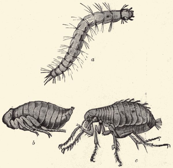
Fig. 68.—The house-flea, Pulex irritans; a, larva; b, pupa; c, adult. (The fleas are probably more nearly related to the Diptera than to any other order of insects.) (After Beneden.)
Lepidoptera: the moths and butterflies.—Technical Note.—Obtain specimens of a few moths, and compare with the butterfly already studied; note especially the character of antennæ. Obtain miscellaneous specimens of larvæ, pupæ, and cocoons of any moths or butterflies. Note the variety in colors, markings, and skin coverings of the larvæ; note the shape and markings of the pupæ. Rear from eggs, larvæ, or pupæ in breeding-cages any moths and butterflies obtainable (for directions for rearing moths and butterflies see Chapter XXXIV), keeping note of the times of molting and of the duration of the various immature stages. If the eggs of silkworms can be obtained the whole life cycle of the silkworm moth can be observed in the schoolroom. The larvæ (worms) feed on mulberry or osage orange leaves, feeding voraciously, growing rapidly and making no attempts to escape. The molting of the larvæ can be observed, the spinning of the silken cocoon, and the final emergence of the moth. The moths after emergence will not fly away, but if put on a bit of cloth will mate, and lay their eggs on it. From these eggs, which should be kept well aired and dry, larvæ will hatch in nine or ten months (if the race is an "annual").
The Lepidoptera (figs. 69-74) include all those insects familiarly known to us as moths and butterflies; they are characterized by their scale-covered wings (fig. 69) and long nectar-sucking proboscis composed of the two interlocking maxillæ. They undergo a complete metamorphosis (fig. 70) and their larvæ are the familiar caterpillars of garden and field. These larvæ have biting mouth-parts and feed on vegetation, some of them being very injurious, for example the army-worms, cut-worms, codlin moth worms, etc. The adult moths and butterflies take only liquid food, or no food at all, and are wholly harmless to vegetation. The structure and life-history of a butterfly has already been studied, and in the more general conditions[Pg 206] of structure and life-history there is much similarity in the many insects of this order. The eggs are usually laid on the food-plant of the larva; the larva feeds on the leaves of this plant, grows, molts several times, and pupates either in the ground or in a silken cocoon or simply attached to a branch or leaf. There are about six thousand species of moths and butterflies known in North America, and they are our most beautiful insects.
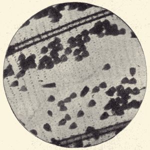
Fig. 69.—A small, partly denuded part, much magnified, of a wing of a "blue" butterfly, Lycæna sp., showing the wing, scales and the pits in the wing-membrane, in which the tiny stems of the scales are inserted. (Photo-micrograph by Geo. O. Mitchell.)
Coleoptera: the beetles.—Technical Note.—Obtain specimens of various beetles, among them some water-beetles and June-beetles with their young stages, if possible; if not, then the young stages and adults of any beetle common in the neighborhood of the school. Of the swimming and diving water-beetles there are three families, viz., the Gyrinidæ or whirligig beetles, with four eyes (each compound eye divided in two), the Hydrophilidæ, or water-scavengers with two eyes and antennæ with the terminal segments[Pg 207] thicker than the others, and the Dytiscidæ or predaceous water-beetles with two eyes and slender thread-like antennæ. Try to find Dytiscidæ, large, oval, shining black beetles; the larvæ are called water-tigers and are long, slim, active creatures with six legs and slender curving jaws (see fig. 76). The June-beetles are the heavy brown buzzing "June-bugs" and their larvæ are the common "white grubs" found underground in lawns and pastures. Have live water-tigers and predaceous water-beetles in the aquarium. Note their feeding and breathing. Compare the external structure of the beetles with that of the other insects, noting especially the biting mouth-parts, and their thickened horny fore wings serving as covers for the folded membranous hind wings.
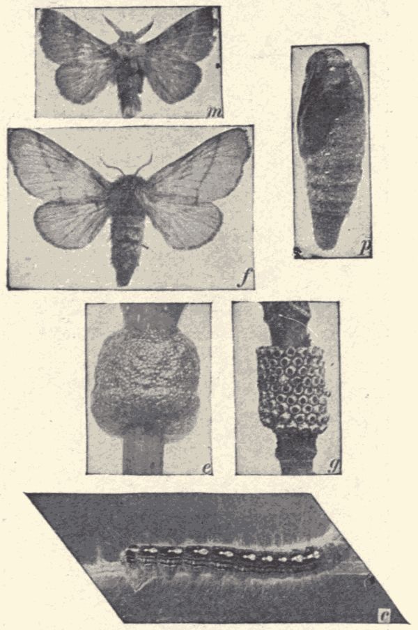
Fig. 70.—The forest tent-caterpillar moth, Clisiocampa disstria, in its various stages; m, male moth; f, female moth; p, pupa; e, eggs (in a ring) recently laid; g, eggs hatched; c, larva or caterpillar. Moths and caterpillar are natural size, eggs and pupa slightly enlarged. (Photograph by M. V. Slingerland.)
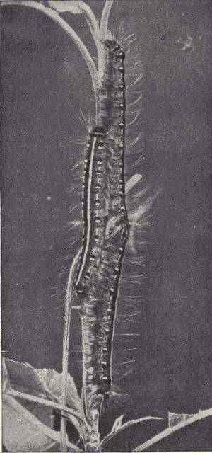
Fig. 71.—A trio of apple tent-caterpillars, Clisiocampa americana, natural size. These caterpillars make the large unsightly webs or "tents" in apple-trees, a colony of the caterpillars living in each tent. (Photograph from life by M. V. Slingerland.)
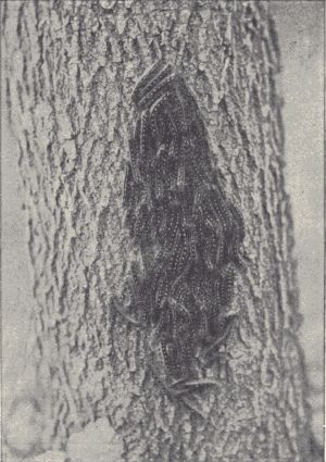
Fig. 72.—A family of forest tent-caterpillars (Clisiocampa disstria), resting during the day on the bark, about one-third natural size. (Photograph from life by M. V. Slingerland.)
The Coleoptera is the largest insect order, probably 100,000 species of beetles being known, of which 10,000 species are found in North America. They pass through a complete metamorphosis (figs. 75 and 76), the larvæ of the various kinds showing much variety in form and habit.[Pg 209] The pupæ are quiescent and are mummy-like in appearance, the legs and wings being folded and pressed to the ventral surface of the body. Among the familiar beetles are the lady-birds, which are beneficial insects feeding on plant-lice and other noxious forms; the beautifully colored tiger-beetles, predaceous in habit; the "tumblebugs" and carrion beetles, which feed on decaying organic matter; the luminous fire-flies with their phosphorescent organs on the ventral part of the abdomen; the striped Colorado potato-beetle and the cucumber-beetles and numerous other destructive leaf-eating kinds; the various weevils[Pg 210] (fig. 78) that bore into fruits, nuts and grains, and the many wood-boring beetles, destructive to fruit-trees as well as to shade- and forest-trees.
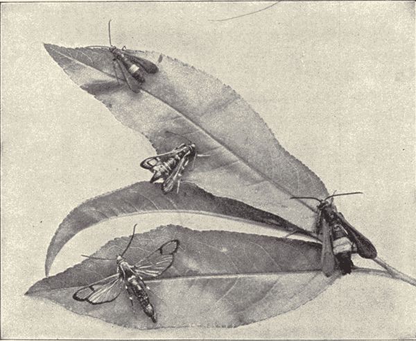
Fig. 73.—Moths of the peach-tree borer, Sanninoidea exitiosa, natural size; the upper one and the one at the right are females. (Photograph by M. V. Slingerland.)
The predaceous water-beetles (Dyticus sp.) are common
in ponds and quiet pools in streams. When at rest they
hang head downward with the tip of the abdomen just
projecting from the water. Air is taken under the tips of
the folded wing-covers (elytra) and accumulates so that
it can be breathed while the beetle swims and feeds under
water. When the air becomes impure the beetle rises to
the surface, forces it out, and accumulates a fresh supply.
The beetles are very voracious, feeding on other insects,
and even on small fish. The eggs are laid promiscuously in
the water, and the elongate spindle-form larvæ (fig. 77)[Pg 211]
[Pg 212]
called water-tigers are also predaceous. They suck the
blood from other insects through their sharp-pointed
sickle-shaped hollow
mandibles. When a larva
is fully grown it leaves
the water, burrows in
the ground, and makes a
round cell within which it
undergoes its transformations.
The pupa state
lasts about three weeks
in summer, but the larvæ
that transform in autumn
remain in the pupa state
all winter.
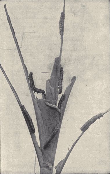
Fig. 74.—Army-worms, larvæ of the moth, Leucania unipuncta, on corn. (Photograph by M. V. Slingerland.)
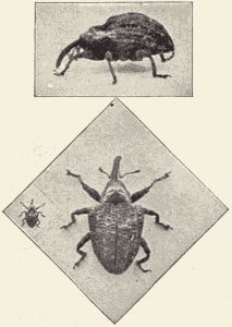
Fig. 75.—The quince-curculio (a beetle),
Conotrachelus cratægi, natural size and
enlarged. (Photograph by M. V.
Slingerland.)
The June-beetles (June-bugs) (Lachnosterna sp.) feed on the foliage of trees. Their eggs are laid among the roots of grass in little hollow balls of earth, and the fat sluggish white larvæ feed on the grass-roots. They sometimes occur in such numbers as to injure seriously lawns and meadows. The larvæ live three years (probably) before pupating. They pupate underground in an earthen cell, from which the adult beetle crawls out and flies up to the tree-tops.
Hymenoptera: the ichneumon flies, ants, wasps, and bees.—Technical Note.—Obtain specimens of wasps, both social (distinguished by having each wing folded longitudinally) and solitary (wings not folded longitudinally), and if possible of both queens (larger) and workers (smaller) of the social kinds; of ants both winged (males or females) and wingless (workers) individuals; also of honey-bees, including a queen, drones, and workers, and some brood comb containing eggs, larvæ, and pupæ. The bee[Pg 213] specimens can be got of a bee raiser. Compare the external structure of ants, bees, and wasps with that of other insects; note the pronounced division of the body into three regions (head, thorax, abdomen); note the character of the mouth-parts having mandibles fitted for biting (ants and wasps) or moulding wax (honey-bees) and having the other parts adapted for taking both solid and liquid food; note the sting (possessed by the females and workers only). Observe the behavior of bees in and about a hive; note the coming and going of workers for food. Observe bees collecting pollen at flowers; observe them drinking nectar. Examine the honey-bee in its various stages, egg, larva, pupa, adult. Note the special structure of the adult worker fitting it to perform its various special labors; the pollen-baskets on the hind legs; the wax-plates on the ventral surface of the abdomen, the wax-shears between tibia and tarsus of hind legs; the antennæ-cleaners on the fore legs; the hooks on front margin of hind wings, etc.
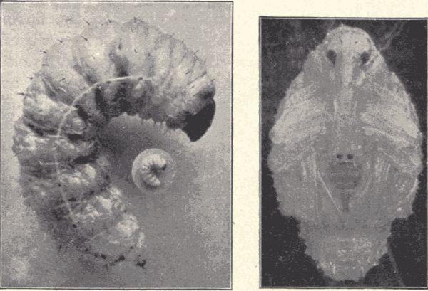
Fig. 76.—Immature stages of the quince curculio, Conotrachelus cratægi; at the left, the larva natural size and enlarged; at the right, the pupa. The beetle lays its eggs in pits on quinces, and the larva lives inside the quince as a grub; the pupa lives in the ground. (Photograph by M. V. Slingerland.)
The Hymenoptera include the familiar ants, bees, and wasps, and also a host of other four-winged, mostly small, insects, many of which are parasites in their larval stage on other insects. All Hymenoptera have a complete[Pg 214] metamorphosis, and their habits and instincts are, as a rule, very highly specialized. The parasitic Hymenoptera such as the ichneumon flies, chalcid flies, etc., are stingless but have usually a piercing ovipositor (the sting being only a modified ovipositor). The general life-history of these ichneumons is as follows: the female ichneumon fly, finding one of the caterpillars or fly or beetle larvæ which is its host, settles on it and either lays an egg or several eggs on it, or thrusting in its ovipositor, lays the eggs in the body; the young ichneumon hatching as a grub burrows into the body of its caterpillar host, feeding on the body-tissues, but not attacking the heart or nervous system, so that the host is not soon killed; the ichneumon pupates either inside the host, or crawls out and, spinning a little silken cocoon (fig. 160), pupates on the surface of the body or elsewhere.
Some of the stingless Hymenoptera are not parasites, but are gall-producers. The female with its piercing ovipositor lays an egg in the soft tissue of a leaf or stem, and after the larva hatches the gall rapidly forms. The larval insect lies in the plant-tissue, having for food the sap which comes to the rapidly growing gall. It pupates in the gall, and when adult eats its way out.
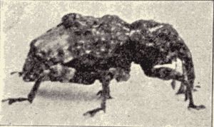
Fig. 78.—The plum curculio,
Conotrachelus nenuphar, a
beetle very injurious to plums.
(Photograph by M. V. Slingerland.)
The ants, bees, and wasps are called the stinging Hymenoptera, although the ants we have in North America have their sting so reduced as to be no longer usable. Among these Hymenoptera are the social or communal insects, viz., all the ants, the bumblebees and honey-bee, and the few social wasps, as the yellow-jacket and black hornet. There are many more species of non-social or solitary bees and wasps than social ones, and their habits and instincts are nearly as remarkable.
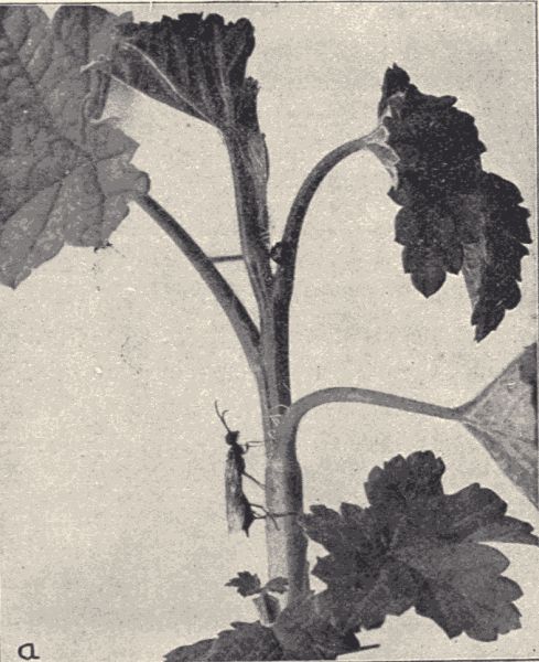
Fig. 79.—The currant-stem girdler, Janus integer, a Hymenopteron at work girdling a stem after having deposited an egg in the stem half an inch lower down. (Photograph by M. V. Slingerland.)
The solitary and digger wasps do not live in communities as the hornets do, but each female makes a nest or several nests of her own, lays eggs and provides for[Pg 216] her own young. The nest is usually a short vertical or inclined burrow in the ground, with the bottom enlarged to form a cell or chamber. In this chamber a single egg is laid, and some insects or spiders, captured and so stung by the wasps as to be paralyzed but not killed, are put in for food. The nest is then closed up by the female, and the larva hatching from the egg feeds on the enclosed helpless insects until full grown, when it pupates in the cell and the issuing adult gnaws and pushes its way out of the ground. Each species of wasp has habits peculiar to itself, making always the same kind of nest, and providing always the same kind of food. Some of these wasps make their nests in twigs of various plants, especially those with pithy centres in the stems. For interesting accounts of the habits of several digger wasps see Peckham's "The Solitary Wasps."
The solitary bees, of which there are similarly many kinds, are like the solitary wasps in general habit, only they provision the nest with a mixture of pollen and nectar got from flowers instead of with stung insects. Sometimes many individuals of a single species of solitary bee will make their nests near together and thus form a sort of community in which, however, each member has its own nest and rears its own young. In the case of certain small mining bees of the genus Halictus, a step farther toward true communal life is taken by the common building and use by several females of a single vertical tunnel or burrow from which each female makes an individual lateral tunnel, at the end of which is a brood-chamber. Perhaps half a dozen females will thus live together, each independent except for the common use of the vertical tunnel and exit.
The bumblebees (Bombus sp.) are truly communal in habit. All the eggs are laid by a queen or fertile female, which is the only member of the colony to live through[Pg 217] the winter. In the spring she finds a deserted mouse's nest or other hole in the ground, gathers a mass of pollen and lays some eggs on it. The larvæ, hatching, feed on the pollen, dig out irregular cells for themselves in it, pupate, and soon issue as workers, or infertile females. These workers gather more pollen, the queen lays more eggs, and several successive broods of workers are produced. Finally late in the summer a brood containing males (drones) and fertile females (queens) is produced, mating takes place, and then before winter all the workers and drones and some of the queens die, leaving a few fertilized queens to hibernate and establish new communities in the spring.
The yellow-jackets and hornets (Vespidæ), the so-called social wasps, have a life-history very like that of the bumblebees. The communities of the social wasps are larger and their nests are often made above ground, being composed of several combs one above the other and all enclosed in a many-layered covering sac open only by a small hole at the bottom. This kind of nest hangs from the branch of a tree and is built of wasp-paper, which is a pulp made from bits of old wood chewed by the workers. The brood-cells are provisioned with killed and chewed insects, the larvæ of both solitary and social wasps being given animal food, while the larvæ of both solitary and social bees are fed flower-pollen and honey. As in the bumblebees, all the members of the community except a few fertilized females die in the autumn, the surviving queens founding new colonies in the spring. The queen builds a miniature "hornet's nest" in the spring, lays an egg in each cell and stores the cells with chewed insects. The first brood is composed of workers, which enlarge the nest, get more food, and relieve the queen of all labor except that of egg-laying. More broods of workers follow until the fall[Pg 218] brood of males and females appears, after which the original process is repeated.
The honey-bees and ants show a highly specialized communal life, with a well-marked division of labor and an individual sacrifice of independence and personal advantage which is remarkable. Their communities are large, including thousands of individuals, and the structural differences among the males, females, and workers are readily recognizable. With the ants the workers may be of two or more sorts, a distinction into large and small workers or worker majors and worker minors being not uncommon.
A honey-bee community, living in hollow tree or hive, includes a queen or fertile female, a few hundred drones or fertile males, and ten to forty thousand workers, infertile females (fig. 80). The number of drones and workers varies, being smallest in winter. Each kind of individual has a certain particular part of the work of the whole community to do; the queen lays all the eggs, that is, is the mother of the entire community; the drones act simply as the royal consorts, fertilizing the eggs; while the workers build the comb, produce the wax from which the cells are constructed, bring in all the food consisting[Pg 219] of flower-pollen and nectar, care for the young bees, fight off intruders, and in fact perform all the many labors and industries of the community except those of reproduction. There is a certain not very well understood and perhaps not very sharply defined division of these labors among the worker individuals, the younger ones acting specially as "nurses," feeding and caring for the young bees (larvæ and pupæ), the older ones making the food-gathering expeditions. The queen lays her eggs one in each of many cells (fig. 81). These eggs hatch in three days, and the young bee appears as a white, soft, footless, helpless grub or larva that is fed at first by the nurses with a highly nutritious substance called bee-jelly which the nurses make in their stomachs and regurgitate for the larva. After two or three days of this feeding the larvæ are fed pollen and honey. After a few days a small mass of this food is put into the cell, which is then "capped" or covered with wax. The larva after using up this food-supply pupates, and lies quiescent in the pupal stage for[Pg 220] thirteen days, when the fully developed bee issues, and breaking through the wax cap of the cell is ready for the labors which are immediately assigned it. The bee with the kind of life-history just described is a worker. It has been demonstrated that the eggs which produce workers and those which produce queens do not differ, but if the workers desire to have a queen produced they tear down two or three cells around some one cell, enlarging this latter into a large vase-shaped cell. When the larva hatches from the egg in this cell it is fed for its whole larval life with bee-jelly. From the pupa into which this larva transforms issues not a worker but a new queen. The eggs which produce drones or males differ from those which produce queens and workers in being unfertilized, the queen having the power to lay either fertilized or unfertilized eggs. When a new queen appears or when several appear at once there is great excitement in the community. If several appear they fight among themselves until only one survives. It is said that a queen never uses its sting except against another queen. The old queen now leaves the hive accompanied by many of the workers. She and her followers fly away together, finally alighting on some tree-branch and massing there in a dense swarm. This is the familiar act of "swarming." Scouts leave the swarm to find a new home, to which they finally conduct the whole swarm. Thus is founded a new colony. "This swarming of the honey-bee is essential to the continued existence of the species; for in social insects it is as necessary that the colonies be multiplied as it is that there should be a reproduction of individuals. Otherwise as the colonies were destroyed the species would become extinct. With the social wasps and with the bumblebees the old queen and the young ones remain together peacefully in the nest; but at the close of the season the nest is abandoned by all as an[Pg 221] unfit place for passing the winter, and in the following spring each young queen founds a new colony. Thus there is a tendency towards a great multiplication of colonies. But with the honey-bee the habit of storing food for winter, and the nature of the habitations of these insects, render it possible for the colonies to exist indefinitely, and thus if the old and young queens remained together peacefully there would be no multiplication of colonies and the species would surely die out in time. We see, therefore, that what appears to be merely jealousy on the part of the queen honey-bee is an instinct necessary to the continuance of the species."
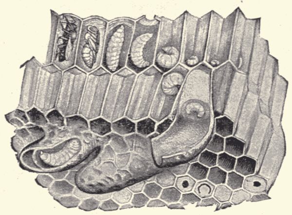
Fig. 81.—Worker brood and queen cells of honey-bee; beginning at the right end of upper row of cells and going to the left is a series of egg, young larvæ, old larvæ, pupa, and adult ready to issue; the large curving cells below are queen cells. (From Benton.)
For the special labors of gathering food, making wax, building cells, etc., the workers are provided with special structures, as the pollen-baskets on the outer surface of the widened tibia of the hind legs, the wax-shears between the tibia and first tarsal joint of the hind legs, the wax-plates on the ventral surface of the abdomen, etc. A great many interesting things connected with the[Pg 222] life and industries of a honey-bee community can be learned by the student from observation, using for a guide some book such as Cowan's "Natural History of the Honey-bee."
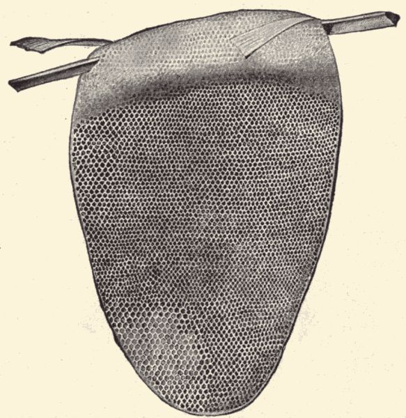
Fig. 83.—Comb of the tiny East Indian honey-bee, Apis florea, one-third natural size. (From Benton.)
The gathering of food from long distances, the details of wax-making and comb-building, of honey-making (for the nectar of flowers is made into honey by an interesting process), the storing of food, how the community protects itself from starvation when winter sets in or food is scarce by killing the useless drones and the immature bees in egg and larval stage, and many other phenomena of the life of the bee community present good opportunities for careful observation and field study. Although the community is a persistent or continuous one, the individuals do not live long, the workers hatched in the spring usually not more than two or three months, and[Pg 223] those hatched in the fall not more than six or eight months. But new ones are hatching while the old ones are dying and the community as a whole always persists. A queen may live several years, perhaps as many as five. She lays about one million eggs a year.
There are more than two thousand known species of ants (fig. 84), all of which live in communities and show a truly communal life. The ant workers are specially distinguished in structure from the males and females by being wingless, and in numerous species there are two sizes or kinds of workers known as worker majors and worker minors. The life-history and communal habits of ants are not so thoroughly known as are those of the honey-bee, but they show even more remarkable specializations. The ant nest or formicary is with most species an elaborate system of underground galleries and chambers, special rooms being used exclusively for certain special purposes, as nurse-rooms, food-storage rooms, etc. The food of ants comprises many animal and vegetable substances, but the favorite food with many species is the "honey-dew" secreted by the plant-lice (Aphididæ) and scale insects (Coccidæ). To obtain this food an ant strokes one of the aphids with its antennæ, when the fluid is excreted by the insect and drunk by the ant. In order to have a certain supply of this food some species of ants care for and defend these defenseless aphids, which have been called the "cattle" of the ants. In some cases they are even taken into the ants' nests and food provided for them. "In the Mississippi Valley a certain kind of plant-louse lives on the roots of corn. Its eggs are deposited in the ground in the autumn and hatch the following spring before the corn is planted. Now the common little brown ant (Lasius flavus) lives abundantly in the cornfields, and is especially fond of the honey secreted by the corn-root louse. So when the plant-lice[Pg 224] hatch in the spring before there are corn-roots for them to feed on, the little brown ants with great solicitude carefully place the plant-lice on the roots of a certain kind of knot-weed which grows in the field and protect them until the corn germinates. Then the ants remove the plant-lice to the roots of the corn, their favorite food-plant. In the arid lands of New Mexico and Arizona the ants rear scale insects on the roots of cactus."
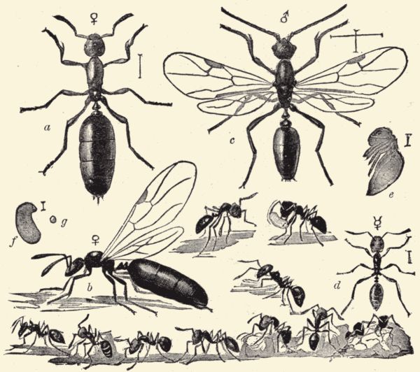
Fig. 84.—The little black ant, Monomorium minutum; a, female, b, female with wings, c, male, d, workers, e, pupa, f, larva, g, egg of worker, all enlarged. (From Marlatt.)
The ants are among the most warlike of insects. Battles between communities of different species are numerous, and the victorious community takes possession of the food-stores of the conquered. Some species of ants live wholly by war and robbery. In the case of the[Pg 225] remarkable robber-ant (Eciton), found in tropical and subtropical regions, most of the workers are soldiers, and no longer do any work but fighting. The whole community lives exclusively by pillage. Some kinds of ants go even farther than mere robbery of food-stores: they make slaves of the conquered ants. There are numerous species of these slave-making ants. They attack a nest of another species and carry into their own nest the eggs and larvæ and pupæ of the conquered community, and when these come to maturity they act as slaves of the victors, collecting food, building additions to the nest, and caring for the young of the slave-makers.
As with the honey-bee the larval ants are helpless grubs and are cared for and fed by nurses. The so-called "ants' eggs," the little white oval masses which we often see being carried in the mouths of ants in and out of an ants' nest, are not eggs, but are the pupæ which are being brought out to enjoy the warmth and light of the sun or being taken back into the nest afterward.
There are in this country numerous species of ants showing much variety of habit and offering excellent opportunities for most interesting field observations. For an account of several of the common species see Comstock's "Manual of Insects," pp. 633-643. Ants may be readily kept in the schoolroom in an artificial nest or formicary and their life-history and habits closely watched. For full directions for making and keeping a simple and inexpensive formicary see Comstock's "Insect Life," pp. 278-281. For an interesting account of some of the habits of the social insects see Lubbock's "Ants, Bees, and Wasps."
Belonging to the branch Arthropoda, with the classes Crustacea and Insecta, are three other classes, of which one, the Onychophora, is represented by a single genus Peripatus (Fig. 85), of extremely interesting animals. However, as these animals are not found in the United States we cannot study them. The other two classes are the Myriapoda, including the centipeds and millipeds or thousand-legged worms, and the Arachnida, including the scorpions, spiders, mites, and ticks. All these animals are often spoken of as insects, but though related to them they are not true insects.
Technical Note.—From under stones or logs obtain specimens of millipeds, or thousand-legged worms (large blackish, cylindrical, worm-like animals with each body-segment back of the fourth bearing two pairs of jointed legs); also specimens of centipeds or hundred-legged worms (flattened, usually brownish or pale worm-like animals with the body-segments bearing only one pair of legs each) in the same places. Examine the external structure; note number of body-rings; division into body-regions; presence of antennæ; character and number of eyes; character of mouth-parts; character and arrangement of legs. In the centipeds the first pair of legs is modified to form a pair of poison-fangs. They appear to belong to the mouth-parts. The internal anatomy will be found to be, if examined, much like that of insects and can be studied from the account of the anatomy of the water-scavenger beetle and butterfly larva. Compare the Myriapods with the Hexapods or true insects. What are the points of resemblance? what are the points of difference?
The Myriapoda are land-animals breathing by means of tracheæ like the insects. In them the body-segments[Pg 227] are nearly uniform in character with the exception of the head, which, as in the insects, bears the mouth-parts and antennæ. There is no grouping of the body-segments into regions except as the head is opposed to the rest of the body. (In a few myriapods there are indications of a division of the hind body into thorax and abdomen.) The presence of true legs on all the segments of the hinder region of the body and the lack of the three-region division of the body are the principal external structural characteristics which distinguish myriapods from insects. The internal anatomy corresponds in general character with that of insects.
The most familiar myriapods are the millipeds, and the lithobians and centipeds. The millipeds are cylindrical in shape, have two pairs of legs on most of the body-segments and are vegetable feeders, though some may feed on dead animal matter. The galley-worms (Julus) (fig. 86), large, blackish, cylindrical millipeds found under stones and logs and leaves and in loose soil, are familiar forms. They crawl slowly and when disturbed curl up and emit a malodorous fluid. They can easily be kept alive in shallow glass vessels with a layer of earth in the bottom, and their habits and life-history may thus be studied. They should be fed sliced apples, green leaves, grass, strawberries, fresh ears of corn, etc. They are not poisonous and may be handled with impunity. They lay their eggs in little spherical cells or nests in the ground. An English species of which the life-history has been studied lays from 60 to 100 eggs at a time. The eggs of this species hatch in about twelve days.
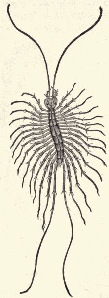
Fig. 87.—The skein centiped,
Scutigera forceps, natural
size, common in houses
and conservatories. (From
Marlatt.)
The lithobians and centipeds are flattened and have but a single pair of legs on each body-ring. They are predaceous in habit, catching and killing insects, snails, earthworms, etc. They can run rapidly, and have the first pair of legs modified into a pair of poison-claws, which are bent forward so as to lie near the mouth. The common "skein" centiped (Scutigera forceps) (fig. 87) is yellowish and has fifteen pairs of legs, long 40-segmented antennæ, and nine large and six smaller dorsal segmental[Pg 229] plates. The true centipeds (Scolopendra) (fig. 88) have twenty-one to twenty-three body-rings, each with a pair of legs, and the antennæ have seventeen to twenty joints. They live in warm regions, some growing to be very large, as long as twelve inches or more. The "bite" or wound made by the poison-claws is fatal to insects and other small animals, their prey, and painful or even dangerous to man. The popular notion that a centiped "stings" with all of its feet is fallacious. It is recorded by Humboldt that centipeds are eaten by some of the South American Indians.
Technical Note.—Obtain specimens of various spiders; the running or hunting spiders may be found on the ground, especially under stones and boards, the web-makers on their snares. Get also spiders' "cocoons" (egg-sacs). Examine the external structure of the spider; note the two body-regions; the number and character of legs; the absence of antennæ; the number and arrangement of the eyes (which are simple, not compound); the mouth-parts, especially the large mandibles; the spinnerets at the tip of the abdomen (examine a cut off spinneret under the microscope to see the spinning-tubes); note the breathing openings or spiracles on under side of abdomen. Obtain also a scorpion if possible, and some ticks and mites. Compare with the spiders and note that in the scorpion the body is plainly seen (especially in the abdomen) to be composed of segments. Note the extreme fusion of the segments and body-regions in the mites and ticks. The common red spider of hothouses and gardens is a mite; ticks may sometimes be found on dogs. Observe various kinds of spider-webs, and try to observe the process of web-making (this can be observed early in the morning or about dusk) by one of the orb-weaving garden-spiders. Live spiders can be kept in the schoolroom and their feeding habits and perhaps web-making habits observed.
The class Arachnida is composed of Arthropods whose body-segments are grouped into two regions, a cephalothorax bearing the mouth-parts, eyes, and legs, and an abdomen. The segments composing these two parts are[Pg 230] so fused that, except in the scorpions, they are usually indistinguishable. There are no antennæ, the eyes are simple, the mouth-parts fitted for biting, and there are four pairs of legs. In their internal anatomy the arachnids show in some forms a peculiar modification of the respiratory organs, the tracheæ being flat and leaf-like and massed together in a few groups rather than being tubular and ramifying through the body.
The dorsal vessel or heart usually has a few blood-vessels or arteries running from it. This class is divided into three orders, the Arthrogastra, or scorpions, the Acarina, or mites and ticks, and the Araneina, or spiders.
The scorpions (fig. 89) have the posterior six segments of the abdomen much narrower than the seven anterior segments and forming a tail which bears at its tip a poison-fang or sting. This sting is used to kill prey, insects and other small animals. The tail can be darted forwards over the body to strike prey which has been previously seized by the large pincer-like maxillary palpi. Scorpions are common in warm regions, about twenty species being[Pg 231] known in southern North America. Their sting though painful is not dangerous to man. The young are born alive and are carried about by the mother for some time after birth.
The mites (figs. 90 and 91) and ticks (fig. 92) are mostly small obscure animals, which live more or less parasitically. The common red spider of house-plants as well as the sugar- and cheese-mites, the dreaded itch-mite and the chigger are familiar examples of these degraded arachnids, and the wood-ticks, dog- and chicken-ticks are common examples of the larger bloodsucking forms. The body in both mites and ticks is very compact, the two body-regions, cephalothorax and abdomen, being closely fused.
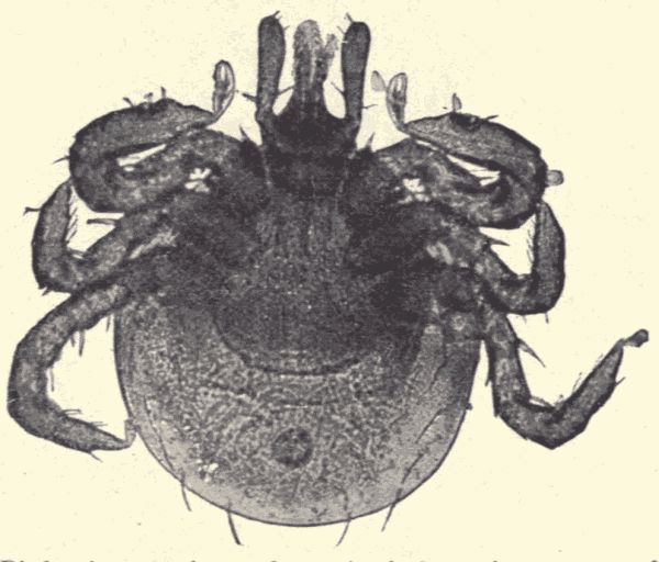
Fig. 91.—Bird mite, species undetermined, from the gnome-owl, Glaucidium gnomus. (Photo-micrograph by Geo. O. Mitchell.)
The spiders have the abdomen distinctly set off from the cephalothorax. The eyes (fig. 93) vary in number and arrangement, the mandibles are large, each being composed of two parts, a basal hair-covered part, the falx, and a terminal smooth, shining, slender, sharp-pointed part,[Pg 232] the fang, which is movably articulated with the falx (fig. 93). In the falx is a poison-sac from which poison flows through the hollow fang and out at its tip. The legs vary in relative length in different spiders, and each is made up of seven joints. The spinnerets (fig. 94), which are situated at the tip of the abdomen, are six in number (a few spiders have only four), and are like little short fingers. They have at their tips many fine little spinning-tubes from each of which a fine silken thread issues when the spider is spinning. These many fine threads fuse as they issue to form a single strong cable or sometimes a flat rather broad band. The spinnerets are movable, and by their manipulation the desired kind of line is produced. The silk comes from[Pg 233] many silk-glands in the abdomen, from each of which a fine duct runs to a spinning-tube.
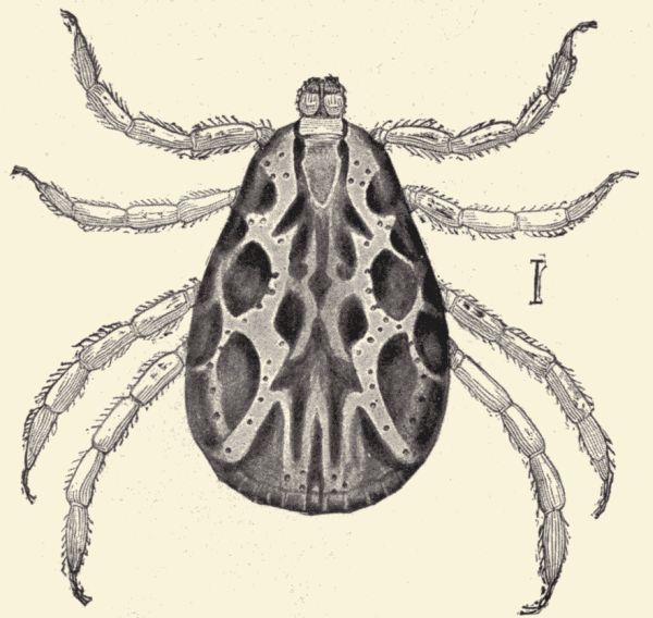
Fig. 92.—The dog or wood tick, Dermacentor americanus male, the most common tick in the Northern States. (After Osborn.)
The spiders may be divided into two groups according to their habits, viz., the wandering or hunting spiders, which do not spin webs to catch their prey, and the sedentary or web-weaving spiders, which spin snares to catch their prey. The wandering spiders can spin silk, however, and often do so to line their burrows, to make nests, or to make egg-sacs.
The hairy tarantulas and the trap-door spiders of similar appearance are among the most interesting of[Pg 234] the hunting spiders. They live in vertical burrows or tunnels in the ground which are lined with silk, and which in the case of the trap-door spider are covered with a door or lid made of silk and soil. The top of this door is always covered with soil or bits of leaves or twigs so that it is nearly indistinguishable from the surface of the ground about it. When the nest is in ground covered with moss the spider covers the door with moss. The tarantulas hunt at night and rest in the burrow in the daytime. They are very large, sometimes having an expanse of legs of 6 inches.
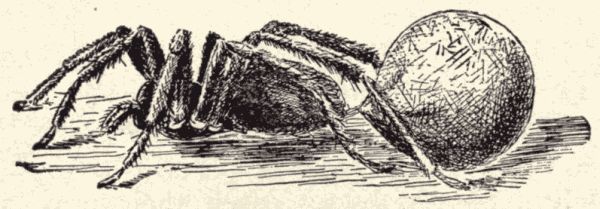
Fig. 97.—A female running spider (Lycosidæ) carrying its egg-sac about attached to its spinnerets. (From Jenkins and Kellogg.)
The common, rather large swift black spiders found[Pg 235] under stones and boards are hunting spiders, belonging to the family Lycosidæ and are called the running spiders (fig. 96). They live in burrows in the ground, coming out to stalk and chase their prey. The eggs are laid in globular egg-sacs which are often carried about, attached to the spinnerets, by the female (fig. 97). The young spiderlings after hatching, in some species, climb on to the mother's back and are carried by her for some time. Other kinds of wandering or hunting spiders are the crab-spiders (Thomisidæ) (fig. 98), which run sidewise or backward as well as forward, and the black and red, fierce-eyed stout-bodied little jumping spiders (Attidæ) (fig. 99), which leap on their prey.
The sedentary or web-weaving spiders are of various kinds. They may be grouped according to their spinning habits into cobweb weavers (Therididæ), small slim-legged spiders which make the familiar unsymmetrical cobwebs of houses and outbuildings; funnel-web weavers (Agalenidæ), larger long-legged spiders of meadow and field which spin a flat or concave horizontal web in the grass with a silken tube leading down to the ground; the curled-thread weavers (Dictynidæ), which use in addition to the usual lines peculiar broad lines made of waved or curled threads in their irregular webs made in fence-corners[Pg 236] and on plants; and finally orb-weavers (Epeiridæ) (fig. 100), the host of variously colored and patterned stout-bodied garden-spiders which spin the beautiful symmetrical circular webs familiar to all (fig. 101). If a complete uninjured orb web be examined it will be found to consist of a small central hub either open or closed, from which run radii to the outer edges of the web. Around the hub is an open or free zone, and farther out a spiral zone, so called because a line running in close spiral turns fills in the space between the radii. This is the real prey-catching part of the snare, and the silken line here is sticky, while the radii and some other parts of the web are made of silk that is not sticky. The web is supported by strong foundation-lines, attached to leaves, stems, or whatever is firm in the neighborhood of the web. The spider either rests on the web, usually in the centre, or lies concealed in a nest or tent near at hand from which a special path-line runs to the centre of the web. The building of one of these orb webs is a great[Pg 237] work, and is done with extraordinary nicety of manipulation by the use of feet and spinnerets. For account of web-making, etc., see McCook's "American Spiders and their Spinning Work."
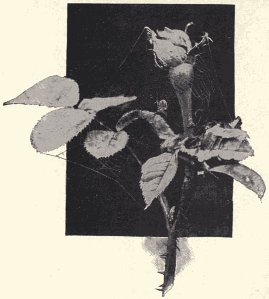
Fig. 101.—Spider and its web in a rose-bush. (Photograph from life by Cherry Kearton; from "Wild Life at Home," by permission of Cassell & Co.).
The habits and instincts of spiders in connection with the care of the young, the building of webs and nests, ballooning by means of silken lines, the active stalking and catching of prey, etc., are very interesting and offer[Pg 238] a good field for independent observation and study by the student.
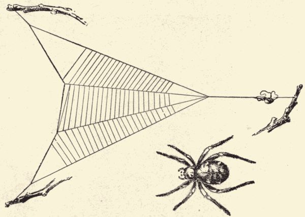
Fig. 102.—The triangle spider, Hyptiotes sp. (California), with its web; the spider rests on the taut guy-line, with a loop of the line held between its fore and hind legs; when an insect gets into the web the spider loosens the hold of its hind feet on the guy-line, thus allowing the web to spring forward sharply and further entangle the prey. (From Jenkins and Kellogg.)
Structure (fig. 103).—Technical Note.—The fresh-water or river mussel lives commonly in the streams and lakes or ponds in the United States. It frequents muddy or sandy bottoms. Specimens can often be secured with a long-handled rake from the shore or picked up in shallow streams with the hand. If possible to keep the animals alive until ready for use, some of their habits may be observed. Place them in a tub or trough with water and mud; when they have settled themselves put some powdered carmine, starch, or similar substance in the water near them, and note the water-currents.
Living mussels which have been placed in a dish with mud several inches deep and covered with water will be seen to travel in a definite direction. The end which is in front is the head end. Note the process of thrusting out and retracting the fleshy foot which extends between the two valves of the shell. Note that the two valves are held together along the upper, or dorsal, surface by a horny structure, the hinge-ligament. Note near the hinge-line a prominence (umbo) in each valve from which extends a series of concentric lines of growth. The umbo is the oldest part of the valve. Note at the lower edge of the valves a soft membrane with a fringe along its free border. This is the edge of the mantle-lobes, flaps of the body-wall which cover the body and which aid in the functions of respiration and nutrition.
Technical Note.—Specimens which are to be dissected should be killed by dropping them for a few seconds into warm water, when the muscles will relax enough so that a chip may be thrust between the valves. If specimens are to be kept for some time before dissecting they should be preserved in alcohol or 4% formalin. In a dead specimen carefully remove the left valve. This is accomplished by slipping in a thin knife-blade close to the inner edge of the left valve and carefully cutting the two large adductor muscles which bind the valves together. The dissection should be made under water.
Before the removal of the valve, as just described, notice a portion of the mantle adhering to the inner face of the valve, along a line of attachment indicated by a crease. This is the pallial line. After the left valve has been removed, the mantle being carefully separated from it, note the large conical projections from the valves, the hinge teeth, which fit into each other. Note the large muscle impression just in front of the hinge-teeth; this is the point of attachment of the anterior adductor muscle, while just behind and adjoining it is the impression of the anterior retractor muscle. Note posterior to the adductor and below the retractor a small impression which affords attachment for the protractor muscles of the foot. At the other end of the valve, note the large impression of the posterior adductor muscle with the impression of the small posterior retractor muscle just above it.
Technical Note.—Lift back the left mantle-lobe, thus exposing the body parts underneath.
Note the projecting muscular foot, the movements of which are governed by the retractor and protractor muscles attached to the impressions just mentioned. Note a pair of flattened plate-like structures composed of thin, ribbed, membranous folds. These are the gills. Note just beneath the anterior adductor muscle a small opening leading into the soft visceral mass of the body. This is the mouth. Note near the mouth two pairs of plate-like structures much smaller than the gills. These[Pg 241] are the labial palpi, and it is by their action that food-particles which have been brought in with the water are conveyed to the mouth. Note at the posterior part of each mantle-lobe a fringed portion which, together with a corresponding part on the other side, forms the inhalant siphon. The cilia of the fringes carry water and food-particles into the space enclosed by the mantle-lobes; this space is the mantle-cavity. After the food has been taken out and the water has passed through the finely striated gills it is collected in a common cavity which extends above the two sets of gills on each side. This space is called the supra-branchial cavity. This cavity is continuous posteriorly with a space between the right and left mantle-lobes, which is connected with the exterior by an opening above the inhalant siphon called the exhalant siphon. The function of the gills is partly to produce currents of water carrying the food to the mouth, and partly respiratory. The mantle is an important organ of respiration.
Make a drawing showing the organs described.
Technical Note.—Carefully cut away the mantle and gills from the left side, and also the labial palpi, being careful not to disturb the visceral mass.
Note two openings along the line where the gills and foot come together. The uppermost is the opening of the ureter giving exit to the excretion from the kidneys; the lower is the opening of the duct from the reproductive organs and is called the genital aperture. The products from both of these organs are carried out through the exhalant siphon.
Note that the mouth leads by a short tube (œsophagus or gullet) into a large cavity, the stomach, which is surrounded by a greenish mass, the digestive gland.
Technical Note.—Carefully cut the delicate covering of the dorsal portion of the visceral mass and expose a cavity.
The cavity thus exposed is the pericardium. Note within the pericardium a long tube extending through it. This is a portion of the alimentary canal, the rectum, which opens posteriorly through the anus into the supra-branchial chamber. Note a muscular sac about the rectum midway of its course through the pericardium. This is the unpaired ventricle of the heart. Attached to each side of the ventricle are thin-walled sacs, the right and left auricles, which are entered by fine blood-vessels, the efferent branchial veins, from the right and left gills. The blood brought through these blood-vessels from the gills flows into the auricles and from them into the unpaired muscular ventricle, from which it is forced anteriorly and posteriorly through two main arteries, the anterior and posterior aortas, to all parts of the body. After bathing the body-tissues the blood is collected into a median longitudinal vein beneath the pericardium called the vena cava. From the vena cava the blood passes through the kidneys and gills to be returned at last to the heart. The mantle acts as an organ for the aeration of the blood, and the blood it receives or at least part of it passes directly back to the heart without passing through the kidneys and gills.
Note the delicate membranous dark-colored sac on the floor of the pericardium, the kidneys or nephridia. These are paired structures which appear as two U-shaped tubes lying side by side. Each consists of a lower portion with thick folded walls, the kidney proper, and an upper thin-walled portion, the ureter. The kidneys open internally through a pair of reno-pericardial openings into the pericardium, while the ureters communicate with the mantle-cavity by an opening on the side of the body beneath the gills as already mentioned. The kidneys are profusely supplied with fine blood-vessels and carry off the waste matter from the blood.
Beneath the posterior adductor muscles note a small white spider-shaped body, the more or less united visceral ganglia of the nervous system. Posteriorly these ganglia give off nerves to the mantle and gills, while anteriorly there proceed two nerves, the cerebro-visceral connectives, running forward, one on either side of the foot close to the visceral mass, to the cerebro-pleural ganglia, paired ganglia lying near the mouth. A delicate commissure running over the gullet connects these ganglia.
Technical Note.—Cut away the skin and outer muscular layer from the left side of the foot.
Note the large stomach-cavity, surrounded by the digestive gland. Trace the convolutions of the alimentary canal through the foot to the anal exit. Note in the anterior portion of the foot a fused pair of ganglia similar to the visceral ganglia. These are the pedal ganglia, which are connected by a pair of delicate commissures, the cerebro-pedal connectives, with the cerebro-pleural ganglia. Note the glandular tissue which fills the cavity of the foot and surrounds the loops of the alimentary canal. This is the reproductive organ, which has its exit beneath the gills on each side of the foot. The sexes of the mussel are separate, but the reproductive organs are very similar.
Life-history and habits.—The eggs (ova) of the female pass first into the supra-branchial chamber, whence, after being fertilized, they drop into the outer pair of gill-chambers. These outer gills serve as brood-pouches, and here it is that the embryonic stages are passed through. The embryo when ready to issue has a soft body enclosed in two triangular valves. At this stage it is called a glochidium. The glochidium on being discharged through the exhalant siphon of the parent falls to the bottom, where it remains for a time, when it attaches itself to some[Pg 244] fish by the lower hook-like projections of the valves and leads a truly parasitic life for two months, after which it undergoes a metamorphosis and falls to the bottom again, there to begin an independent existence. Mussels often congregate in favorite mud or sand banks. Their food consists primarily of small organisms, both plants and animals, which are taken from the water entering the mantle-cavity. Mussels move about slowly over the muddy bottom of the stream by means of the muscular foot.
The branch Mollusca includes the fresh-water mussels, the clams, oysters, snails, and slugs, the cuttlefishes, and all that host of animals we call "shells" or shell-fish, which we know familiarly only by the shell which they make, live in, and leave at death to tell the tale of their existence. Not all the molluscs, however, form shells, that is, external shells which serve as houses. The familiar slugs do not, nor do a number of ocean forms called nudibranchs, which are somewhat like the land-slugs, only much prettier and more attractive. All the cuttlefishes and octopi are also without the hard calcareous shell. But most of the molluscs are shell-bearing animals. The shell may be bivalved, as in the mussel and clam, or univalved, that is, composed of a single piece which may be spirally twisted, as with the snail, or otherwise curiously shaped. The variety in the form, colors, and markings of the shells indicates the great diversity among molluscs. Molluscs live on land, in fresh water and in the ocean. No depths of the ocean abysses are too great for the octopi, no coast but has its many shells, hardly a pond or stream is without its mussels and pond-snails, and in all regions the land-snails and slugs abound.
Body form and structure.—The molluscs are not to be mistaken for any other of the lower animals; they have a structure peculiarly their own. In them the body is not articulated or segmented as with the worms and arthropods, nor radiate as in the echinoderms, nor plant-like as with the sponges and polyps. (Where the typical molluscan body is well developed it is composed of four principal parts: a head, with the mouth, feelers, eyes, and other organs of special sense; a trunk containing the internal organs; a foot which is a thick muscular mass not at all foot- or leg-like in shape, but which is the organ of locomotion by means of which the mollusc crawls; and a mantle which is a fold of the skin enclosing most of the body and which produces the shell. Such a typical molluscan body is possessed by most of the snails. But in most of the other molluscs one or more of these four body-regions are so fused with some other region as to be indistinguishable. In the mussels and clams the head is not at all set off from the rest of the body, the cuttlefishes and octopi have no foot, the slugs have no shell. In the case of some of the molluscs without external shell there are inside the body the rudiments or vestiges of a shell.
With regard to the internal organs we note the constant presence of three pairs of ganglia, viz., the brain, lying above the pharynx, which sends nerves to the feelers, eyes, and auditory organs; the pedal ganglion, which sends nerves to the foot, and the visceral ganglion, which sends nerves to the viscera. This is a condition of the nervous system characteristic of all molluscs. The heart is a well-developed pulsating sac in the upper part of the body composed of either two or three chambers, and there is a well-defined closed system of arteries and veins, specially complete in the cuttlefishes and octopi. This highly developed condition of the circulatory system also distinguishes the molluscs from the other invertebrates.
Development.—Reproduction among the molluscs is always sexual. Multiplication by budding or by the parthenogenetic production of eggs is not known to occur. The eggs are usually laid in a mass held together by a gelatinous substance. In most species the young mollusc on hatching from the egg does not resemble its parent, but is a free-swimming larva called a veliger. It is provided with cilia for organs of locomotion. It must undergo a radical change in order to reach the adult stage. Thus metamorphosis occurs in this branch as well as among the Arthropods and Echinoderms. In the development of some molluscs, however, there is little or no metamorphosis, the young being hatched in a condition much resembling, except in size, the parent.
Some of the special characteristics of structure, life-history, and habits of the molluscs will be noted in our consideration of the various kinds.
Classification.—The branch Mollusca is divided into five classes, three of which include the more familiar kinds. These three classes are the Pelecypoda, including the mussels, cockles, clams, scallops, oysters, etc., molluscs with a shell composed of two pieces, one on each side of the body and hinged together; the Gastropoda, including the snails, slugs, periwinkles, whelks, and a host of other univalved shell-fish, that is, molluscs which have a shell composed of a single piece; and the Cephalopoda, including the squids, cuttlefishes, octopi, and the pearly nautilus.
Clams, scallops, and oysters (Pelecypoda).—Technical Note.—Shells of scallops, oysters, and sea-mussels should be had for examination; also specimens of Teredo or Pholas in alcohol or formalin, and pieces of pile bored by Teredo. Make drawings of various bivalve shells, and of Teredo.
The fresh-water mussel which we have studied is an example of the bivalve molluscs. The members of this[Pg 247] class show a range in size from the little fresh-water Cyclas about 1 cm. long to the giant clam of the Indian and Pacific islands "which is sometimes 60 cm. (2 feet) in length and 500 pounds in weight." They show also some variety in the form and appearance of the shell, but not anything like the degree of variety shown by the shells of the Gastropods.
The edible clams are of several different species. The hard-shell clam (Venus mercenaria), or "quohog" as it is often called, is found along the Atlantic coast from Texas to Cape Cod. It is "common on sandy shores, living chiefly on the sandy and muddy plots, just beyond low-water mark.... It also inhabits estuaries, where it most abounds. It burrows a short distance below the surface, but is frequently found crawling at the surface with the shell partly exposed." The shells of this edible clam are white. The soft-shell clam (Mya arenaria), "the clam par excellence, which figures so largely in the celebrated New England clam-bake, is found in all the northern seas of the world.... All along the coasts of the eastern States, every sandy shore, every mud flat, is full of them, and from every village and hamlet the clam-digger goes forth at low tide to dig these esculent bivalves. The clams live in deep burrows in the firm mud or sand, the shells sometimes being a foot or fifteen inches beneath the surface. When the flats are covered with water his clamship extends his long siphons up through the burrow to the surface of the sand, and through one of these tubes the water and its myriads of animalcules is drawn down into the shell, furnishing the gills with oxygen and the mouth with food, and then the water charged with carbonic acid and fæcal refuse is forced out of the other siphon. When the tide ebbs the siphons are closed and partly withdrawn." Ocean clams and mussels have furnished food for man for ages, and[Pg 248] along coasts are found here and there great mounds made of heaps of clam-shells which have become covered over with soil and vegetation. Such mounds are the old feasting-places of the early coast inhabitants, and the archæologist often finds in these "kitchen-middens," as they are called, various relics of the early natives of the continent.
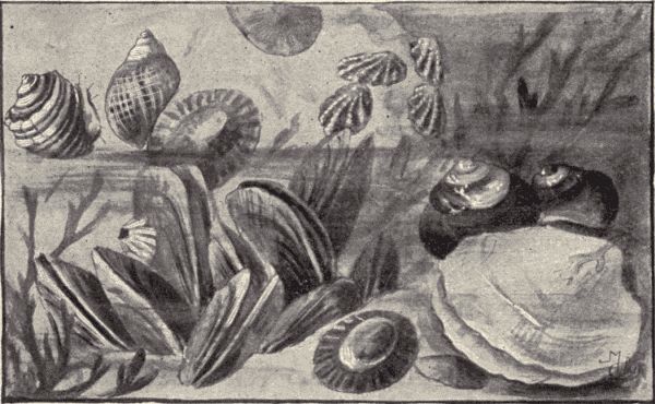
Fig. 104.—A group of marine Pacific Coast molluscs; in upper left-hand corner, Purpura saxicola; next to the right, Littorina scutulata; farthest to right, limpets, Acmara spectrum; left-hand lower corner, Mytilus californianus; in right-hand lower corner the black shells just above the large clam-shell, Chlorostomum funebrale. (From living specimens in a tide pool in the Bay of Monterey, California.)
Even more widely known that the clams are the oysters (Ostrea virginiana), also members of this class of molluscs. The oyster is carefully cultivated by man in many countries. It has its two shells or two shell-halves dissimilar, one valve being hollowed out to receive the body, while the other is nearly flat. The oyster is attached to the sea-bottom by the outside of the hollowed-out valve. When first hatched the young oyster swims freely by means of its cilia; after a few days it attaches itself to[Pg 249] some solid object and grows truly oyster-like. Much care has to be taken in cultivating oysters to furnish proper conditions for growth and development. The young oysters when first attached are called "spat"; when a little older this "spat," now called "seed," may be transplanted to new beds, which are stocked in this way. In fact some beds have constantly to be thus restocked, the young oysters produced on them not finding good places to attach themselves, and so swimming away. Sometimes pieces of slate, pottery, etc., are strewed about the oyster-beds to serve as "collectors," that is, as places for the attachment of the young oysters. The extent of the acreage of the American oyster-beds is larger than that of any other country. "The Baltimore oyster-beds on the Chesapeake River and its tributaries cover 3,000 acres, and produce an annual crop of 25,000,000 bushels."
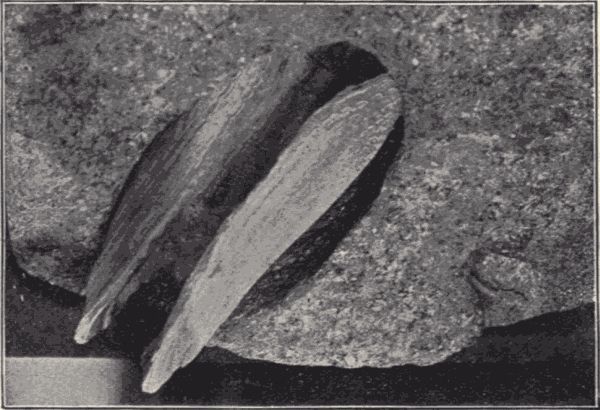
Fig. 105.—Dactylus sp., a mollusc, excavating granite. (Photograph by C. H. Snow; permission of Amer. Soc. Civil Engineers.)
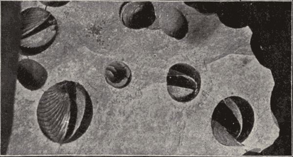
Fig. 106.—Pholas sp., a mollusc, burrowing in sandstone. (Photograph by C. H. Snow; permission of Amer. Soc. Civil Engineers.)
The "pearl-oyster" is not a true oyster, that is, not a member of the family to which the edible oysters belong,[Pg 250] but it is a member of the same class, that is, it is a bivalve mollusc. Pearls are obtained from a number of different "pearl-oysters," but the finest pearls and mother-of-pearl come from the tropical species Meleagrina margaritifera. This pearl-oyster "has an extensive distribution, being found in Madagascar, the Persian Gulf, Ceylon, Australia, Philippine Islands, South Sea Islands, Panama, West Indies, etc." Mother-of-pearl is simply the inner lining of the shell, which is composed of numerous thin layers of carbonate of lime so arranged that the edges of the successive layers produce many fine striæ very close together. The beautiful iridescence of this inner shell-lining is caused by the complicated diffraction and reflection (interference effects) of the light by the fine striæ and the translucent superposed thin plates of shell material. Pearls are simply isolated deposits of shell material usually around some particle of foreign substance which has found lodging in the mantle-cavity. Sometimes small objects are purposely introduced into the shell in order to stimulate the formation of pearls. The pearl-fishers go out in boats and dive to the bottom, filling baskets with pearl-oysters. These are piled up in a bin and left to die and[Pg 251] decompose. "When the flesh is pretty thoroughly disintegrated, it is washed away with water, great care being taken that none of the pearls loose in the flesh are lost. When the washing is concluded the shells themselves are examined for pearls which may be attached to the interior of the valves." The principal pearl-fishery is that on the coast of Ceylon; pearl-fishing has been carried on here for over 2000 years.
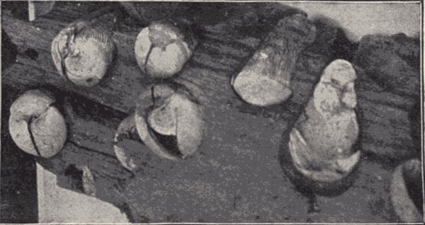
Fig. 107.—Martesia xylophaga, a Pholad, in Panama mahogany. (Photograph by C. H. Snow; permission of Amer. Soc. Civil Engineers.)
The ship-worm (Teredo) is an interesting member of this class of bivalve molluscs, because of its unusual habits, and strangely modified body form. The teredo is long and worm-like in general appearance, with a small bivalve shell at one end and two elongated siphons at the other. The young teredo is a free-swimming ciliated embryo like the young of the other bivalve molluscs, but it soon settles on a piece of submerged wood, usually the pile of a wharf, or the bottom of a ship, and burrows into this wood. As it grows it enlarges and deepens its tube-like burrow, and lines it with a calcareous deposit. The burrow may be a foot long or longer, and when thousands of teredos attack a pile or the bottom of a ship, the wood soon becomes riddled with holes. These boring molluscs[Pg 252] do great damage to wharves and ships. In Holland where they were first discovered they caused such injuries to the piles and other submerged wood which supported the dikes and sea-walls that they seriously threatened the safety of the country.

Fig. 108.—The giant yellow slug of California, Ariolimax californica. This slug reaches a length when outstretched of 12 inches. (From living specimen.)
Snails, slugs, nudibranchs and "sea-shells" (Gastropoda).—Technical Note.—Pond-snails can be readily found clinging to submerged stems, leaves, or pieces of wood in almost any pond. Collect some and carry alive, in a jar of water, to the schoolroom. Observe the habits of these live snails in the school aquarium. Note the movements, the coming to the surface to breathe, the eating (by scraping the surface of the leaves with the "radula" or tongue; provide fresh bits of cabbage or lettuce-leaves), the use of the feelers. Make drawings illustrating these habits. Examine the shell; note that it is univalved, that is, composed of one piece. Do the whorls of all the shells turn the same way? Make a drawing of the shell, naming such parts as the apex, spire (all the whorls taken together), the aperture, the columella (the axis of the spire), the lip (outer edge of the aperture), the lines of growth (parallel to the tip), the suture (the spiral groove on the outside). Examine the snail; note the character of the foot; note the protrusible tentacles or feelers, the eyes (dark spots at bases of the tentacles), the mouth, the respiratory opening (on right side of body in the edge of the mantle which protrudes beneath the lip when the snail's body is extended), the radula or ribbon-like tongue with fine teeth. Compare with the body of the mussel.
Slugs may be found during the day concealed under boards or elsewhere; they are nocturnal in habit. If specimens can be obtained, compare with the pond-snails, noting the absence of a shell, and the fleshy mantle on the dorsal surface near the head; note the presence of two pairs of tentacles (the eyes being at the tips of the[Pg 253] second or hinder pair), and the respiratory pore. Note the streak of mucus left by the slugs in crawling about.
Some sea-shells can be got from private collections of "curios" to illustrate the variety of form of the univalve shells.
Perhaps one-half of all the known species of molluscs are snails and slugs (fig. 108). Snails are either aquatic or terrestrial in habit, but in either case they (the true pulmonate snails) breathe not by means of gills, as do most of the other molluscs, but by means of a so-called "lung." This lung is a sac with an external opening on the right side of the body and with its inner surface richly furnished with fine blood-vessels. The exchange of gases between the blood and the outer air takes place through the thin walls of the blood-vessels. Most snails which live in the water, as the pond-snails and the river-snails, have to come occasionally to the surface to breathe. These fresh-water and land-molluscs which possess a lung-sac instead of gills constitute the order Pulmonata. The pulmonate pond- and land-snails and slugs are vegetable feeders and where they occur in large numbers do much injury to vegetation. While the common pond-snails have but one pair of feelers, at the base of which are found the eyes, most of the land-snails and slugs have two pairs of "horns," the eyes being on the tips of the second pair. The lung-sac, besides serving as a breathing organ, also enables the snail to rise or sink according as the animal varies the size of the sac and consequently the amount of air in it. All the Pulmonata are hermaphroditic, each individual producing both sperm- and egg-cells. The eggs of the pond-snail "are laid in gelatinous transparent capsules, half an inch to an inch in length, flattened and linear or oblong in outline. After a few snails have been kept a short time in a small vessel of water with their appropriate food, these egg-capsules may be looked for on the bottom and sides of the vessel or closely adherent[Pg 254] to the stems or leaves of plants placed in the water. They are so transparent as to be easily overlooked." Young snails may be reared from these eggs.
There are other snails common in ponds, also called, like the pulmonate forms, pond-snails, which have gills and no lung-sac. These pond-snails belong to a different order of molluscs, and live on the bottom of the pond, crawling about in the soft mud and feeding on animal instead of vegetable food.
The shells of the various kinds of snails vary much. In many of the land-snails the spiral is not spire-shaped or conical, but is flat. In some the whorls of the spiral run from left to right (dextral) when the shell is looked at with apex held toward one, while in others the whorls run from right to left (sinistral).
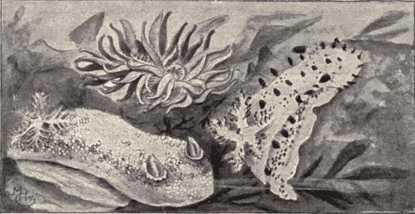
Fig. 109.—Three Pacific Coast nudibranchs; Doris tuberculata (in lower left-hand corner), Echinodoris sp. (upper one), and Triopha modesta (at right). (From living specimens in a tide-pool on the Bay of Monterey, California.)
Of the hosts of marine Gastropods we can notice only a few kinds. The nudibranchs (fig. 109) are a group of beautiful forms in which the shell is wholly wanting and the mantle is usually absent. The gills are thus exposed and are usually in the shape of delicate freely projecting[Pg 255] tufts arranged in rows along the back. The body is often strikingly and variedly colored. These soft, naked "sea-slugs" live near the shore, creeping about among the rocks and seaweeds. About a thousand species of nudibranchs are known.
Among the shell-forming marine Gastropods there is great variety in the size and shape and coloring of the shells. Many are beautifully colored and patterned; others are oddly and fantastically shaped. The cowries, or porcelain shells, familiar in collections of ocean curiosities, have a large body whorl and a very short flat spire, and the brightly colored shell looks as if enamelled. Some of the coast tribes of Africa once used, and perhaps still use to some extent, cowries as money. The limpets (fig. 104) are among the most abundant of the seashore molluscs, their low, broadly conical shells being plentifully scattered over the rocks between tide-lines. The "oyster-drills" are Gastropods with odd spiny shells which do much harm in oyster-beds by settling down on the oysters, boring holes through the shells and eating the soft parts within. The helmet-shells, from which shell cameos are cut, are composed of layers of shell material of different colors. Among the specially beautiful shells are the cone-shells, the olive-shells, the ivory-shells, etc.
Squids, cuttlefishes, and octopi (Cephalopoda).—Technical Note.—Small squids preserved in alcohol or formalin can be had of all dealers in biological supplies (see p. 453), and specimens should be examined.
The squids (fig. 110), cuttlefishes, octopi or "devil-fishes," and the three living species of Nautilus constituting the class Cephalopoda are very different from the other molluscs in appearance, and are in fact different in important structural characters. They can move swiftly, have strangely modified organs of prehension, strong biting mouth-parts, and eyes of very complex organization.[Pg 256] They are the most highly organized molluscan forms, and their predaceous habits and the great size to which some of them attain have given them distinction among the fierce and dangerous creatures of the sea. They are all strictly marine in habitat, and are all carnivorous. Most of them have no shell, or where the shell is present it is internal in all but a very few forms. The tentacle-like arms or feet surrounding the mouth which occur in all the Cephalopods are provided with sucking organs or suckers, in some cases with a horny toothed rim. These long, powerful, grasping, tentacular feet, with the suckers and five hooks, are very effective means of securing prey, and the pair of strong, sharp, cutting mandibles or beaks are equally effective in tearing to pieces. The eyes of the Cephalopods are almost as highly developed as those of the vertebrates. They are unusually large and staring, and add much to the terrifying appearance of the "devil-fishes." Cephalopods have the power of quickly changing color, because of the presence in the skin of many pigment-cells which can expand so as nearly to touch each other, thus producing a uniform tint over the whole body, or which can contract so as to destroy this uniformity of color. There are several sets of these color-carrying cells or chromatophores, each set of a color different from the others. The purpose of this change of color is protective, the animal being thereby able to make its color so harmonize with that of its immediate surroundings as to become indistinguishable.
There are two principal groups of Cephalopods, viz., the Decapods and the Octopods. The Decapods, as their name indicates, have ten feet or arms surrounding the mouth, and in them the body is usually elongate, containing a horny "pen" or calcareous "bone." This group includes the cuttlefishes or sepias, from which are obtained sepia ink and the cuttlefish bone used to feed[Pg 257] canary birds. The ink is a secretion which the cuttlefish discharges when attacked to create a cloud in the water and thus escape unperceived. The squids (Loligo) commonly used as bait by fishermen belong to the Decapoda. The two extra feet or arms which the Decapods have in addition to the eight possessed by the Octopods, differ from the others in being longer and slenderer and having suckers only on the distal extremities which are expanded into "clubs" (fig. 110).
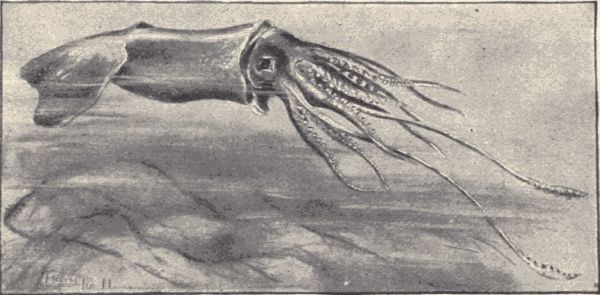
Fig. 110.—The giant squid, Ommatostrephes californica. (From specimen with body (exclusive of tentacles) four feet long, thrown by waves on shore of the Bay of Monterey, California.)
The Octopods have a short, sac-like, sub-spherical body and neither external nor internal shell. To this group belong the famous devil-fishes (Octopus), whose strange and terrifying appearance combined with their frequently great size has furnished the basis for many a weird tale of the sea. Octopi have been killed having tentacles more than 30 feet in length. The largest members of the class, however, are probably the giant squids (belonging to the Decapoda) specimens of which have been captured with a body-length of twenty feet, and arms thirty-five feet long.
The beautiful paper sailor or argonaut (Argonauta argo),[Pg 258] which secretes a thin shell (not homologous with the shell of the other molluscs) to protect her eggs, is a member of the Octopod group. In fine weather the argonauts sail in fleets on the surface of the ocean.
The pearly nautilus (Nautilus pompilius) is a Cephalopod with four gills instead of two, as with the Decapoda and Octopoda, and is the only existing member of what was in the earlier times of the earth's history a large group of animals. The nautili live in rather shallow water usually creeping over the bottom feeding on small marine animals. They make a many-chambered spiral shell with its inner surface lined with beautiful pearly nacre.
The branch Chordata includes all the backboned animals or vertebrates, comprising the fishes, salamanders, frogs and toads, lizards, crocodiles, turtles and snakes, birds, and all the quadrupeds or mammals, and includes also a few small unfamiliar ocean animals which do not look at all like the backboned animals, but which agree with them in possessing a peculiar structure called the notochord. This notochord consists of a series or cord of cells extending longitudinally through the body from head to tail, above the alimentary canal and below the spinal nerve-cord. In all the vertebrates excepting a few low forms, the notochord while present in the young, is replaced in the adult by a segmented bony or cartilaginous axis, the spinal or vertebral column. But in the ascidians or sea-squirts (called also tunicates) it persists throughout life. In addition to this characteristic notochord, nearly all the Chordata are marked by the presence, either in embryonic or larval stages only, or else persisting throughout life, of a number of slits or clefts in the walls of the pharynx which serve for breathing, and which are called gill-slits.
Structure of the vertebrates.—As the backboned or vertebrate animals make up almost the whole of the branch Chordata, and as the few other chordates are animals the special structures of which we shall not undertake to study in this book, we may note here some of the other more obvious structural characteristics of the true[Pg 260] vertebrates. The possession of a backbone or bony (sometimes cartilaginous) spinal column is the characteristic by which we distinguish them from the invertebrate or backboneless animals. Furthermore, all of the vertebrates possess an internal skeleton which is in most cases composed of bone, and is firm and strong. In some of the lower fishes, as the sharks and sturgeons, the skeleton is made up of cartilage, tough but not hard. The vertebrate skeleton consists typically of an axial portion comprising the spinal column and head, and of two pairs of appendages or limbs, variously developed as fins, wings, legs and arms. In some vertebrates these limbs are represented by mere rudiments, and in the lowest fish-like forms, the lancelets and lampreys, there is not the slightest trace of limbs. A part of the central nervous system, the spinal cord, runs longitudinally through the body on the dorsal side of the alimentary canal; the circulatory system is closed, the blood being always confined in the heart and in vessels called arteries, veins, and capillaries, and the blood is red in color owing to the presence of numerous red corpuscles or blood-cells. The nervous system is highly developed, with a large brain in all the typical forms, and with complex and usually highly efficient special sense-organs. Respiration is carried on by means of external gills, or by internal lungs which communicate with the outside through the mouth and nostrils. To the lungs and gills the blood is brought to be "purified," i.e., to give up its carbonic-acid gas and to take up oxygen.
Classification.—The Chordata are variously divided by zoologists into eight or ten classes, of which (in the eight-class system) the five classes[15] Pisces (fishes),[Pg 261] Batrachia (batrachians), Reptilia (reptiles), Aves (birds), and Mammalia (mammals), belong to the true vertebrates. These classes will be considered in the five following chapters.
The remaining three classes include a number of strange marine forms which until recent years were considered as worms, but which are now known to be the nearest living allies of the earliest or primitive vertebrates. The relationship of these forms to early types is manifest, not in the appearance or structure of the adult stage, but only during embryonic or larval stages.
The ascidians.—The sea-squirts, or Ascidians, common on the seashore, compose one class of these primitive chordate animals. They possess a simple, sac-like body (fig. 111), fastened to the rocks by one end, the other being[Pg 262] provided with two openings, one for the ingress and the other for the exit of water, a strong current of which flows constantly through the body. By means of this current the ascidian obtains food. Usually sea-squirts live together in large colonies, and in some cases a number of individuals enclose themselves in a common gelatinous mass, forming what is called a compound ascidian.
The ascidian when born is a tiny, free-swimming, tadpole-like creature with a slender finned tail. It swims about freely for only a few hours, however, soon attaching itself to a rock, and in its further development becoming degenerate. It loses its tail and with it the short notochord possessed by the larva; the eye and the auditory organ are lost, and the nervous system and alimentary canal become much reduced and simplified. Sea-squirts in their adult stage are very simple degenerate animals, with low functional development, yet their embryonic and larval conditions show a considerable degree of structural specialization, and the presence of the notochord in these early stages reveals their affinity with the backboned animals.
Technical Note.—The species of sunfish named, or some closely related species, can be obtained in any brook or stream in the United States. Gibbosus lives in all streams north of Dubuque, Chicago, Pittsburg, and along the eastern coast north of Charleston. Closely allied species live in all the other parts of the country except in the higher Rocky Mountains west of Bismarck, Pueblo, and Santa Fe. One species is found in the streams of California, but none occurs in Washington or Oregon. In the few places where a sunfish cannot be had, any species of bass or perch may be used. Sunfish live in ponds and sluggish streams in deep holes under a log or at the foot of a stump. They take eagerly a hook baited with a worm, or they may be caught in nets. When sunfish cannot be kept fresh for study in class, specimens may be preserved in alcohol or 4% formalin. But if possible to keep some alive for a time in a jar or tub with plenty of fresh water, the colors of the living fish, together with its manner of swimming and mode of breathing, can be observed.
External structure[16] (fig. 112).—Examine the general configuration and make-up of the body. Note the deep, laterally flattened trunk and paddle-like tail. The head is closely fitted to the trunk without any neck. Note that[Pg 264] the body is thickly covered with firm, hard scales, arranged like the shingles on a roof. Remove one of these scales and examine it under a hand lens. What sort of an edge has it? Such a scale is said to be ctenoid.
The body of the sunfish terminates behind in the caudal fin, a series of cartilaginous rays connected by thin skin and attached to a bony plate at the end of the backbone. Along the median dorsal line will be noted another fin composed anteriorly of spines and posteriorly of soft rays jointed and branched. This is the dorsal fin. How many spines has it? Anterior to the caudal fin on the ventral surface is a median unpaired anal fin. How many spines has it? Anterior to the anal fin are the ventral fins, while on the sides of the body back of the head in a line with the mouth are found the pectoral fins. The ventral fins, attached to a rudimentary pelvis, correspond to the hind legs of the other vertebrates. The pectoral fins, attached to the shoulder girdle, correspond to the arms. In front of the anal fin note a small pit-like opening, the opening from the kidneys and reproductive organs, and just anterior to this a large aperture, the anus. At the anterior end of the head note the broad mouth, surrounded by a complicated system of bones. Note the large eyes surrounded by a series of small bones, the orbital chain. Just anterior to the eyes are two pairs of openings, one pair of each side opening into a closed sac. What are these openings? Note the presence of various bones on the side of the head, each covered with a thin layer of skin. These are membrane bones, characteristic of fishes. Are there any external ears in the fish? Examine the inside of the mouth. Is there a tongue? If so, of what character? Are there teeth? If so, where are they situated?
Note along each side extending to the base of the tail a line of modified scales, on each scale a little mucous tube, the whole series constituting the lateral line. These[Pg 265] scales are intimately associated with a large nerve (the vagus), and probably serve an important part, not yet clearly understood, in the life of the fish.
Lift up the flap in front of one of the pectoral fins. This is the opercular flap which covers the gills that lie beneath. Bend this forward and find four gill-arches, each with its double fringe of gills. Note the gill-rakers, short and blunt, on the first gill-arch. Note also on the under side of the flaps turned back, delicate red gill-like structures covered by a membrane. These are the false gills or pseudo-branchiæ, larger in most fishes than in the sunfish. The gills in the fish subserve the same function as the gills of the crayfish, that of purifying the blood by eliminating carbonic-acid gas from it and taking up oxygen from the air mixed with or dissolved in the water. Organs subserving the same purposes in different kinds of animals as, for example, the gills in fish and in crayfish, are called analogous structures. But there is an important morphological difference between the fish's gills and the gills of the crayfish. In the latter animal they are outgrowths of the basal segments of the walking-legs; in the fish they are outgrowths from the alimentary canal. The internal gills of the young toad (tadpole) arise in the same way as those of a fish. Structures which are identical in their origin, like the gills of tadpole and fish, are called homologous structures.
Make a drawing of the sunfish from a lateral aspect, showing the external parts named.
Internal Structure.—Technical Note.—Insert one point of the scissors a little to one side of the anus and cut dorsally on the left side of the body to the backbone. Now cut anteriorly from the anus along the ventral wall to where the jaws unite, and cut, also anteriorly, along the dorsal wall until the left side of the body can be removed. Bend the opercular flap backward over the eye and pin the entire fish, uncut side down, to the bottom of the dissecting-pan, covering it with water.
The above operation will have severed the large powerful muscles forming the body-wall and extending along the sides. Note a membranous sac completely filling a large dorsal cavity. This is the swim-bladder, a float filled with air which tends to give the fish the same weight as the water it displaces. It arises as a diverticulum from the alimentary canal, but soon becomes permanently shut off from it. Beneath the swim-bladder is a large cavity filled with various organs, collectively known as the viscera. In vertebrate animals the cavity which contains the viscera is generally called the peritoneal cavity. It is lined by the peritoneum, a delicate membrane, part of which is deflected as the mesentery over the alimentary canal and the other organs, thus suspending them all from the dorsal wall. Note in the anterior end of the peritoneal cavity a large bi-lobed gland, the liver, red in fresh, yellowish in alcoholic specimens. Its function, like that of the liver of the toad, is to store up nutriment for the blood and to secrete a digestive fluid called bile. Behind the liver note a long, convoluted tube. What is this tube? Unfold this tube, separating it from its enveloping membrane, the mesentery. Thrust a probe down the throat and note that it passes into a thick-walled sac, the stomach. The mouth and gill-slits open into the front part of the alimentary canal called the pharynx, which leads by a short tube, the œsophagus, into the stomach. Note the large, thickened portion of the alimentary canal leading from the stomach. This is the pylorus, and to its walls are attached a number of finger-like projections, the pyloric cæca. The pyloric cæca secrete a fluid which is poured into the alimentary canal and which assists in the process of digestion somewhat as does the secretion from the pancreas of the toad. From the pylorus, passing backwards in one or two loops, is the small intestine. Trace this to its exit. Lying within the mesentery near[Pg 267] the posterior end of the body-cavity note a small red glandular mass, the spleen.
At the anterior end of the body in front of the liver and between the sets of gills note the small pericardial cavity within which is contained the heart. The pericardial cavity is separated from the peritoneal cavity by a thick muscular wall against which the liver abuts. The heart consists of four parts. The posterior part is a thin-walled reservoir, the sinus venosus, into which blood enters through the jugular vein from the head and through the cardinal vein from the kidney. From the sinus venosus it passes forward into a large chamber, the auricle. Next it flows into the ventricle, where, by the contraction of the walls, rhythmical pulsations force it into the conus arteriosus, thence into the ventral aorta, and lastly into the gills, where it is purified. After passing through the capillaries in the fine gill-filaments it is again collected, now pure, by paired arteries from each pair of gills, which arteries unite to form the dorsal aorta extending backward just below the backbone to the end of the tail. From the dorsal aorta a pair of arteries, the subclavian, are given off to the pectoral fins. At this point two other arteries branch off ventrally, the first being the cardiac artery, which distributes blood to the stomach and pyloric cæca. The second divides into several long mesenteric arteries supplying blood to all parts of the intestine and spleen. In the caudal region blood is taken up through the caudal vein and carried forward to the kidneys. These strain out the impurities arising from waste of tissues, after which the blood is carried back to the sinus venosus through the cardinal vein. From the intestine it is gathered into the large portal vein as in the toad. The portal vein carries blood to the liver, where nutriment may be stored up, and from thence it flows back[Pg 268] to the sinus venosus through a very short thin-walled vessel, the hepatic sinus.
The kidneys, more or less united in one mass, lie in the posterior part of the body-cavity along the dorsal wall. Note running from each side of the kidney a ureter which unites with its fellow and opens into a small urinary bladder which discharges through a small opening immediately back of the anus.
The reproductive organs lie below the swim-bladder near the posterior end of the body-cavity. If the fish are caught in the spring, the greater part of the body-cavity of the female is found to be filled with small eggs. When mature, these eggs are deposited by the mother fish in the gravel of the stream-bed where they are fertilized by the sperm-cells poured over them by the male and left floating in the water.
The nervous system of fishes is best studied in a specimen treated with nitric acid. Carefully remove the roof of the skull, thereby exposing the brain. Most anteriorly make out, as in the toad, the paired olfactory lobes. These are attached by long stalks to the cerebrum or forebrain, which is followed by two large hollow lobes, the midbrain or optic lobes. Behind the midbrain is the cerebellum. Following the cerebellum is the elongate medulla oblongata, which tapers backward into the spinal cord. How far backward does the spinal cord extend? On each side of the brain-case about opposite the cerebellum are located the auditory organs, each consisting of three semicircular canals which lie in different planes, and of the vestibule. These parts are filled with liquid, and suspended in the liquid in the vestibule are small calcareous bodies called otoliths or ear-stones. Running out beneath from the midbrain are the optic nerves, which cross, the left one connected with the right eye, the right one with the left eye. From each side of the medulla oblongata[Pg 269] there is given off a large nerve, the vagus, which sends branches to the lateral line organs on either side, and extends backward to the stomach and viscera.
For further study of the nervous system see Parker's "Zootomy," pp. 122-130.
Make a drawing of the nervous system as worked out.
Technical Note.—To make a good skeleton immerse a fresh or preserved specimen for some time in a hot soap solution. When the muscles have commenced to soften remove the body from the solution, pick the flesh away, and leave to dry.
Note that the main axis of the skeleton is composed of vertebræ placed end to end. How many vertebræ are there? What vertebræ bear ribs? The ribless ones beyond the body-cavity are called caudal vertebræ. Note the interspinal bones which support the fins, with large muscles on either side to control their action. Note that the group of bones supporting the pectoral fin is attached to the back of the brain-case and makes up the shoulder girdle. The ventral fins are attached to a rudimentary pelvic girdle, attached in front to the shoulder girdle, as the shoulder girdle is in turn attached to the skull. It will be seen that the sunfish has no neck and we may say, also, no back. Its skeleton consists only of a tail attached to the skull. The brain-case is made up of a number of bones closely joined together. From it is suspended the lower jaw, which comprises a number of bones but loosely attached to each other. Overlying these is the system of membrane bones already mentioned, including the opercle or gill-cover.
For a detailed study of the fish-skeleton see Parker's "Zootomy," pp. 86-101, or Parker and Haswell's "Zoology," vol. ii. pp. 183-195.
Life-history and habits.—The sunfish or "pumpkin-seed" lives in quiet corners of the brooks and rivers, preferably under a log or at the root of an old stump. It[Pg 270] is a beautiful fish, shining "like a coin fresh from the mint." Its body is mottled golden, orange and blue, with metallic lustre, darker above, pale or yellowish below. Its fins are of the same color. The tip of its opercle is prolonged like an ear and jet black in color, with a dash of bright scarlet along its lower edge. Nearly all the thirty species of sunfish found in the United States have this black ear, but some have it long, some short, and in some it is trimmed with yellow or blue instead of scarlet.
The sunfish lays its eggs in the spring in a rude nest it scoops in the gravel, over which it stands guard with its bright fins spread, looking as big and dangerous as possible. When thus employed it takes the hook savagely, perhaps regarding the worm as a dangerous enemy. The young fishes soon hatch, looking very much like their parents, although more transparent and not so brightly colored. They grow rapidly, feeding on insects and other small creatures, and reach their growth in two or three years. They do not wander far and never willingly migrate. Students should verify this account on the different species. A more exact study of the nests of the different species and the fishes' defence of them would be a valuable addition to our knowledge. The most striking traits of the habits of this fish are its vivacity and courage; it reveals its great muscular strength when captured. The sexes are similar in appearance and both defend the nest alike.
Fishes constitute the largest class of vertebrate animals and are to be found everywhere in ponds, streams, or ocean. About 15,000 species of fish are known, of which 3,000 live in North America. The largest of all fishes is the basking shark (Cetorhinus), which reaches a length of thirty-six feet. The smallest is the dwarf goby (Mistichthys), less than half an inch long, found in Luzon, one of the Philippine Islands. Between these extremes is every variety in size, form, and relative proportions. The body, for example, may be greatly elongated and almost cylindrical as in the eels; or long and flattened from side to side as in the ribbon-fishes; or the head may be very large, wider and higher than the rest of the body as in the anglers, or may have a great beak as in the sword-fish.
Body form and structure.—When we consider the fish as a whole, we find first a body formed for progression in the water, the typical fish being pointed at each end (the shorter point in front), and having the sides flattened, the back and belly rather narrow, and the motive power located in the fin on the tail. From this typical form diverge all conceivable variations, adaptations to every sort of fish life.
Most fishes have the body covered with scales, although many have the skin naked or covered with small scales so hidden in the skin as to be hardly visible. The scales are small horny or bony plates which fit into small pockets or folds of the skin, and are usually arranged shingle-fashion, overlapping each other. They are of various shapes, mostly classified as of three kinds, namely, squarish enamelled scales called ganoid, roundish smooth-edged called cycloid, and roundish tooth-edged called ctenoid.
The skeleton of the fish is relatively complex. Its
bones are comparatively soft, having little lime in them,
indeed in many cases they are mere cartilage. The
vertebral column is made of twenty-four vertebræ in the
typical fishes, the number in the others being variously
increased, or sometimes diminished. These vertebræ are
of two classes, abdominal or body, and caudal or tail
vertebræ. The former have a neural arch which encloses[Pg 271]
[Pg 272]
the spinal cord and from which projects a spine. Below,
the processes spread apart, surrounding the kidneys and
partly enclosing the air-bladder. To these processes ribs
are loosely attached. The caudal vertebræ have no ribs
and leave no room below for viscera. Their lower arch
(hæmal), similar to the dorsal (neural) arch, surrounds a
blood-vessel. The fins of a fish are composed of bony
rods or rays joined by membrane. Some of these rays
may be unbranched and unjointed, being then known as
spines, and usually occupy the front part of the fin.
Other rays are made up of little joints and are usually
branched toward their tip. Such ones are called soft
rays. Soft rays make up the greatest part of most fins.
The vertical fins are on the middle line of the body.
These are the dorsal above, anal below, and caudal forming
the end of the tail. The paired pectoral and ventral
fins are ranged one on each side corresponding to the
arms and legs of higher animals. The pectoral fin or
arm is fastened to a series of bones called the shoulder
girdle. These bones do not correspond to those in the
shoulder girdle of the higher animals, and the various
parts in the two structures are differently named. The
uppermost bone of the shoulder girdle is usually attached
to the skull. To the lowermost is attached the rudimentary
pelvis, which supports the hinder limb or ventral fin.
Usually the pelvis is farther back and loose in the flesh,
but sometimes it is placed far forward, being occasionally
attached at the chin.
The head contains the various bones of the cranium, usually closely wedged together and not easily distinguished. The jaws are each made of several pieces; the lower one is suspended from the skull by a chain of three flat bones. The jaws may bear any one of a great variety of forms of teeth or no teeth at all, and any of the bones of the mouth-cavity and throat may have teeth as well.[Pg 273] On the outside of the head are numerous bones called membrane bones, because they are made up of ossified membrane. The most important of these is the opercle or gill-cover. Within are the tongue with the five gill-arches attached to it below and to the floor of the skull above, the last arch being usually modified to form the pharyngeal jaw.
The stomach may be a blind sac with entrance and exit close together, or it may have the form of a tube or siphon. At its end are often found the large glandular tubes called pyloric cæca which secrete a digestive fluid; and to its right side is attached the red spleen. The liver is large, having usually, but not always, a gall-bladder; it pours its secretion into the upper intestine. In fishes which feed on plants the intestine is long, but it is short in those which eat flesh, because flesh is digested in the stomach, not in the intestines. The kidney is usually a long slender forked gland showing little variation. The egg-glands differ greatly in different sorts of fishes, the size and number of eggs varying equally. The air-bladder is a lung which has lost both lung structure and respiratory function, being simply a sac filled with gas secreted from the blood, and lying in the upper part of the abdominal cavity. It is subject to many variations. In the gar pike, bow-fin and the lung-fishes of the tropics, the air-bladder is a true lung used for breathing and connected by a sort of glottis with the œsophagus. In others it is rudimentary or even wholly wanting, while in still others its function as an air-sac is especially pronounced, and in many it is joined through the modified bones of the neck to the organ of hearing.
The blood of the fish is purified by circulation through its gills. These are a series of slender filaments attached to bony arches. Among them the blood flows in and out, coming in contact with the water which the fish takes in[Pg 274] through its mouth and which passes across the gills to be expelled through the gill-openings. The blood is received from the body into the first chamber of the heart, a muscular sac called the auricle. From here it passes into the ventricle, a chamber with thicker walls, the contraction of which sends it to the gills, thence without return to the heart it passes over the body. The circulation of blood in fishes is slow, and the blood, which receives relatively little oxygen, is cold, being but little warmer than the water in which the individual fish lives.
Inside the cranium or brain-case is the brain, small and composed of ganglia which are smooth at the surface and contain little gray matter. At the posterior end of the brain is the thickened end of the spinal cord, called the medulla oblongata. Next overlapping this is the cerebellum, always single. Before this lie the largest pair of ganglia, the optic lobes or midbrain, round, smooth, and hollow. From the under side of these, nerves run to the eyes with or without a chiasma or crossing. In front of the optic lobes and smaller than them is the cerebrum or forebrain, usually of two ganglia but sometimes (in the sharks) united into one. In front of these are the small olfactory lobes which send nerves to the nostrils.
The sense organs are well developed. The sense of touch has in some fishes special organs for its better effectiveness. For instance certain fin-rays in some fishes, or, as in the catfish, slender, fleshy, whip-like processes on the head, are developed as feelers or special tactile organs. Other fishes, the sucker and loach for example, have specially sensitive lips and noses with which they explore their surroundings. The sense of taste does not seem to be well developed in this group. Taste-papillæ are often present in small numbers on the tongue or on the palate. The sense of smell is good. The olfactory organs, one on each side of the head, are[Pg 275] hollow sac-like depressions, closed at the rear. In most cases each sac has two openings or nostrils. The sense of hearing is not very keen. The ears are fluid-filled sacs buried in the skull, and without external or (except in a few cases) internal opening. Fishes are far more sensitive to sudden jars or sudden movements than to any sound. They possess what is generally believed to be a special sense organ not found in other animals. This is the lateral line which extends along the sides of the body and which consists of a series of modified scales (each one with a mucous channel) richly supplied with nerves. The eyes are usually large and conspicuous. They differ mainly from the eyes of other vertebrates in their myopic spherical crystalline lens, made necessary by the density of the medium in which fishes live. There are usually no eyelids, the skin of the body being continuous but transparent over the eyes. Being near-sighted, fishes do not discriminate readily among forms, their special senses fitting them in general to distinguish motions of their enemies or prey rather than to ascertain exactly the nature of particular things.
The colors of fishes are in general appearance protective. Thus most individuals are white on the belly, mimicking the color of the sky to the enemy which pursues them from below. Seen from above most of them are greenish, like the water, or brownish gray and mottled, like the bottom. Those that live on sand are sand-colored, those on lava black, and those among rose-red sea-weeds bright red. In many cases, especially among kinds that are protected by their activity, brilliant colors and showy markings are developed. This is especially true among fishes of the coral reefs, though species scarcely less brilliant are found among the darters of our American brooks.
Among fresh-water fishes bright colors, crimson,[Pg 276] scarlet, blue, creamy white, are developed in the breeding season, the then vigorous males being the most highly colored. Many of the feeble minnows even become very brilliant in the nuptial season of May and June. Color in fishes is formed by minute oil-sacs on the scales, and it often changes quickly with changes in the nervous condition of the individuals.
Development and life-history.—The breeding habits of fishes are extremely varied. Most fishes do not pair, but in some cases pairing takes place as among higher animals. Ordinarily fishes lay their eggs on the bottom in shallow water, either in brooks, lakes, or in the sea. The eggs of fishes are commonly called spawn, and egg-laying is referred to as spawning. The spawn of some fishes is esteemed a special food delicacy. Spring is the usual time of spawning, though some fishes spawn in summer and some even in winter; generally they move from their usual haunts for the purpose. The eggs of the different species vary much in size, ranging from an inch and a half in diameter (barn-door skate) down to the tiniest dots, like those of the herring. The number of eggs laid also varies greatly. The trout lays from 500 to 1,000, the salmon about 10,000, the herring 30,000 to 40,000, and some species of river fish 500,000, while certain flounders, sturgeons, and others each lay several millions of eggs. The adults rarely pay any attention to the eggs, which are hatched directly by the heat of the sun or by heat absorbed from the water. The length of incubation varies much. When the young fish leaves the egg-shell it carries, in the case of most species, a part of the yolk still hanging to its body. Its eyes are very large, and its fins are represented by thin strips of membrane. It usually undergoes no great changes in development from the first, resembling the adult except in size. But some of the ocean fishes[Pg 277] show a metamorphosis almost as striking as that of insects or toads or frogs.
Some fishes build nests. Sticklebacks build elaborate nests in the brooks and defend them with spirit. Sunfishes do the same, but the nests are clumsier and not so well cared for.
The salmon is the type of fishes which run up from the sea to lay their eggs in fresh water. The king salmon of the Columbia River, for example, leaves the sea in the high waters of March and ascends without feeding for over a thousand miles, depositing its spawn in some small brook in the fall. After making this long journey to lay the eggs, the salmon become much exhausted, battered and worn, and are often attacked by parasitic fungi. They soon die, probably none of them ever surviving to lay eggs a second time.
Classification.—A fish is an aquatic vertebrate, fitted to breathe the air contained in water, and never developing fingers and toes. Accepting this broad general definition we find at once that there are very great differences among fishes. Some differ more from others than the ordinary forms differ from rabbits or birds. So although we have entitled this chapter as if all fishes belonged to the class Pisces, we cannot arrange them satisfactorily in less than three classes.
The lancelets (Leptocardii).—The lowest class of fish-like animals is that of the lancelets, the Leptocardii. These little creatures, translucent, buried in the sand, of the size and form of a small toothpick, are fishes reduced to their lowest terms. They have the form, life, and ways of a fish, but no differentiated skull, brain, heart, or eyes. Moreover they have no limbs, no jaws, no teeth, no scales. The few parts they do have are arranged as in a fish, and they show something in common with the fish[Pg 278] embryo. Lacking a distinct head, the lancelets are put by some zoologists in a group called the Acrania, as opposed to the Craniata, which includes all the other vertebrates. Lancelets have been found in the North Atlantic and Mediterranean, on the west coast of North America, on the east coast of South America and on the coasts of Japan, Australia, New Zealand, the East Indies and Malayan Islands. The best-known members of the group belong to the genus Amphioxus. There are but one to two other genera in the class.
The lampreys and hag-fishes (Cyclostomata).—The next class of fish-like animals is that of the lampreys (fig. 113) and hag-fishes, the Cyclostomata. The lampreys and hags are easily distinguished from the true fishes by their sucking mouth without jaws, their single median nostril, their eel-like shape and lack of lateral appendages or paired fins. The hag-fishes (Myxine), which are marine, attach themselves by means of a sucker-like mouth to living fishes (the cod particularly), gradually scraping and eating their way into the abdominal cavity of the fish. These hags or "borers" "approach most nearly to the condition of an internal parasite of any vertebrate." The lampreys, or lamprey-eels as they are often called because of their superficial resemblance to true eels, are both marine and fresh-water in their habitat, and most of them attach themselves to live fishes and suck their blood. They also feed on crustacea, insects, and worms. The[Pg 279] brook-lamprey, Lampetra wilderi, is never parasitic. It reaches its full size in larval life and transforms simply for spawning. The sea- and lake-lampreys ascend small fresh-water streams when ready to lay their eggs, few living to return. Sometimes small piles of stones are made for nests. The young undergo a considerable metamorphosis in their development. The largest sea-lampreys reach a length of three feet. The common brook-lampreys are from eight to twelve inches long only.
The true fishes (Pisces).—All the other fish-like animals are grouped in the class Pisces. They are characterized, when compared with the lower fish-like forms just referred to, by the presence of jaws, shoulder girdle, and pelvic girdle. The class includes both the cartilaginous and bony fishes, and is divided into three sub-classes, namely, the Elasmobranchii, including the sharks, rays, skates, torpedoes, etc., the Holocephali, including the chimæras (a few strange-bodied forms), and the Teleostomi, including all the other fishes, as the trout, catfishes, darters, bass, herring, cod, mackerel, sturgeons, etc., etc.
The sharks, skates, etc. (Elasmobranchii).—The sharks and skates are characterized by the possession of a skeleton composed of cartilage and not bone, as in the bony fishes; they have no operculum; their teeth are distinct, often large and highly specialized, and their eggs are few and very large. There are two principal groups among Elasmobranchii, viz., the sharks, which usually have an elongate body, and always have the gill-openings on the sides, and the rays or skates, which have a broad flattened body with the gill-openings always on the under side. All the members of both groups are marine. The sharks are active, fierce, usually large fishes, which live in the surface-waters of the ocean and make war on other marine animals, all of the species except half a dozen being fish-eaters. The shark's mouth is on the[Pg 280] under side of the usually conical head, and the animal often turns over on its back in order to seize its prey. The largest American sharks, and the largest of all fishes, are the great basking-sharks (Cetorhinus), which reach a length of nearly forty feet. They get their name from their habit of gathering in numbers and floating motionless on the surface. They feed chiefly on fishes.
The hammer-headed sharks (Sphyrna) are odd sharks which have the head mallet or kidney shaped, twice as wide as long, the eyes being situated on the ends of the lateral expansions of the head. The man-eating or great white sharks (Carcharodon) are nearly as large as the basking-sharks, and are extremely voracious. They will follow ships for long distances for the refuse thrown overboard. They do not hesitate to attack man. Among the more familiar smaller sharks are the dog-fishes and sand-sharks of our Atlantic coast.
The rays and skates are also carnivorous, but are with few exceptions sluggish, lying at the bottom of shallow shore-waters. They feed on crabs, molluscs, and bottom-fishes. The small common skates, "tobacco-boxes" (Raja erinacea) (fig. 114), about twenty inches long, and the larger "barn-door skates" (R. lævis), are numerous along the Atlantic coast from Virginia northward. Especially interesting members of this group, because of the peculiar character of the injuries produced by them, are the sting-rays and torpedoes or electric-rays. The sting-rays (Dasyatis) have spines near the base of the tail which cause very painful wounds. The torpedoes (Narcine) have two large electrical organs, one on each side of the body just behind the head, with which they can give a strong electric shock. "The discharge from a large individual is sufficient to temporarily disable a man, and were these animals at all numerous they would prove dangerous to bathers." Very different from the typical[Pg 281] rays in external appearance are the saw-fishes (Pristis pectinatis) which belong to this group. The body is elongate and shark-like, and has a long saw-like snout. This saw, which in large individuals may reach a length of six feet and a breadth of twelve inches, makes its owner formidable among the small sardines and herring-like fishes on which it feeds. The saw-fishes live in tropical rivers, descending to the sea.
The bony fishes (Teleostomi).—The bony or true fishes are distinguished from the lampreys and sharks and rays by having in general the skeleton bony, not cartilaginous, the skull provided with membrane bones, and the eggs small and many. In this group are included all the fishes of our fresh-water lakes, ponds, and streams as well as most of the marine forms. Fish life, being spent under[Pg 282] water, is not familiar to most of us, and beginning students are rarely helped enough in getting acquainted with the different kinds and the interesting habits of fishes. But they offer a field of study which is really of unusual interest and profit. We can refer in the following paragraphs to but few of the numerous common and readily found kinds, and to these but briefly.
Closely related to the sunfish, studied as example of the bony fishes, are the various kinds of bass, as the "crappie" (Pomoxis annularis), the calico bass (P. separoides), the rock-bass (Ambloplites rupestris) and the large-mouthed and small-mouthed black bass (Micropterus salmoides and M. dolomieu respectively). All the members of this sunfish and bass family are carnivorous fishes especially characteristic of the Mississippi valley.
Another family of many species especially common in the clear, swift, and strong Eastern rivers is that of the darters and perches. The darters are little slender-bodied fishes which lie motionless on the bottom, moving like a flash when disturbed and slipping under stones out of sight of their enemies. Some are most brilliantly colored, surpassing in this respect all other fresh-water fishes.
Unlike the sunfishes and darters are the catfishes, composing a great family, the Siluridæ. The catfish (Ameiurus) gets its name from the long feelers about its mouth; from these feelers also come its other names of horned pout, or bull-head. It has no scales, but its spines are sharp and often barbed or jagged and capable of making a severe wound.
Remotely allied to the catfish are the suckers, minnows, and chubs, with smooth scales, soft fins and soft bodies and the flesh full of small bones. These little fish are very numerous in species, some kinds swarming in all fresh water in America, Europe, and Asia. They usually swim in the open water, the prey of every carnivorous[Pg 283] fish, making up by their fecundity and their insignificance for their lack of defensive armature. In some species the male is adorned in the spring with bright pigment, red, black, blue, or milk-white. In some cases, too, it has bony warts or horns on its head or body. Such forms are known to the boys as horned dace.
Most interesting to the angler are the fishes of the salmon and trout (fig. 115) family, because they are gamy, beautiful, excellent as food and above all perhaps because they live in the swiftest and clearest waters in the most charming forests. The salmon live in the ocean most of their life, but ascend the rivers from the sea to deposit their eggs. The king salmon (Oncorhynchus tschawytscha) of the Columbia goes up the great river more than a thousand miles, taking the whole summer for it, and never feeding while in fresh water. Besides the different kinds of salmon, the black-spotted or true trout, the charr or red-spotted trout of various species, the whitefish (Coregonus), the grayling (Thymallus signifer) and the famous ayu of Japan belong to this family.
In the sea are multitudes of fish forms arranged in many families. The myriad species of eels agree in having no ventral fins and in having the long flexible body of the snake. Most of them live in the sea, but the single[Pg 284] genus (Anguilla) or true eel which ascends the rivers is exceedingly abundant and widely distributed. Most eels are extremely voracious, but some of them have mouths that would barely admit a pin-head. The codfish (Gadus callarias) is a creature of little beauty but of great usefulness, swarming in all arctic and subarctic seas. The herring (Clupea harengus), soft and weak in body, are more numerous in individuals than any other fishes. The flounders (fig. 116) of many kinds lie flat on the sea-bottom. They have the head so twisted that the two eyes occur both together on the uppermost side. The members of the great mackerel tribe swim in the open sea, often in great schools. Largest and swiftest of these is the sword-fish (Xiphias gladius), in which the whole upper jaw is grown together to form a long bony sword, a weapon of offence that can pierce the wooden bottom of a boat.
Many of the ocean fishes are of strange form and appearance. The sea-horses (Hippocampus sp.) (fig. 117) are odd fishes covered with a bony shell and with the head having the physiognomy of that of a horse. They are little fishes rarely a foot long, and cling by their[Pg 285] curved tails to floating seaweed. The pipefish (Syngnathus fuscum) is a sea-horse straightened out. The porcupine-fishes and swellfishes (Tetraodontidæ) have the power of filling the stomach with air which they gulp from the surface. They then escape from their pursuers by floating as a round spiny ball on the surface. The flying-fishes (Exocœtus) leap out of the water and sail for long distances through the air, like grasshoppers. They cannot flap their long pectoral fins and do not truly fly; nevertheless they move swiftly through the air and thus escape their pursuers. In its structure a flying-fish differs little from a pike or other ordinary fish.
For an account of the fishes of North America see Jordan's "Manual of Vertebrates," eighth edition, pp. 5-173, and Jordan and Evermann's "Fishes of North and Middle America," where the 3,127 species known from our continent are described in detail with illustrative figures.
Habits and adaptations.—The chief part of a fish's life is devoted to eating, and as most fishes feed on other fishes, all are equally considerably occupied in providing for their own escape.
In general the provisions for seizing prey are confined to sharp teeth and the strong muscles which propel the caudal fin. But in some cases special contrivances appear. In one large group known collectively as the[Pg 286] "anglers" the first spine of the dorsal fin hangs over the mouth. It has at its tip a fleshy appendage which serves as a bait. Little fishes nibble at this, the mouth opens, and they are gone. In the deep seas, many fishes are provided with phosphorescent spots or lanterns which light up the dark waters, and enable them to see their prey. In storms these lantern-fishes sometimes lose their bearings and are thrown upward to the surface.
In general the more predatory in its habits any fish is the sharper its teeth, and the broader its mouth. Among brook-fishes the pickerel has the largest mouth and the sharpest teeth. It has been called a "mere machine for the assimilation of other organisms." The trout has a large mouth and sharp teeth. It is a swift, voracious, and predatory fish, feeding even on its own kind. The sunfish is less greedy and its mouth and teeth are smaller, though it too eats other fish.
As means of escape, most fishes depend on their speed in swimming. But some hide among rocks and weeds, disguising themselves by a change in color to match their surroundings. Others, like the flounders and skates, lie flat on the bottom. Still others retreat to the shallows or the depths or the rock-pools or to any place safer than the open sea. Some are protected by spines which they erect when attacked. Some erect these spines only after they have been swallowed, tearing the stomach of their enemy and killing it, but too late to save themselves. Again in some species the spines are armed with poison which benumbs the enemy. Sometimes an electric battery about the head or on the sides gives the biting fish a severe shock and drives him away. Such batteries are found in the electric rays or torpedo, in the electric eel of Paraguay, the electric catfish of the Nile, the electric stargazer and other fishes.
Some fishes are protected by their poor and bitter flesh.[Pg 287] Some have bony coats of mail and sometimes the coat of mail is covered with thorns, as in the porcupine-fish. This fish and various of its relatives have the habit of filling the stomach with air when disturbed, then floating belly upward, the thorny back only within reach of its enemies.

Fig. 118.—The remora, or cling fish, Remoropsis brachyptera. Note sucker on top of head. (After Goode.)
Many species (cling fishes) attach themselves to the rocks by a fleshy sucking-disk. Some (Remora) (fig. 118) cling to larger fishes by a strange sucking-disk on the head, a transformed dorsal fin, being thus shielded from the attacks of fish smaller than their protectors. Some small fishes seek the shelter of the floating jellyfishes, lurking among their poisoned tentacles. Others creep into the masses of floating gulf-weed. Some creep into the shell of clams and snails. In the open channel of a sponge, the mouth of a tunicate and in similar cavities of various animals, little fishes may be found. A few fishes (hag-fishes) are parasitic on others, boring their way into the body and devouring the muscles with their rasp-like teeth.
Some fishes are provided with peculiar modifications of the gills which enable them to breathe for a time out of water. Such fish have the pectoral fins modified for a rather poor kind of locomotion on land, thus enabling them to move from pond to pond or from stream to stream. In cold climates the fishes must either migrate to warmer latitudes in winter, as some do, or withstand variously the cold, often freezing weather. Some fish can be frozen[Pg 288] solid, and yet thaw out and resume active living. Some lie at the bottoms of deep pools through the colder periods, while many others, such as the minnows, chubs, and other kinds common in small streams, bury themselves in the mud, and lie dormant or asleep through the whole winter. On the other hand in countries where the long intense rainless summers dry up the pools, some fishes have the habit of burying themselves in the mud, which, with slime from the body, forms about them a sort of tight cement ball in which they lie dormant until the rains come. "Thus a lung-fish (called Protopterus), found in Asia and Africa, so completely slimes a ball of mud around it that it may live for more than one season, perhaps many; it has been dug up and sent to England, still enclosed in its round mud-case, and when it was placed in warm water it awoke as well as ever."
Food-fishes and fish-hatcheries.—Most fishes are suitable for food, though not all. Some are too small to be worth catching or too bony to be worth eating. Some of the larger ones, especially the sharks, are tough and rank. A few are bitter and in the tropics a number of species feed on poisonous coelenterates about the coral reefs, becoming themselves poisonous in turn. But a fish is rarely poisonous or unwholesome unless it takes poisonous food. Where fishes of a kind specially used for food gather in great numbers at certain seasons of the year, fishing is carried on extensively and with an elaborate equipment. Such fisheries, some of which have been long known, are scattered all over the world. Along the shores of the Mediterranean Sea, and on the coasts of Norway, France, the British Isles and Japan are numerous great fishing-places. But "nowhere are there found such large fisheries as those along the northern Atlantic coasts of our own continent, extending from Massachusetts to Labrador. Especially on the banks of Newfoundland are codfish,[Pg 289] herring, and mackerel caught." Among our fresh-water fisheries the great salmon fisheries of the Penobscot and Columbia rivers and of the Karluk and other rivers of Alaska are the best known. The whitefish of our Great Lakes is also one of the important food-fishes of the world.
In many places fishes are raised in so-called hatcheries, not usually for immediate consumption but for the purpose of stocking ponds and streams either in the neighborhood of the hatchery or in distant waters which the special species cultivated has not been able naturally to reach. The eggs of some fishes are large and non-adherent, two features which greatly favor artificial impregnation and hatching. In the hatcheries the eggs are put first into warm water, where development begins; they are then removed into cool water, which arrests development without injury, making shipment possible. The eggs of salmon and trout in particular can be sent long distances to suitable streams or ponds. The eggs of the shad have been thus carried from the East to the streams of California and trout have been distributed to many streams in our country which by themselves they could never have reached.
The salmon is a conspicuous example of those fishes which can be artificially propagated. The eggs of the salmon are large, firm, and separate from each other. If the female fish be caught when the eggs are ripe and her body be pressed over a pan of water the eggs will flow out into the water. By a similar process the milt or male sperm-cells can be procured and poured over the eggs to fertilize them. The young after hatching are kept for a few days or weeks in artificial pools, till the yolk-sacs are absorbed and they can take care of themselves. They are then turned into the stream, where they drift tail foremost with the current and pass downward to the sea. All trout may be treated in similar fashion, but there are[Pg 290] many food-fishes which cannot be handled in this way. In some the eggs are small or soft, or viscid and adhering in bunches. In others the life-habits make artificial fertilization impossible. Such species are artificially reared only by catching the young and taking them from one stream to another. To this type belong the black bass, the sunfish, the catfish and other familiar forms.
The structure, life-history, and habits of the garden-toad (Bufo lentiginosus) have already been studied (see Chapter II and Chapter XII).
The class Batrachia includes the animals familiarly known as cœcilians, sirens, mud-puppies, salamanders, toads, and frogs. Although differing plainly from fishes in appearance and habits, the batrachians are really closely related to them, resembling them in all but a few essential characters. Among the distinctive characters of batrachians may be noted the absence of fins supported by fin-rays, the presence usually of well-developed legs for walking or leaping, and the absence or reduction of certain bones of the head connected with the gills and lower jaw and which are well developed in the fishes. The batrachians stand in somewhat intermediate position between the fishes and the reptiles, showing some of the characters of both. They are, like fishes and reptiles, cold-blooded. In their adult condition some are terrestrial and some aquatic as to habitat, but all have an aquatic larval life. The water-inhabiting young breathe at first by means of gills, later lungs begin to develop, and for a time both gills and lungs are used in respiration. Finally in the adult condition in almost all of the forms the gills[Pg 292] are wholly lost and breathing is done by the lungs and skin solely. Correlated with the change of habits from larval to adult stage there is usually a well-marked metamorphosis in post-embryonic development. This metamorphosis is specially striking among the frogs and toads. None of the aquatic forms is marine, salt water always killing eggs, larvæ or adults. Batrachians are found all over the world, although there are few in the extreme North. They are most abundant in warm and tropical lands.
Body form and organization.—The body varies from a long and slender, truly snake-like form as in the tropical cœcilians through the usual salamander (fig. 119) shape, where it is more robust but still elongate and tailed, to the heavy, squat, tailless condition of the toads. Legs, with five digits, are usually present, and are used for swimming, walking, or leaping. The legs are longest and best developed in the short tailless frog and toad forms which are mostly terrestrial, and are short and weak in the tailed salamander forms, many of which are aquatic. The skin is almost always naked, showing a marked difference from the scaled condition of reptiles and most of the fishes, and its cells secrete a slimy, sticky, usually whitish fluid, which in some cases is irritating, or even poisonous.[Pg 293] The skin is sometimes thrown up into folds or ridges, and in some species is elevated to form a kind of fin on the tail or back. This unpaired fin differs from the dorsal fin (and other fins) of fishes in not being supported by rayed processes of the skeleton. There are in some batrachians traces of an exoskeleton in the presence of scale-like structures in the skin or in the horny nails on the digits, but these cases are rare. The skin contains pigment-cells and many of the batrachians are brilliantly colored and patterned; some of the pigment is carried by special contractile or expansile cells, the chromatophores (see account of chromatophores of the Cephalopoda, p. 256), so that the animal can change its tint and markings more or less rapidly. All the batrachians possess external gills in their aquatic larval stage, and in a few forms, as the sirens and mud-puppies, gills are retained all through life. These gills are branched folds of the skin abundantly supplied with blood-vessels.
In the organization of the batrachian body the usual vertebrate characters appear, the body-organs being arranged with reference to a supporting and protecting internal bony skeleton. The head is plainly set off from the rest of the body and bears the mouth and the organs of hearing and sight. Certain so-called lateral sense organs, the function of which is not exactly known, occur arranged in three lines on each side of the body of some of the forms. Both pairs of limbs are present and functional in almost all of the species. In the cœcilians the limbs are wholly wanting; in the sirens only the fore legs are present.
Structure.—The most obvious skeletal differences among batrachians are those due to variations in external form. While there are as many as 100 vertebræ in some of the elongate long-tailed salamanders (even 250 in the strange snake-like cœcilians), there are but 10 (the last[Pg 294] or tenth being the rod-shaped bone called the urostyle) in the short, tailless frogs and toads. To any of the vertebræ except the first (the single cervical vertebra) and the last, ribs may be attached and the cœcilians have about as many pairs of ribs as vertebræ. In the frogs and toads, however, the ribs are lost. In any case they are never fastened by their lower ends to the breast-bone.
The alimentary canal is usually not much longer than the body and is plainly divided into mouth, pharynx, œsophagus, small intestine, large intestine or rectum, and anal opening. The teeth when present occur on both the jaws and the palate. They are small, sharp, point backward and are fused to the bones. They are wholly wanting in the toad and in some other allied forms. The tongue may be wanting, or may be immovably fixed to the floor of the mouth, or as in the frogs, fastened at its front end but free behind, so that the hinder end can be protruded far from the mouth for the purpose of catching insects.
The organs of respiration are gills, external and internal, lungs, trachea or windpipe, and the skin. In the earliest larval stages all batrachians have gills; later, in most cases, the gills become reduced and disappear, while at the same time lungs are developing. In some salamanders the lungs never develop, but the animals, in their adult stage, breathe wholly by means of the skin. In a few cases, as in the siren and mud-puppies, gills are retained through the whole life, although lungs are also present in the adult stage. The lungs are two in number, a right and a left lung, and are simple sacs with the walls more or less folded or thrown into ridges and richly supplied with blood-vessels. The front end of the lungs opens directly into the pharynx or, in the more elongate batrachians, is connected with it by a tubular trachea or windpipe. In the frogs and toads there are vocal cords[Pg 295] stretched across the short windpipe; the vibration of these cords produces the croaking.
The heart is always three-chambered, consisting of the right and left auricles and a single ventricle. The circulation of the more generalized salamanders like the mud-puppies is essentially like that of a fish. In the frogs and toads there is a distinct advance beyond this condition. The red corpuscles of the blood are oval in shape and are the largest found among any of the vertebrates.
In the nervous system the small size of the hindbrain or cerebellum is noticeable. The sense organs are fairly well developed. The skin of the whole body is provided with tactile nerve-endings. There are special taste organs on the lining membrane of the tongue and mouth-cavity. The eyes have no lids in some of the lower forms; most of the frogs and toads have an upper lid but no under one, although a thin membrane, called the nictitating membrane, arises from the lower margin of the eye and can be drawn up over it. The ears have no external parts, other than the thin tympanic membranes. The nostrils of frogs and toads can be closed by the contraction of certain special muscles.
Life-history and habits.—The sexes are distinct, and in most cases the young hatch from eggs. A few of the salamanders give birth to free young. The eggs are usually in strings or chains enclosed in a clear gelatinous substance; these chains of eggs are either simply dropped into the water or are fastened to water-plants. The young, called tadpoles (fig. 120), in their earlier larval stages are extremely fish-like in character, long-bodied, tailed, swimming freely about by means of the fin-like flattened tail, and breathing by means of external gills. Nor do they show any sign of legs. As the tadpoles grow and develop the legs begin to appear, the hind legs first in the frogs and toads, the fore legs first in the salamanders; lungs develop[Pg 296] and the gills disappear (except in the cases of the few forms which retain gills through life). The tail shortens and finally disappears in the frogs and toads; with the salamanders the tail-fin only is lost. At the same time the change from water to land is made. Further growth is very slow; frogs are not really adult, that is, capable of producing young, until they are five years old, and they may continue to increase in size until they are ten years old.
The food of the adult batrachians is almost exclusively small animals, particularly insects and worms. Crustaceans, snails, and young fish are also eaten. The tadpoles also eat vegetable matter. Almost all batrachians are nocturnal in habit, remaining concealed by day. In the zones in which cold winters occur they hibernate or pass the winter in a torpid condition, or state of "suspended animation," or, as it is said, they sleep through the winter. Frogs burrow into the mud at the bottom of ponds at the approach of winter and come forth early in[Pg 297] the spring to lay their eggs. Most batrachians are very tenacious of life, being able to withstand long periods of fasting and serious mutilation, and most of them can regenerate certain lost parts, such as the tail or legs.
Classification.—The living Batrachia are divided into three orders, viz., the Urodela, including the sirens, mud-puppies, salamanders, and newts, batrachians which retain the tail throughout life, having generally two pairs of limbs of approximately equal size, and sometimes possessing gills or gill-slits in the adult condition; the Anura, or frogs and toads, with no tail in the adult condition, with short and broad trunk, with hind limbs greatly exceeding the fore limbs in size, and never with gills or gill-slits in the adult stage; and the Gymnophiona, or cæcilians, snake-like batrachians having neither limbs nor tail, with a dermal exoskeleton and without gills or gill-slits in the adult.
Mud-puppies, salamanders, etc. (Urodela).—Technical Note.—If possible obtain specimens of mud-eels (Siren), common in the South, or mud-puppies (Necturus), common in the central North, as examples of batrachians with gills persisting in the adult stage. One or more species of Amblystoma may be found in almost any part of the country, and larvæ of large size may be found with the external gills. For an example of the general long-tailed or Urodelous type of batrachian any salamander or newt occurring in the vicinity of the school may be used. The little green triton or eft (Diemictylus viridiscens) of the eastern States, or its larger brown-backed congener of the Pacific coast (D. torosus) is common in water, while another eft, the little red-backed salamander, (Plethodon) is common in the woods under logs and stones. The external characters of the body should be compared with those of the toad. The skeleton should be prepared by macerating away the flesh (for directions, see p. 452), and the presence of the many caudal vertebræ and the ribs, the equality in size of the legs, and other points should be noted. Compare with skeleton of toad. Make drawings. It will be well, also, to dissect out and examine the various internal organs of the salamander, comparing them with the same organs in the toad. The salamander, indeed, is in many ways better than the toad as an example of the class. Its body is less adaptively modified and shows the essentially fish-like character of the batrachian structure.
The batrachians which retain external gills in the adult stage are the members of two families of which the American representatives are known as mud-eels (Siren) and mud-puppies or water-dogs (Necturus). The mud-eels, which are found "in the ditches in the swamps of the southern States from South Carolina to the Rio Grande of Texas and up the Mississippi as high as Alton, Illinois," are blackish in color, have no hind legs and are long and slender, with the tail shorter than the rest of the body. They reach a length of nearly three feet. The mud-puppies, found in the Great Lakes and in the rivers of the upper Mississippi valley, are brown with colored spots, and are about two feet long when full grown. They have both fore and hind legs.
A few salamanders, while not possessing external gills when adult, have a spiracle or small circular opening in the side of the neck which leads into the throat. The best-known American salamander of this kind is the large heavy-bodied blackish water-dog or "hellbender" (Cryptobranchus) of the Ohio River. It is about two feet long, and is "a very unprepossessing but harmless creature." It has a conspicuous longitudinal fold of skin along each side of the body. The largest known batrachian, the giant salamander of Japan (Megalobatrachus), reaching a length of three feet, is related to the water-dog.
Of all the salamanders the most interesting are the blunt-nosed salamanders (Amblystoma). A dozen or more species of Amblystoma occur in North America, of which tigrinum, a dark-brown species with many irregular yellow blotches sometimes arranged in cross-bands, is the most widespread. The larvæ of some Amblystoma retain their gills until they have reached a large size, and in one or two species the usual metamorphosis is very long delayed and the salamanders produce young while in the[Pg 299] larval condition, that is, while retaining the gills and a compressed fin-like tail. In the case of a certain Mexican species (A. maculatum) it is believed that the final metamorphosis never occurs. The Mexicans call these gilled larval Amblystoma axolotls, and use them for food. For a long time naturalists supposed the Amblystoma larvæ which produce young to be the adults of a species of salamanders which retained their gills through life, like the sirens and mud-puppies, and classified them in a distinct genus.
Of the various common salamanders or newts some are found in streams, ponds, and ditches, and some under logs and stones in the woods. The aquatic forms have the tail compressed (flattened from side to side), while the land forms have the tail cylindrical, tapering to a point. Most of the land-salamanders produce their young alive, while the water forms lay eggs which are usually attached to a submerged plant-stem. The salamanders are, almost without exception, found only in the northern hemisphere.
Frogs and toads (Anura).—There are about a dozen species of frogs in the United States. The largest of these, and indeed the largest of all the frogs, is the well-known bullfrog (Rana catesbiana), which reaches a length (head to posterior end of body) of eight inches. It is found in ponds and sluggish streams all over eastern[Pg 300] United States and in the Mississippi valley. It is greenish in color with the head usually bright pale green. Its croaking is very deep and sonorous. The pickerel-frog (R. palustris), which is bright brown on the back with two rows of large oblong square blotches of dark brown on the back, is found in the mountains of eastern United States. The little pale reddish-brown wood-frog (R. sylvatica) with arms and legs barred above is common in damp woods and is "an almost silent frog." The peculiar and infrequently seen frogs known as the "spade-foots" (Scaphiopus) are subterranean in habit and usually live in dry fields or even on arid plains and deserts. They pass through their development and metamorphosis very rapidly, appearing immediately after a rain and laying their eggs in temporary pools. At this time of egg-laying they utter extraordinarily loud and strange cries. Some frogs in other parts of the world live in trees, and the eggs of one species are deposited on the leaves of trees, leaves which overhang the water being selected so that the issuing young may drop into it.
The true tree-frogs or tree-toads (Hylidæ) constitute a family especially well represented in tropical America. They have little disk- or pad-like swellings on the tips of their toes to enable them to hold firmly to the branches of the trees in which they live. Some, like the swamp tree-frog and the cricket-frog, are not arboreal in habit, remaining almost always on the ground. The common tree-frog of the eastern States (Hyla versicolor) is green, gray, or brown above with irregular dark blotches, and yellow below. It croaks or trills, especially at evening and in damp weather. Pickering's tree-frog (Hyla pickeringii) makes the "first note of spring" in the eastern States. This tree-frog is the one most frequently heard in the autumn too, but "its voice is less vivacious than in the spring and its lonely pipe in dry woodlands is[Pg 301] always associated with goldenrods and asters and falling leaves." The tree-frogs of North America lay their eggs in the water on some fixed object as an aquatic plant, in smaller packets than those of the true frogs, and not in strings as do the toads.
The toads (Bufonidæ) differ from the true frogs in having no teeth and in not having, as the frogs do, a cartilaginous process uniting the shoulder-bones of the two sides of the body. The absence of this uniting process makes the thoracic region capable of great expansion. There are only a few species of toads in North America, but one of these species, the common American toad (Bufo lentiginosus), is very abundant and widespread. It appears also in two or three varieties, the common toad of the southern States differing in several particulars from that of the northern. The toad is a familiar inhabitant of gardens, and does much good by feeding on noxious insects. It is most active at twilight. Its eggs are laid in a single line in the centre of a long slender gelatinous string or rope, which is nearly always tangled and wound round some water-plant or stick near the shore on the bottom of a pond. The eggs are jet black and when freshly laid are nearly spherical. At the time of egg-laying the toads croak or call, making a sort of whistling sound and at the same time pronouncing deep in the throat "bu-rr-r-r-r." The toad does not open its mouth when croaking, but expands a large sac or resonator in its throat. The toad-tadpoles are blacker than those of frogs or salamanders, and undergo their metamorphosis while of smaller size than those of frogs. When they leave the water they travel for long distances, hopping along so vigorously that in a few days they may be as far as a mile from the pond where they were hatched. They conceal themselves by day, but will appear after a warm shower; this sudden appearance of many small toads[Pg 302] sometimes gives rise to the false notion that they have fallen with the rain.
Cœcilians (Gymnophiona).—The third order of batrachians, the cœcilians, includes about twenty species of slender worm- or snake-like limbless forms which are confined to the tropics. Some of them are wholly blind and the others have only rudimentary eyes. In them the skin is folded at regular intervals so that the body appears to be rigid or segmented, and in some species there are small concealed horny scales in the skin.
Technical Note.—Garter snakes may be found almost anywhere during the spring and summer months. If possible each student should have a specimen, but in case it is difficult to get enough snakes two students can use a single specimen. If garter snakes are rare, take any other snake. Snakes will live a long time without feeding and specimens should be kept alive until ready to use. Kill with chloroform as directed for the toad (p. 5). After completing the study of the external characters place each specimen in a dissecting-pan and with a pair of scissors cut through the scales on the ventral side, passing backwards from the eighteenth to the fortieth. Pin back the edges of the cut and thus expose the heart. Through its lower end, the ventricle, insert a large canula; inject with a fairly large syringe the glue mass which is described on p. 452. This injection will fill the entire arterial system. To inject the venous system make another cut through the ventral scales, cutting forward from the anal scale through about forty of them. Note the injected mass in some of the vessels already filled. Take one of the large vessels still containing blood and pass two ligatures beneath it. Get ready a small canula and cut a slit in the vessel, elevating the head so that the blood will run out as much as possible. Now wash the blood off, insert the canula in the slit and tie one ligature about the vessel containing the canula; have the other ready to tie after the vein has been injected. Use a new color for the venous system. Leave specimen in cold water for a time until the injection is hard. Then continue the cut from the anal plate forward to the lower jaw and pin out the edges of the cut on both sides in the dissecting-pan.
Structure (fig. 122).—Note that the snake is covered with horny scales somewhat as the fish is. How do these scales differ from those of the fish? In snakes the scales[Pg 304] are not bony, but are true skin structures. Note the modification of the scales on the head, back, and ventral surface. Those on the dorsal surface often have minute ridges, the keels. How do the ventral scales differ from the dorsal ones and others? By a system of muscles these ventral scales are rhythmically moved and as their posterior edges are pushed back against some resisting object the body glides forward. On the head note the pair of eyes. Are there eyelids? In front of each eye note an opening. What are these openings? Thrust a bristle into the opening and see where it enters the mouth-cavity through the internal nares. Does the snake have external ears? Observe the very long jaws and note that they are loosely hinged. Examine the inside of the mouth. Are there teeth? If so where are they situated, and how arranged? Note that all of the teeth point backwards. Food is not chewed. When some object of prey, a frog, or mouse, for example, is seized, the teeth hold it fast to the roof of the mouth and by a backward and forward movement of the lower jaws it is gradually drawn into the large œsophagus. What is the character and situation of the tongue? Just behind the tongue note the narrow slit, glottis, opening into the windpipe, or trachea. Back of the trachea opens the œsophagus.
When the snake is laid open the elongate heart will be conspicuous in the anterior third of the body. Insert a blowpipe or quill into the glottis just back of the tongue, and inflate the lung, which is a long, thin-walled bag extending from the region of the heart posteriorly for two-thirds of the length of the body. There is but one developed lung, the right; note at the anterior end of the lung a small mass of tissue, the atrophied left lung. Running forward from the lung is a long tube composed of incomplete cartilaginous rings, connected by membrane, the trachea. Note the long straight alimentary[Pg 305] canal. Distinguish the œsophagus, stomach, intestine, rectum and the anus.
In the region of the lung is an elongated dark-red glandular mass, the liver. The secretion from the liver passes down through the long hepatic duct to the oval-shaped green gall-bladder and into the intestine.
Technical Note.—The bile-duct may be injected through the gall-bladder with some colored injecting mass.
Note that the duct running off from the gall-bladder to the intestine passes through a pink glandular organ, the pancreas. At the anterior end of the pancreas is a dark-red nodular structure, the spleen. The alimentary canal, the liver and the spleen are all suspended from the dorsal wall of the body-cavity by a delicate sheet of tissue. What is this? This condition we have also noted in the toad and fish.
Toward the posterior end of the body cavity are two long, dark-red glands, the kidneys, which are the principal excretory organs of the body. Through a long, slender tube (the ureter) each of the kidneys passes off its wastes. Where do the ureters open?
Anterior to the kidneys are the reproductive organs. The eggs, produced by the female snake, after being fertilized, pass backward through the egg-tubes. During the breeding season these tubes are much distended. This is due to the presence of the developing eggs, for the young snakes are hatched in the egg-tubes.
A successful injection as directed in the first technical note will have filled both arterial and venous systems. How does the general shape of the snake's heart compare with that of the toad? The heart consists of two ventricles, incompletely separated, and two auricles. In the snake the conus arteriosus is very much shortened and is not visible. Note two large vessels arising from the[Pg 306] median portion of the ventricle. The one on the left side is the left aortic artery or left aortic arch, while the right gives off two branches. Where does the anterior one of these run? The main branch, or right aortic arch, passes back to meet its fellow, the left aortic artery, forming with it the dorsal aorta, which runs posteriorly to the end of the tail. Note the various branches given off by the dorsal aorta and trace some of them. Arising from the ventricles beneath the two aortic arches is the pulmonary artery, which goes to the lung. There the blood is purified, after which it is taken up by the pulmonary vein and carried back to the left auricle, whence it passes into the ventricle to be mixed with the impure blood from the right auricle. From the arteries the blood flows to all parts of the body through fine capillaries, bathing the tissues, giving off oxygen and taking up the carbonic acid gas. From these capillaries it passes into veins and so back to the heart; from the anterior end of the body through the jugular veins and from the posterior portion of the body through the postcaval vein. Flowing forward from the tail in the caudal vein, the blood enters the capillaries of the kidneys, where the waste matter is taken from it. This part of the circulatory system is known as the renal-portal circulation. From the kidneys the blood flows through the postcaval vein anteriorly to the heart.
The blood which passes out from the dorsal aorta to all parts of the alimentary canal is again collected into veins which unite to form the mesenteric vein. This vein runs to the liver, where it breaks up into capillaries. Thence the blood is carried into the postcaval vein, which leads directly to the heart. This part of the circulatory system which collects blood from the alimentary canal and carries it to the liver is called the hepatic-portal system.
Just in front of the heart will be noted a nodular structure, the thyroid gland, while a little in advance of the[Pg 307] thyroid may be seen a long glandular mass, the thymus gland. The functions of these glands are not certainly understood.
Remove the alimentary canal and muscles from a part of the body and note that the axial skeleton, like that of the other vertebrates studied, consists of a series of vertebræ placed end to end. Are there arms or legs? Are shoulder and pelvic girdles present? How many of the vertebræ bear ribs? The ribs connect at their lower ends with the ventral scales. Note the great number of the vertebræ and ribs as compared with those of the toad or fish. What are those vertebræ called which bear no appendages or ribs? Examine carefully the elongated skull of the snake, especially the modified jaws. A detailed study of the skeleton may be made by referring to the account of the skeleton of the lizard in Parker's "Zootomy," pp. 130 et seq.
The nervous system may be worked out in a specimen which has been immersed in 20 per cent nitric acid. The description of the nervous system of the toad (see pp. 12-13) will suffice for a guide to the study of the nervous system of the snake. The special sense organs, as eyes and ears, should be examined and compared with those of the fish and toad.
Life-history and habits.—The garter snakes are more or less aquatic in habit and are good swimmers. They are often found far from water, but in greatest abundance where the cat-tails and rushes grow thickest. They feed on frogs, salamanders, and field-mice, which they swallow whole. All the garter snakes are ovoviviparous, i.e., hatch eggs within the body-cavity. The eggs, often as many as eighteen or twenty, are enclosed within widened portions of the oviducts during embryonic existence; when the young are born they are able to shift for themselves. During cold weather the garter snake hibernates, hiding[Pg 308] then in some gopher-hole, or, in the warmer climates, under some log or stone, there to lie dormant until the warm days of spring come, when it resumes activity.
The garter snake sheds its skin at least once a year, sometimes oftener. This process may be observed in snakes kept in confinement. For some time before molting the animal remains torpid, the eyes become milky, and the skin loses its lustre. After a few days it conceals itself, the skin about the lips and snout pulls away and the animal slips out of its entire skin. The snake not only sheds the skin of the body but also the covering of the eyes. Snakes have no eyelids, as we have already noted, that which represents the eyelid being a transparent membrane which covers the eyeball.
No species of the garter snake group is poisonous. Sometimes a garter snake may appear to be vicious, but its teeth are very short and at best it can only make a small scratch scarcely piercing the skin.
The class Reptilia includes the lizards, snakes, tortoises, turtles, crocodiles, and alligators. Although popularly associated in the common mind with the batrachians, the reptiles are really more nearly related to the birds than to the salamanders and frogs. In general shape they more nearly resemble the batrachians, but in the structural condition of the internal body organs they are more like the birds. They are cold-blooded, and breathe exclusively by means of lungs, the forms which live in water coming to the surface to breathe. They are covered with horny scales or plates, which with the entire absence of gills after hatching readily distinguish them from all the batrachians. While most reptiles live on land, some inhabit fresh water and some the ocean. As the young[Pg 309] have the same habitat and general habits as the adult, there is no such metamorphosis in their life-history as is shown by the batrachians. The reptiles are widespread geographically, occurring, however, in greatest abundance in tropical regions, and being wholly absent from the Arctic zone. They are not capable of such migrations as are accomplished by the birds and many mammals, but withstand severely hot or cold seasons by passing into a state of suspended animation or seasonal sleep or torpor.
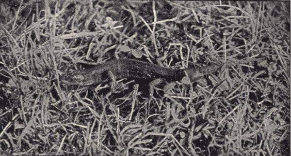
Fig. 123.—A lizard in the grass. (Photograph from life by Cherry Kearton; permission of Cassell & Co.)
Body form and organization.—The chief variations in body form among the reptiles are manifest when a turtle, lizard, and snake are compared. In the turtles, the body is short, flattened, and heavy, and provided always with four limbs, each terminating in a five-toed foot; in the lizards the body is more elongate and with usually four legs, but sometimes with two only, or even none at all; while in the snakes the long, slender, cylindrical body is legless or at most has mere rudiments of the hinder limbs. With the reptiles locomotion is as often effected by the bending or serpentine movements of the trunk as by the use of legs. Among lizards and snakes the body is covered with horny epidermal scales or plates, while[Pg 310] among the turtles and crocodiles there may be, in addition to the epidermal plates, a real deposit of bone in the skin whereby the effectiveness of the armor is increased. The epidermal covering of snakes and lizards is periodically molted, or, as we say, the skin is shed. The bright colors and patterns of snakes and of many lizards are due to the presence and arrangement of pigment-cells in the skin. Among some reptiles, notably the chameleons, the colors and markings can be quickly and radically changed by an automatic change in the tension of the skin.
Structure.—In reptiles, as in batrachians, the chief variations in the body skeleton are correlated with differences in external body form. In the short compact body of the turtles and tortoises the number of vertebræ is much smaller than in the snakes. Some turtles have only 34 vertebræ; certain snakes as many as 400. The reptilian skull, in the number and disposition of its parts and in the manner of its attachment to the spinal column, resembles that of the birds, although the cranial bones remain separate, not fusing as in the birds. In the snake the two halves of the lower jaw are not fused in front but are united by elastic ligaments, which condition, together with the extremely mobile articulation of the base of the jaws, allows the snakes to open their mouths so as to take in bodies of great size. All of the reptiles, except the turtles, are provided with small teeth which serve, generally, for seizing or holding prey and not for mastication. The poisonous snakes have one or more long, sharp, and grooved or hollow fangs (fig. 131). In the legless reptiles both shoulder and pelvic girdles may be wholly lacking; in the limbed forms both girdles are more or less well developed.
The tongue of many reptiles, notably the snakes, is bifid or forked, and is an extremely mobile and sensitive organ. The œsophagus is long and in the snakes can be stretched very wide so as to permit the swallowing of large animals whole. Reptiles breathe solely by lungs, of which there is a pair. They are simple and sac-like, the left lung being often much smaller than the other. In turtles and crocodiles the lungs are divided internally by septa into a number of chambers. Because of the rigidity of the carapace or "box" of turtles the air cannot be taken in the ordinary way by the use of the ribs and rib-muscles, but has to be swallowed. The reptilian heart consists of two distinct auricles and of two ventricles, which in most reptiles are only incompletely divided, the division into right and left ventricles being complete only among the crocodiles and alligators, the most highly organized of living reptiles.
The organs of the nervous system reach a considerable
degree of development in the animals of this class. The
brain in size and complexity is plainly superior to the
batrachian brain and resembles quite closely that of birds.
Of the organs of special sense those of touch are limited
to special papillæ in the skin of certain snakes and many
lizards. Taste seems to be little developed, but olfactory
organs of considerable complexity are present in most
forms, and consist of a pair of nostrils with olfactory
papillæ on their inner surfaces. The ears vary much in
degree of organization, crocodiles and alligators being the
only reptiles with a well-defined outer ear. This consists
of a dermal flap covering a tympanum. Eyes are always
present and are highly developed. They resemble the
eyes of birds in many particulars. All reptiles, excepting
the snakes and a few lizards, have movable eyelids, including
a nictitating membrane like that of the birds.
With the snakes the eye is protected by the outer skin,
which remains intact over it, but is transparent and
thickened to form a lens just over the inner eye. Turtles
and lizards have a ring of bony plates surrounding the[Pg 311]
[Pg 312]
eyes similar to that of the birds. In addition to the usual
eyes there is in many lizards a remarkable eye-like organ,
the so-called pineal eye, which is situated in the roof of
the cranium, and is believed to be the vestige of a true
third eye, which in ancient reptiles was probably a well-developed
organ.
Life-history and habits.—Most reptiles lay eggs from which the young hatch after a longer or shorter period of incubation. Usually the eggs are simply dropped on the ground in suitable places (although certain turtles dig holes in which to deposit them), where they are incubated by the general warmth of the air and ground. However, some of the giant snakes, the pythons for instance, hold the eggs in the folds of the body. In the case of some snakes and lizards the eggs are retained in the body of the mother until the young hatch; such reptiles are said to be ovoviviparous, because the young, although born alive, are in reality enclosed in an egg until the moment of birth. Among reptiles the newly hatched young resemble the parents in most respects except in size, yet striking differences in coloration and pattern are not rare. But there is in this class no metamorphosis such as characterizes the post-embryonic development of the batrachians.
The food of reptiles consists almost exclusively of animal substance, although some species, notably the green turtles and certain land-tortoises, are vegetable-feeders. The animal-feeders are mostly predaceous, the smaller species catching worms and insects, while the larger forms capture fishes, frogs, birds, and their eggs, small mammals, and other reptiles.
Classification.—The living Reptilia are divided into four orders, of which one includes only a single genus, Hatteria, a peculiar lizard found in New Zealand. The other three are the Squamata, which includes the lizards[Pg 313] and snakes,[17] distinguished by the scaly covering of the body, the Chelonia, which includes the tortoises and turtles, distinguished by the shell of bony plates which encloses the body, and the Crocodilia, which includes the crocodiles and alligators, whose bodies are covered with rows of sculptured bony scutes.
Tortoises and turtles (Chelonia).—Technical Note.—Obtain specimens of some pond- or land-turtle common in the vicinity of the school. The red-bellied and yellow-bellied terrapins (Pseudemys) or the painted or mud-turtles (Chrysemys) are common over most of the United States. (Pseudemys is found south of the Ohio River and Chrysemys north of it.) They may be raked up from creek-bottoms or fished for with strong hook and line, using meat as bait. They will live through the winter if kept in a cool place, without food or special care of any kind. Observe their swimming and diving, the retraction of head and limbs into the shell, the use of the third eyelid (nictitating membrane), and the swallowing of air.
Examine the external structure of a dead specimen (kill by thrusting a bit of cotton soaked with chloroform or ether into the windpipe; see opening just at base of tongue). Note shell consisting of a dorsal plate, the carapace, and ventral plate, the plastron, and the lateral uniting parts, the bridge. Note legs, and head with horny beak but no teeth. Compare with snake. The examination of the internal structure of the turtle can be readily made by sawing through the bridge on either side and removing the plastron. Note the ligaments which attach the plastron to the shoulder and pelvic girdles. Note muscles covering these bones. Note just behind the shoulder girdle the heart (perhaps still pulsating) and the dark liver on each side of it. Work out the alimentary canal, the trachea and lungs, and other principal organs, comparing them with those of the snake. The skeleton can be studied by dissecting and boiling and brushing away the flesh which still adheres to the bones. The comparison of the skeleton of the turtle with that of the snake is very instructive; marked differences in the skeletons of the two kinds of reptiles are obviously correlated with the differences in habits and shape of body. Note in the skeleton of the turtle especially the shoulder and pelvic girdles and limbs (absent in the snake) and small number of vertebræ and ribs.
Among the common turtles and tortoises of the United States are several species of soft-shelled turtles (Trionychidæ)[Pg 314] with carapace not completely ossified and both carapace and plastron covered by a thick leathery skin which is flexible at the margins; the snapping-turtle (Chelydra serpentina), common in streams and ponds, with shell high in front and low behind and head and tail long and not capable of being withdrawn into the shell; the red-bellied and yellow-bellied terrapins (Pseudemys), red and yellow, with greenish-brown and black markings, common on the ground in woods and among rocks and also near water and sometimes in it; the pond- or mud-turtle (Chrysemys), also brightly colored and usually confined to ponds and pond-shores; and the box-tortoise (Cistudo carolina), common in woods and upland pastures and readily recognizable by its ability to enclose itself completely in its shell by the closing down of the lids of the plastron. All of these fresh-water and land-turtles except the soft-shelled turtles belong to one family, the Emydidæ, but have somewhat diverse habits. Most of them are carnivorous, but few catch any very active prey. While some are strictly aquatic, others are as strictly terrestrial, never entering the water. The eggs of all are oblong and are deposited in hollows, sometimes covered in sand. The newly hatched young are usually circular in shape, and vary in color and pattern from the parents.
The "diamond-back terrapin" (Malaclemmys palustris), used for food, is a salt-water form "inhabiting the marshes along the Atlantic coast from Massachusetts to Texas. About Charleston [and Baltimore] they are very abundant and are captured in large numbers for market, especially at the breeding season, when the females are full of eggs. Further north they are dug from the salt mud early in their hibernation and are greatly esteemed, being fat and savory."
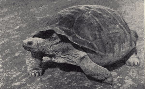
Fig. 124.—The giant land-tortoise of the Galapagos Islands, Testudo sp. These tortoises reach a length of four feet. (Photograph from life by Geo. Coleman.)
Strongly contrasting with the usually small land- and[Pg 315] fresh-water turtles are the great sea-turtles, such as the leather-back, the loggerhead and the green turtles. Some of these animals reach a length of six feet and more and a weight of nine hundred pounds, and have the feet compressed and fin-shaped for swimming. They live in the open ocean, coming on land only to lay their eggs, which are buried in the sand of ocean islands. These egg-laying visits are almost always made at night, and the turtles are then often caught by "turtlers." The flesh of most of the sea-turtles is used for food, and from the shell of certain species, notably the "hawk-bill" (Eretmochelys imbricata) the beautiful "tortoise-shell" used for making combs and other articles is obtained. The common green turtle (Chelonia mydas) of the Atlantic coast is the species most prized for food. It is a vegetarian, feeding on the roots of Zostera, the plant known in New England as eel-grass, though farther south it is called turtle-grass.[Pg 316] When grazing the turtles eat only the roots, the tops thus rising to the surface, where they indicate to the turtler the animal's whereabouts. The turtler, armed with a strong steel barb attached to a rope and loosely fitted to the end of a pole, carefully rows up to the unsuspecting animal, and with a strong thrust plunges the barb through its shell, withdraws the pole, and, grasping the rope, now firmly attached to the turtle's back, lifts the animal to the surface. Here, with assistance, he turns it into the boat, where it is rendered helpless by being thrown on its back and by having its flippers tied. These turtles are also caught on their breeding-grounds, being found on the sand at night by the turtler, turned over on their backs, and left thus securely caught until assistance comes to help get them into the boats.
Snakes and lizards (Squamata).—Technical Note.—A snake has already been dissected and studied. It will be instructive to compare the external structures, at least, of a lizard with that of the snake. Specimens of some species of the common swift (Sceloporus) are obtainable almost anywhere in the United States. The "pine-lizards" of the east belong to this genus. Lizards may be sought for in woods, along fences, and especially on warm rocks.
The group of lizards is a very large one, about 1,500 species being known, but it is represented in the United States by comparatively few species. Lizards are especially abundant in the tropics of South America. The strange and fantastic appearance presented by some of them has made certain species the object of much interest[Pg 317] and often fear on the part of the natives of tropical lands. In those regions are current extraordinary stories and beliefs regarding the habits and attributes of certain lizards like the basilisk and chameleon. Lizards are all more or less elongate and some are truly snake-like in form. The legs, though usually present and functional, are in many cases much reduced, and in some forms, as the glass-snake, either one or both pairs are so rudimentary as to have no external projection whatever. Although lizards are often regarded as being poisonous, only one genus, Heloderma, the Gila Monster, is really so. All others are perfectly harmless as far as poison is concerned, and most of them are unusually timid. They vary in size from a few inches to six feet in length. Most of them are terrestrial, some arboreal, and some aquatic.

Fig. 126.—The Gila monster, Heloderma horridum, the only poisonous lizard. (Photograph from life by J. O. Snyder.)
Among the lizards of this country the swifts and ground-lizards are familiar everywhere. In certain regions the glass-snake or joint-snake (Opheosaurus ventralis) is common. This animal, popularly considered to be a snake, has no external limbs, and its tail is so brittle, the vertebræ composing it being very fragile, that part of it may break off at the slightest blow. In time a new tail is regenerated. It lives in the central and northern part of the United States, and burrows in dry places. In the western part of the country horned toads (Phrynosoma) are common, about ten different species being known. These are lizards with shortened and depressed body and well-developed legs. The body is covered with protective[Pg 318] spiny protuberances, and in individual color and pattern resembles closely the soil, rocks, and cactus among which the particular horned toad lives. All the species of Phrynosoma are viviparous, seven or eight young being born alive at a time.
In New Mexico, Arizona, and northern Mexico the only existing poisonous lizards, the Gila Monster (Heloderma) (fig. 126) is found. This is a heavy, deep-black, orange-mottled lizard about sixteen inches long. There is much variance of belief among people regarding the Gila Monster, but recent experiments have proved the poisonous nature of the animal. The poison which is secreted by glands in the lower jaw flows along the grooved teeth into the wound. A beautiful and interesting little lizard found in the South is the green chameleon (Anolis principalis). Its body is about three inches long with a slender tail of five or six inches. The normal color of the chameleon is grass-green, but it may "assume almost instantly shades varying from a beautiful emerald to a dark and iridescent bronze color."
In the tropics many of the lizards reach great size and are of strange shape and patterns. The flying dragons (Draco) have a sort of parachute on each side of the body composed of a fold of skin supported by five or six false posterior ribs. These lizards live in the trees of the East Indies and "fly" or sail from tree to tree. They are very beautifully colored. The iguanas (Iguana) of the tropics of South America are commonly used for food. They live mostly in trees, and reach a length of five or six feet. The monitor (Varanus niloticus) is a great water-lizard that lives in the Nile, and feeds on crocodiles' eggs, of which it destroys great numbers. It is the principal enemy of the crocodile. When full grown it reaches a length of six feet or even more.
About 1,000 living species of snakes are known.[Pg 319] Usually they have the body regularly cylindrical, and without distinct division into body regions. Legs are wanting, locomotion being effected by the help of the scales and the ribs. No snake can move forward on a perfectly smooth surface and no snake can leap. In some forms, such as the pythons, external rudiments of the hind limbs are present, but do not aid in locomotion. The mouth is large and distensible so that prey of considerably greater size than the normal diameter of the snake's body is frequently swallowed whole. The sense of taste is very little if at all developed, as the food is swallowed without mastication. The tongue, which is protrusible and usually red or blue-black, serves as a special organ of touch. Hearing is poor, the ears being very little developed. The sense of sight is also probably not at all keen. Snakes rely chiefly on the sense of smell for finding their prey and their mates. The colors of snakes are often brilliant, and in many cases serve to produce an effective protective resemblance by harmonizing with the usual surroundings of the animal. The food of snakes consists almost exclusively of other animals, which are caught alive. Some of the poisonous snakes kill their prey before swallowing it, as do some of the constrictors. While most snakes live on the ground, some are semi-arboreal and others spend part or all of their time in water. Cold-region snakes spend the winter in a state of suspended animation; in the tropics, on the contrary, the hottest part of the year is spent by some species in a similar "sleep."
There are so many common snakes in the United States that only a few of the more familiar forms can be mentioned. The non-poisonous species of America belong to the family Colubridæ, while all but one of the poisonous species belong to the family Crotalidæ, characterized by the presence of a pair of erectile poison-fangs on the upper jaw. Among the commonest of the Colubridæ are the[Pg 320] garter snakes (Thamnophis) (fig. 127), always striped and not more than three feet long. The most widespread species is Thamnophis sirtalis, rather dully colored with three series of small dark spots along each side. The common water-snake (Natrix sipedon) is brownish with back and sides each with a series of about 80 large square dark blotches alternating with each other. It feeds on fishes and frogs, and although "unpleasant and ill-tempered" is harmless. One of the prettiest and most gentle of snakes is the familiar little greensnake (Cyclophis æstivus), common in the East and South in moist meadows and in bushes near the water. It feeds on insects and can be easily kept alive in confinement. A familiar larger snake is the blacksnake or blue racer (Bascaniom constrictor), "lustrous pitch black, general color greenish below and with white throat." It is "often found in the neighborhood of water, and is particularly partial to thickets of alders, where it can hunt for toads, mice, and birds, and being an excellent climber it is often seen among the branches of small trees and bushes, hunting for young birds in the nest." The chain-snake (Lampropeltis getulus) of the southeast and the king-snake (also a Lampropeltis) (fig. 128) of the central[Pg 321] States are beautiful lustrous black-and-yellow spotted snakes which feed not only on lizards, salamanders, small birds and mice but also on other snakes. The king-snake should be protected in regions infested by "rattlers." The spreading adder or blowing viper (Heterodon platirhinos), a common snake in the eastern States, brownish or reddish with dark dorsal and lateral blotches, depresses and expands the head when angry, hissing and threatening. Despite the popular belief in its poisonous nature this ugly reptile is quite harmless. It specially infests dry sandy places.
With the exception of the coral or beadsnake (Elaps fulvius), a rather small jet-black snake with seventeen broad yellow-bordered crimson rings, found in the southern States, the only poisonous snakes of the United States are the rattlesnakes and their immediate relatives, the copperhead and water-moccasin. These snakes all have a large triangular head, and the posterior tip of the body is, in the rattlesnakes, provided with a "rattle" composed of a series of partly overlapping thin horny capsules or cones of shape as shown in figure 130. These horny pieces are simply the somewhat modified successively formed epidermal coverings of the tip of the body,[Pg 322] which instead of being entirely molted as the rest of the skin is, are, because of their peculiar shape, loosely attached to one another, and by the basal one to the body of the snake. The number of rattles does not correspond to the snake's years for several reasons, partly because more than one rattle can be added to the tail in a year, and especially because rattles are easily and often broken off. As many as thirty rattles have been found on one snake. There are two species of ground-rattlesnakes or massasaugas (Sistrurus) in the United States and ten species of the true rattlesnakes (Crotalus). The centre of distribution of the rattlesnakes is the dry tablelands of the southwest in New Mexico, Arizona, and Texas. But there are few localities in the United States outside the high mountains in which "rattlers" do not occur or did not occur before they were exterminated by man. The copperhead (Agkistrodon contortix) is light chestnut in color, with inverted Y-shaped darker blotches on the sides, and[Pg 323] seldom exceeds three feet in length. It occurs in the eastern and middle United States from Pennsylvania and Nebraska southward. It is a vicious and dangerous snake, striking without warning. The water-moccasin (Agkistrodon piscivorous) is dark chestnut-brown with darker markings. The head is purplish black above. It is found along the Atlantic and Gulf coasts from North Carolina to Mexico, extending also some distance up the Mississippi valley. It is distinctively a water-snake, being found in damp swampy places or actually in water. It reaches a length of over four feet and is a very venomous snake, striking on the slightest provocation. The common harmless water-snake is often called water-moccasin in the southern States, being popularly confounded with this most dangerous of our serpents. The poison of all of these snakes is a rather yellowish, transparent, sticky fluid secreted by glands in the head, from which it flows through the hollow maxillary fangs. The character and position of the fangs are shown in figure 131. Remedial measures for the bite of poisonous snakes are, first, to stop, if possible, the flow of blood from the wound to the heart, by[Pg 324] compressing the veins between the wound and heart, then to suck (if the lips are unbroken) the poison from the wound, next to introduce by hypodermic injection permanganate of potash, bichloride of mercury or chromic acid into the wound, and finally perhaps to take some strong stimulant as brandy or whiskey.
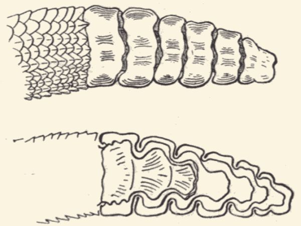
Fig. 130.—The rattles of the rattlesnake; the lower figure shows a longitudinal section of the rattle.
Of the kinds of snakes not found in this country perhaps the most interesting are the gigantic boa constrictors, anacondas, and pythons. Pythons are found in India, the islands of the Malay archipelago, and Australia, while the boas and anacondas live in the tropics of America. The largest pythons reach a length of thirty feet and some of the boas are nearly as large. These snakes feed on small mammals such as fawns, kids, water-rats, etc., and birds. The prey is swallowed whole, being first encircled and crushed to death in folds of the body. After a meal the python or boa lies in a sort of torpor for some time. A famous snake is the deadly cobra-da-capello of India. These snakes are so abundant and the bite is so nearly certainly fatal that thousands of persons are killed each year in India by it. Other extremely poisonous snakes are the vipers (Vipera cerastes), which live in the hot deserts of northern Africa. Over each eye there is a scaly[Pg 325] spine or horn, from which the name horned viper is derived. The most poisonous snake of South Africa is the large and ugly puff-adder, which puffs itself up when irritated. An interesting group of snakes is that of the Hydrophidæ or sea-snakes, which swim on the surface of the ocean by means of their flattened and oar-like tails. These forms live in the tropical portions of the Indian and Pacific oceans, ranging as far north as the Gulf of California, and spend their whole life in the water, "out of which they appear to be blind and soon die." They are extremely venomous, but are all of small size, rarely two feet long.
Crocodiles and alligators (Crocodilia).—The crocodiles and alligators are reptiles familiar by name and appearance, though seen in nature only by inhabitants or visitors in tropical and semitropical lands. In the United States there are two species of these great reptiles, the American crocodile (Crocodilus americanus), living in the West Indies and South America and occasionally found in Florida, and the American alligator (Alligator mississippiensis), common in the morasses and stagnant pools of the southern States. The alligator differs from the crocodiles in having a broader snout. It is rarely more than twelve feet long. The best-known crocodile is the Nile crocodile, which is not limited to the Nile, but is found throughout Africa. In the Ganges of India is found another member of this group of reptiles called the gavial. It is among the largest of the order, reaching a length of twenty feet. The crocodiles, alligators, and gavials comprise not more than a score of species altogether, but because of their wide distribution, great size, and carnivorous habits they are among the most conspicuous of the larger living animals. They live mostly in the water, going on land to sun themselves or to lay their eggs. They move very quickly and swiftly in water[Pg 326] but are awkward on land. Fish, aquatic mammals and other animals which occasionally visit the water are their prey. The gavial and Nile crocodile are both known to attack and devour human beings, and these species annually cause a considerable loss of life. But few such fatalities, however, are accredited to the American alligator.
Technical Note.—The English sparrow may be found now in cities and villages all over the United States. It has become a veritable pest, and the killing of the few needed for the laboratory may be looked on as desirable rather than deplorable, as is the killing of birds in almost all other cases. The males have a black throat, with the other head-markings strong and contrasting (black, brown, and white), while the females have a uniform grayish and brownish coloration on the head.
Specimens are best taken alive, as shooting usually injures them for dissection. One can rely on the ingenuity of the boys of the class to procure a sufficient number of specimens. Observations on the habits of the birds should be made by the pupils as they go to and from school. For dissection use fresh specimens if possible. If desirable a pigeon or dove may be used in place of the sparrow.
External structure.—Note in the sparrow the same general arrangement of body parts as in the toad, the body being divided into head, upper limbs, trunk, and lower limbs. In the toad, however, all of the limbs are fitted for walking and jumping, whereas in the sparrow the anterior pair of appendages, the wings, are modified to be organs of flight, and the posterior limbs are specially adapted for perching. Note that the sparrow is covered with feathers, some long, some short, in some places thick and in others thin, but all fitting together to form a complete covering for the body. Note also that the anterior end of the head is prolonged into a hard bony structure,[Pg 328] the bill, covered with horny substance. This horny substance together with the feathers and horny covering of the feet are modified portions of the skin. Note the long quill-feathers attached to the posterior edge of the wing. By these the bird sustains its flight. Other long quill-feathers are attached to the posterior end of the body, forming the tail. By a system of muscles connected with these feathers they act together, serving as a rudder during flight and as a balancing contrivance when perching. Note just above the bill two openings protected by tufts of feathers. What are these openings? How are they connected with the mouth? Note the large eyes, and at the inner angle of each the delicate nictitating membrane which can be drawn over the ball. Does the bird have external ears? Lift the feathers just above the tail (the upper tail-coverts) and note a small median gland, the oil-gland, from which the bird derives the oil with which it oils its feathers. Beneath the tail note the opening from the alimentary canal and from the kidneys and reproductive organs. This is called the cloacal opening.
Examine in detail some of the feathers. In one of the quill-feathers note the central stem or shaft composed of two parts, a basal hollow quill, which bears no web and by which the feather is inserted in the skin, and a longer, terminal, four-sided portion, the rachis, which bears on either side a web or vane. Each vane is composed of many narrow linear plates, the barbs, from which rise (like miniature vanes) many barbules. Each barbule bears many fine barbicels and hamuli or hooklets. The barbs of the feather are interlocked. How is this effected? The feathers which overlie the whole body and bear the color pattern are called contour-feathers. How do they differ from or correspond with the quill-feathers in structure? Soft feathers called down-feathers or plumules, cover the body more or less completely, being, however,[Pg 329] mostly hidden by the contour-feathers; the barbs of these are sometimes not borne on a rachis, but arise as a tuft from the end of the quill. Certain other feathers which have an extremely slender stem and usually no vane, except a small terminal tuft of barbs, are called thread-feathers, or filoplumules. They are rather long, but are mostly hidden by the contour-feathers. In certain birds they stand out conspicuously, as the vibrissæ about the nostrils.
In the determination of birds by the use of a classificatory "key" (see p. 359) it is necessary to be familiar with the names applied to the various external regions of the body and plumage, and with the terms used to denote the special varying conditions of these parts. By reference to figure 133 the names of the regions or parts most commonly referred to may be learned. A full account of all of the external characters with definitions of the various terms used in referring to them may be found in Coues's "Key to North American Birds."
Technical Note.—Pull the feathers from the body, being careful not to tear the skin.
In the fish and toad, already studied, the head is closely joined to the trunk. How is it with the bird? Observe that the knee of the sparrow is covered by feathers and that it is the ankle which extends down as the bare unfeathered part to the digits. How many digits have the feet of the bird? How are they arranged?
Internal structure (fig. 132).—Technical Note.—With a pair of scissors cut just beneath the skin anteriorly from the cloacal opening to the angle of the lower jaw. Pin the sparrow on its back by the wings, feet, and bill. Push back the skin from both sides and pin out.
Note the large powerful pectoral muscles. Note a hard
median projection of bone, the sternum, which is a large[Pg 330]
[Pg 331]
keel-shaped bone with lateral expansions to which are
attached the ribs. Where are the largest and most
powerful muscles of the toad located? Where are they
in the fish? In the bird the most powerful muscles are
these pectoral muscles, which move the wings in flight.
Technical Note.—Cut the pectoral muscles from the left side of the sternum, push back and pin to one side. With a strong pair of scissors cut through the ribs on the left side of the sternum and through the overlying bones. Lift the whole sternum, with the right pectoral muscle attached, to the left side of the pan and pin it down. Cut through the membrane which covers the viscera and cover the dissection with water.
In this operation note the V-shaped wishbone in front of the sternum. It is composed of the two clavicles with their inner ends fused. Note the stout coracoid bones extending from the anterior end of the sternum to the shoulder.
Note near the middle of the body the heart with the large blood-vessels proceeding from it. Behind the heart lies the large reddish-brown liver, and on the left side below the liver is the large gizzard or muscular stomach. Note the viscera folded over themselves in the body-cavity. Push them temporarily aside and note in the dorsal region under the heart large pinkish spongy sacs, the lungs. These are attached by short tubes, the bronchi, to the long cartilaginous trachea. At the union of the bronchi with the trachea is a small expansion with cartilaginous walls, within which are stretched small bands of muscles. This organ is the syrinx, the song- or voice-apparatus of the bird. It should be cut open and carefully examined. Trace the trachea forward to its anterior end. It opens by a glottis into the larynx, a slightly swollen chamber with cartilaginous walls. Note the U-shaped hyoid bone surrounding the front of the glottis. Through a blowpipe or quill inserted into the glottis blow air into the trachea and observe the inflation of the lungs and of[Pg 332] certain large air-sacs in the abdomen, which communicate with them.
Beneath the trachea note the long œsophagus. Inflate the œsophagus with a blowpipe and note how distensible is its lower end near the breast. This distensible portion is called the crop. If the alimentary canal be drawn out straight the œsophagus will be found to run as an almost straight tube down the left side of the body to the gizzard. This latter organ has very thick muscular walls and in it the food is ground up among the small bits of gravel it contains. Extending from the gizzard near the entrance of the œsophagus note the long pyloric loop of the intestine called duodenum. Within this loop is a long pinkish gland, the pancreas, which empties by a duct into the duodenum. Into the duodenum also the overlying liver empties its secretion of bile from the median-placed gall-bladder. From the duodenum the small intestine or ileum extends with many convolutions to its exit through the cloacal aperture. On the intestine near the cloacal opening note a pair of glandular structures, the cæca. The short part of intestine between the cæca and cloaca is called the rectum. On the left side of the body beneath the gizzard note a dark glandular structure, the spleen.
Make a drawing of the dissection as so far worked out.
Technical Note.—Remove the alimentary canal, cutting it free posteriorly at the cæca and anteriorly just above the muscular gizzard. Cut open the gizzard and note its structure. The contained sand and gravel grains are picked up by the bird as it eats.
On either side of the throat note the well-defined thyroid gland; in young sparrows will be noted on each side of the neck a mass of tissue, the remains of the thymus gland, which disappears in the adult.
Cut transversely through the lower end of the heart and note that the ventricles are wholly distinct, whereas in the toad and snake they are incompletely separated. In the[Pg 333] bird there is a complete double circulation. Its blood is not mixed, the pure with the impure, as in the toad and snake. Blood passing through the right auricle and ventricle goes to the lungs; on its return to the heart purified, it enters the left auricle and left ventricle thence to pass out over the body through the arteries.
Note the large aorta given off from the left ventricle. Note the two large branches, the innominate arteries, given off by it near its origin. Each innominate divides into three smaller arteries, a carotid, branchial, and pectoral. The aorta itself turns toward the back and continues posteriorly through the body as the dorsal aorta. To the right auricle come three large veins, the right and left præcavæ and the postcava. Each præcava is formed by three veins, the jugular from the head, the branchial from the wing, and the pectoral from the pectoral muscles. The postcava comes from the liver. From the right ventricle go the short right and left pulmonary arteries to the lungs, and from the lungs the blood is brought to the left auricle through the right and left pulmonary veins.
Technical Note.—For a detailed study of the circulation of the bird the teacher should inject the blood system of some larger bird, as a pigeon or fowl, for a class-demonstration. (For a guide, use Parker's "Zootomy," p. 209, or Martin and Moale's "How to Dissect a Bird," pp. 135-140 and pp. 148, 149.)
In the posterior dorsal region of the body-cavity will be found large three-lobed organs fitting into the spaces between the bones of the back on either side. These are the kidneys, and from their outer margins on each side a ureter runs posteriorly into the cloaca. Overlying the anterior ends of the kidneys are the reproductive organs. In the male these glands consist of firm, whitish, glandular bodies. From each runs a long convoluted vas deferens, which enters the cloaca. This tube corresponds to the egg-duct of the female. In the female the right egg-gland[Pg 334] and egg-duct or oviduct are wanting. The left egg-gland appears as a glandular mass; during the breeding season yellow ova or eggs in various stages of development project from its surface. The oviduct opens by a funnel-shaped mouth near the egg-gland and runs thence to the cloaca. The eggs pass from the egg-gland into the body-cavity, where they are caught in the upper end of the oviduct and carried down and out through the cloacal opening. It is in the oviduct that the egg derives its accessory covering, which consists of a white or albuminous portion, together with several enveloping membranes and the hard shell enclosing all.
Remove the top of the skull and note the large brain. What portions of the brain make up the greater part of it? Note the differences between this brain and that of the toad. Trace the principal cranial nerves. Work out the spinal cord and principal spinal nerves. For an account of the nervous system of the sparrow see Martin and Moale's "How to Dissect a Bird," pp. 150-163.
Technical Note.—For a study of the skeleton of the sparrow a specimen should be cleaned by boiling in a soap-solution (see p. 452).
In the sparrow's skeleton note the compactness of the skull and the fusion of its bones. Observe the long cervical vertebræ which support the skull, also the thoracic vertebræ bearing the ribs and sternum. How many of each of these kinds of vertebræ are there? The vertebræ posterior to the thorax are more or less fused together to form the sacrum, which, with the pelvic girdle, supports the leg-bones. The bones of the tail consist of a number of very small vertebræ, some of which are fused together. Note the correspondence between the bones of the leg and those of the wing. What are the names of each of the bones of each limb, and what are the corresponding bones in the two limbs? The wings and legs being modified for different uses, their various bones have assumed different relations to each other and to the body, for they are bent at directly opposite angles and the attachment of muscles is different. Compare the skeleton of the bird with that of the toad. (For a detailed account of the skeleton of the bird see Parker's "Zootomy," pp. 182-209, or Martin and Moale's "How to Dissect a Bird," pp. 102-125.)
Life-history and habits.—The English sparrow was first introduced into the United States in 1850, and since that time has rapidly populated most of the cities and towns of the country. On account of its extreme adaptability to surroundings, its omnivorous food-habits and its fecundity it survives where other birds would die out. It also crowds out and has caused the disappearance or death of other birds more attractive and more useful. The sparrow annually rears five or six broods of young, laying from six to ten eggs at each sitting. Had it no enemies a single pair of sparrows would multiply to a most astonishing number. The sparrow has, however, a number of enemies, most common among them perhaps being the "small boy," but birds and mammals play the chief part in the destruction. The smaller hawks prey upon them, and rats and mice destroy great numbers of their young and of their eggs whenever the nests can be reached. The sparrow is omnivorous and when driven to it is a loathsome scavenger, though at other times its tastes are for dainty fruits. Its senses of perception are of the keenest; it can determine friend or foe at long range. The nesting habits are simple, the nests being roughly made of any sort of twigs and stems mixed with hair and feathers and placed in cornices or trees. A maple-tree in a small Missouri town contained at one time thirty-seven of these nests.
Birds are readily and unmistakably distinguishable from all other kinds of animals by their feathers. They are further distinguished from the reptiles on one hand by their possession of a complete double circulation and by their warm blood (normally of a temperature of from 100-112° F.), and from the mammals on the other by the absence of milk-glands. There are about 10,000 known species of living birds; they occur in all countries, being most numerous and varied in the tropics. Birds are exceptionally available animals for the special attention of beginning students, because of their abundance and conspicuousness and the readiness with which their varied and interesting habits may be observed. The bright colors and characteristic manners which make the identification of the different kinds easy, the songs and flight, and the feeding, nesting and general domestic habits of birds are all excellent subjects for personal field-studies by the students. We shall therefore devote more attention to the birds than to the other classes of vertebrates, just as we selected the insects among the invertebrates for special consideration.
Body form and structure.—The general body form and external appearance of a bird are too familiar to need description. The covering of feathers, the modification of the fore limbs into wings, and the toothless, beaked mouth are characteristic and distinguishing external features. The feathers, although covering the whole of the surface of the body, are not uniformly distributed, but are grouped in tracts called pterylæ, separated by bare or downy spaces called apteria. They are of several kinds, the short soft plumules or down-feathers, the large stiffer contour-feathers, whose ends form the outermost covering of the body, the quill-feathers of the wings and tail, and[Pg 337] the fine bristles or vibrissæ about the eyes and nostrils called thread-feathers. The fore limbs are modified to serve as wings, which are well developed in almost all birds. However, the strange Kiwi or Apteryx of New Zealand with hair-like feathers is almost wingless, and the penguins have the wings so reduced as to be incapable of flight, but serving as flippers to aid in swimming underneath the water. The ostriches and cassowaries also have only rudimentary wings and are not able to fly. Legs are present and functional in all birds, varying in relative length, shape of feet, etc., to suit the special perching, running, wading, or swimming habits of the various kinds. Living birds are toothless, although certain extinct forms, known through fossils, had large teeth set in sockets on both jaws. The place of teeth is taken, as far as may be, by the bill or beak formed of the two jaws, projecting forward and tapering more or less abruptly to a point. In most birds the jaws or mandibles are covered by a horny sheath. In some water and shore forms the mandibular covering is soft and leathery. The range in size of birds is indicated by comparing a humming-bird with an ostrich.
Many of the bones of birds are hollow and contain air. The air-spaces in them connect with air-sacs in the body, which connect in turn with the lungs. Thus a bird's body contains a large amount of air, a condition helpful of course in flight. The breast-bone is usually provided with a marked ridge or keel for the attachment of the large and powerful muscles that move the wings, but in those birds like the ostriches, which do not fly and have only rudimentary wings, this keel is greatly reduced or wholly wanting. The fore limbs or wings are terminated by three "fingers" only; the legs have usually four, although a few birds have only three toes and the ostriches but two.
As birds have no teeth with which to masticate their food, a special region of the alimentary canal, the gizzard, is provided with strong muscles and a hard and rough inner surface by means of which the food is crushed. Seed-eating birds have the gizzard especially well developed, and some birds take small stones into the gizzard to assist in the grinding. The lungs of birds are more complex than those of batrachians and reptiles, being divided into small spaces by numerous membranous partitions. They are not lobed as in mammals, and do not lie free in the body-cavity, but are fixed to the inner dorsal region of the body. Connected with the lungs are the air-sacs already referred to, which are in turn connected with the air-spaces in the hollow bones. By this arrangement the bird can fill with air not only its lungs but all the special air-sacs and spaces and thus greatly lower its specific gravity. The vocal utterances of birds are produced by the vocal cords of the syrinx or lower larynx, situated at the lower end of the trachea just where it divides into the two bronchial tubes, the tracheal rings being here modified so as to produce a voice-box containing two vocal cords controlled by five or six pairs of muscles. The air passing through the voice-box strikes against the vocal cords, the tension of which can be varied by the muscles. In mammals the voice-organ is at the upper or throat end of the trachea.
The heart of birds is composed of four distinct chambers, the septum between the two ventricles, incomplete in the Reptilia, being in this group complete. There is thus no mixing of arterial and venous blood in the heart. The systemic blood-circulation being completely separated from the pulmonic, the circulation is said to be double. The circulation of birds is active and intense; they have the hottest blood and the quickest pulse of all animals. In them the brain is compact and large, and more highly[Pg 339] developed than in batrachians and reptiles, but the cerebrum has no convolutions as in the mammals. Of the special senses the organs of touch and taste are apparently not keen; those of smell, hearing, and sight are well developed. The optic lobes of the brain are of great size, relatively, compared with those of other vertebrate brains, and there is no doubt that the sight of birds is keen and effective. The power of accommodation or of quickly changing the focus of the eye is highly perfected. The structure of the ear is comparatively simple, there being ordinarily no external ear, other than a simple opening. The organs of the inner ear, however, are well developed, and birds undoubtedly have excellent hearing. The nostrils open upon the beak, and the nasal chambers are not at all complex, the smelling surface being not very extensive. It is probable that the sense of smell is not, as a rule, especially keen.
Development and life-history.—All birds are hatched from eggs, which undergo a longer or shorter period of incubation outside the body of the mother, and which are, in most cases, laid in a nest and incubated by the parents. The eggs are fertilized within the body of the female, the mating time of most birds being in the spring or early summer. Some kinds, the English sparrow, for example, rear numerous broods each year, but most species have only one or at most two. The eggs vary greatly in size and color-markings, and in number from one, as with many of the Arctic ocean birds, to six or ten, as with most of the familiar song-birds, or from ten to twenty, as with some of the pheasants and grouse. The duration of incubation (outside the body) varies from ten to thirty days among the more familiar birds, to nearly fifty among the ostriches. The temperature necessary for incubation is about 40° C. (100° F.). Among polygamous birds (species in which a male mates with several or many[Pg 340] females) the males take no part in the incubation and little or none in the care of the hatched young; among most monogamous birds, however, the male helps to build the nest, takes his turn at sitting on the eggs, and is active in bringing food for the young, and in defending them from enemies. The young, when ready to hatch, break the egg-shell with the "egg-tooth," a horny pointed projection on the upper mandible, and emerge either blind and almost naked, dependent upon the parents for food until able to fly (altricial young), or with eyes open and with body covered with down, and able in a few hours to feed themselves (precocial young).
More details regarding the eggs, nest, and young of birds will be given later in this chapter.
Classification.—The class Aves is usually divided into numerous orders, the number and limits of these as published in zoological manuals varying according to the[Pg 341] opinions of various zoologists. The rank of an order in this group is far lower than in most other classes. In other words, the orders are very much alike and are recognized mainly for the convenience in breaking up the vast assemblage of species. In North America practically all the ornithologists have agreed upon a scheme of classification, which will therefore be adopted in this book. According to this classification the eight hundred (approximately) known species of North American birds represent seventeen orders. Certain recognized orders, for example, the ostriches, are not represented naturally in North America at all. As birds can usually be readily identified, the species being easily distinguished by general external appearance, and as there are many excellent book-guides to their classification, the beginning student can specially well begin with them his study of systematic zoology, which concerns the identification and classification of species. In a later paragraph are given therefore some suggestions for field and laboratory work in the determination of local bird-faunæ. In the following paragraphs each of the American orders is briefly discussed, as is also the foreign order of ostriches.
The ostriches, cassowaries, etc. (Ratitæ).—The ostriches, familiar to all from pictures and to some from live individuals in zoological gardens and menageries, or stuffed specimens in museums, together with a few other similar large species, are distinguished from all other birds by having the breast-bone flat instead of keeled. There are about a score of species of ostriches and ostrich-like birds all confined to the southern hemisphere. In them the wings are so reduced that flight is impossible, but the legs are long and strong, and they can run as swiftly as a galloping horse. They are said to have a stride of over twenty feet. They use their legs also as weapons, kicking viciously when angered. The true[Pg 342] ostriches (Struthio camelus) (fig. 135) live in Africa. They are the largest living birds, reaching a height of nearly seven feet and weighing as much as two hundred pounds. They are hunted for their feathers, and are now kept in captivity and bred in South Africa and California for the same purpose. About five million dollars' worth of ostrich-feathers are used each year. The eggs, which are from five to six inches long and nearly five inches thick, are laid in shallow hollows scooped out in the sand of the desert. The male undertakes most of the incubation, although when the sun is hot no brooding is necessary.[Pg 343] The young (fig. 136) hatch in from seven to eight weeks, and can run about immediately.
The rheas, found in South America, and the cassowaries of Australia are the only other living ostrich-like birds. Their feathers are of much less value than those of the true ostrich.
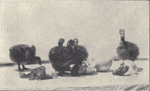
Fig. 136.—Young ostriches just from egg; on ostrich farm at Pasadena, California. (Photograph from life.)
The loons, grebes, auks, etc. (Pygopodes).—The
loons, grebes, and auks are aquatic birds, living in both
ocean and fresh waters. Their feet are webbed or lobed,
and their legs set so far back that walking is very difficult
and awkward. But all the birds of this order are excellent
swimmers and divers. They are distinctively the
diving birds. They have short wings and almost no tail.
The dab-chick or pied-billed grebe (Podilymbus podiceps)
is common in ponds over all the country. Its eggs are
laid in a floating nest of pond vegetation and are often
covered with decaying plants. The horned grebe
(Colymbus auritus) is common west of the Mississippi in
lakes and ponds. The loon or great northern diver[Pg 344]
[Pg 345]
(Gavia imber), found all over the United States in winter,
is the largest of this group, reaching a length (from bill to
tip of tail) of three feet. It is black above with many
small white spots, and with a patch of white streaks on
each side of the neck and on the throat; it is white on
breast and belly. The female is duller, being brownish
instead of black.
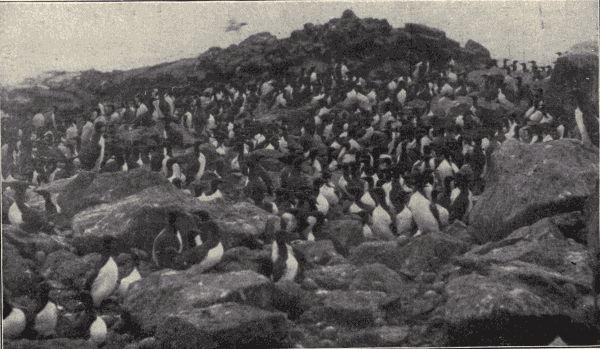
Fig. 137.—Murres, Uria troile californica, on Walrus Island, (Pribilof Group) Behring's Sea. Note the eggs scattered about over the bare rocks. (Photograph from life by the Fur Seal Commission.)
The auks, guillemots, puffins, and murres (fig. 137) are ocean birds which gather, in the breeding season, in countless numbers on the bleak rocks and inaccessible cliffs of the northern oceans. Each female lays a single egg (in some cases two or at most three) on the bare rock or in a crevice or sort of burrow. These birds mostly fly well, but are especially at home in the water, feeding exclusively on animal substances found there. A famous species is the great auk (Alca impennis), which has become extinct in historical times. The last living specimen was seen in 1844.
The gulls, terns, petrels, and albatrosses (Longipennes).—The Longipennes are water-birds, mostly maritime, with webbed feet and very long and pointed wings. They are all strong flyers, and most of them are beautiful birds. Their prevailing colors are white, slaty or lead-blue, black, and, in the young, mottled brownish. They subsist chiefly on fish, but any animal substance will be eagerly picked up from the water; some of the gulls forage inland. Occasionally great flocks may be seen following a plow near the shore and feeding on the grubs and worms exposed in the freshly-turned soil. Some of the gulls, like the great black-backed gull (Larus marinus), attain a length of two and one-half feet. The terns (Sterna) are mostly smaller than the gulls, have a bill not so heavy and not hooked, and have the tail forked.
The fulmars, shearwaters, petrels, and albatrosses are[Pg 346] strictly maritime. The albatrosses are very large, the largest being three feet long with a spread of wing of seven feet. They are often found flying easily over the open ocean at great distances from land. Like the auks and puffins, the fulmars and shearwaters gather in extraordinary numbers on rocky ocean islets or cliffs of the coast to breed.
The cormorants, pelicans, etc. (Steganopodes).—The Steganopodes are water-birds with full-webbed feet, and prominent gular pouch, swimmers rather than flyers like the Longipennes. The cormorants (Phalacrocorax) inhabit rocky coasts and are green-eyed, large, heavy, black birds with greenish-purple and violet iridescence; they are among the most familiar of seashore birds. They feed chiefly on fish and dive and swim under water with great ability. Cormorants are rather gregarious, keeping together in small groups when fishing, migrating often in great flocks, and in the breeding season gathering in immense numbers on certain rocky cliffs or islets. They build their nests of sticks and sea-weed; the eggs are three or four, and usually bluish green with white, chalky covering substance.
The pelicans are large, long-winged, short-legged water-birds with enormous bill and large gular sac which is used as a dip-net to catch fish. There are three species in North America, the white pelican (Pelecanus erythrorhynchus) occurring over most of the United States, the brown pelican (P. fuscus) of the Gulf of Mexico, and the California brown pelican (P. californicus) of the Pacific coast.
An interesting member of this order is the famous frigate or man-of-war bird (Fregata aquila), with very long wings and tail and feet extraordinarily small. The frigates have the greatest command of wing of all the birds. They cannot dive and can scarcely swim or walk.
The ducks, geese, and swans (Anseres).—The familiar wild ducks, of which there are forty species in North American fresh and salt waters; the geese, of which there are sixteen species, and the three species of wild swans constitute the order Anseres. The bill in these birds is more or less flattened and is also lamellate, i.e. furnished along each cutting-edge with a regular series of tooth-like processes; the feet are webbed, and the body is heavy and flattened beneath. Of the fresh-water or inland ducks, the more familiar are the mallard (Anas boschas), a large duck with head (male) and upper neck rich glossy green; the blue-winged teal (Querquedula discors) and green-winged teal (Nettion carolinense); the shoveller (Spatula clypeata) with spoon-shaped bill; the beautiful crested wood-duck (Aix sponsa); the expert diver, the plump little ruddy duck (Erismatura rubida), and others. Of the coastwise ducks, the canvas-back (Aythya vallisneria) is famous because of its fine flavor, while among the strictly maritime ducks the eiders (Somateria), which live in Arctic regions, are well known for their fine down. Of the geese, the commonest is the well-known Canada goose (Branta canadensis), while the pure-white snow-goose (Chen hyperborea), with black wing-feathers and red bill, is not unfamiliar. The wild swans (Olor) are the largest birds of the order, and are less familiar than the ducks and geese.
The ibises, herons, and bitterns (Herodiones).—The tall, long-necked, long-legged, wading birds, known as herons and ibises, compose a small order, the Herodiones, of which but few representatives are at all familiar. Perhaps the most abundant species is the green heron (Ardea virescens) or "fly-up-the-creek," one of the smaller members of the order. The crown, back, and wings are green, the neck purplish cinnamon, and the throat and fore neck white-striped. This bird is commonly[Pg 348] seen perching on an overhanging limb, or flying slowly up or down some small stream. The great blue heron (Ardea herodias) is common over the whole country. It is four feet long and grayish blue, marked with black and white. It may be seen standing alone in wet meadows or pastures, or flying heavily, with head drawn back and long legs outstretched. It breeds singly, but oftener in great heronries, in trees or bushes. Its large bulky nests contain three to six dull, greenish-blue eggs about two and one-half inches long. The white egrets of the Southern States are shot for their plumes and have been locally exterminated in some places. The night-herons (Nycticorax) differ from the other forms in having both the neck and legs short. The bittern (Botaurus lentiginosus), Indian hen, stake-driver, or thunder-pumper, as it is variously called, is a familiar member of the order, found in marshes and wet pastures, and known by its extraordinary call, sounding like the "strokes of a mallet on a stake." In color it is brownish, freckled and streaked with tawny whitish and blackish. Its nest is made on the ground; its eggs, from three to five in number, are brownish drab and about two inches long.
The cranes, rails, and coots (Paludicolæ).—The cranes, of which three species are known in North America, are large birds with long legs and neck, part of the head being naked or with hair-like feathers. The rare whooping crane (Grus americana) is pure white with black on the wings, and is fifty inches long from tip of bill to tip of tail. The sand-hill crane (G. mexicana) is slaty gray or brownish in color, never white, and although rare in the East is quite common in the South and West. Cranes build nests on the ground, and lay but two eggs, about four inches long, brownish drab in color with large irregular spots of dull chocolate-brown.
The rails are smaller than the cranes, with short wings and very short tail. They live in marshes and swamps, and in flying let the legs hang down. Their legs are strong, and for escape they trust more to speed in running than to flight. They are hunted for food. The most abundant rail is the "Carolina crake" or "sora" (Porzana carolina), small and olive-brown with numerous sharp white streaks and specks. Many of these birds are shot each year during migration in the reedy swamps of the Atlantic States. The American coot or mud-hen (Fulica americana), dark slate-color with white bill, is one of the most familiar pond-birds over all temperate North America. Its nest consists of a mass of broken reeds resting on the water; the eggs number about a dozen, and are clay-color with pin-head dots of dark brown.
The snipes, sandpipers, plover, etc. (Limicolæ).—The large order Limicolæ, the shore-birds, includes the slender-legged, slender-billed, round-headed, rather small wading birds of shores and marshes familiar to us as snipes, plovers, sandpipers, curlews, yellow-legs, sandpeeps, turnstones, etc. Most of them are game-birds, such forms as the woodcock and Wilson's or English snipe being much hunted. The food of these birds consists of worms and other small animals, which are chiefly obtained by probing with the rather flexible, sensitive, and usually long bill in the mud or sand. The killdeer (Ægialitis vocifera), familiar to all in its range by its peculiar call and handsome markings, the upland or field plover (Bartramia longicauda), with its long legs and melodious quavering whistle, the tall, yellow-shanked "telltale" or yellow-legs (Totanus melanoleucus) of the marshes and wet pastures, are among the most widespread and familiar species of the order. On the seashore the dense flocks of white-winged, whisking sandpipers and the quickly running groups of plump ring-necked[Pg 350] plover are familiar sights. One of the largest birds of this order is the long-billed curlew (Numenius longirostris) of the upland pastures. The bill of the curlew is long and curved downwards. The nests of these shore-birds are made on the ground and are usually little more than shallow depressions in which the few spotted eggs (four is a common number) are laid. The young are precocial.
The grouse, quail, pheasants, turkeys, etc. (Gallinæ).—The Gallinæ include most of the domestic fowls, as the hen, turkey, peacock, guinea-fowls, and pheasants, and the grouse, quail, partridges, and wild turkeys. The chief game-birds of most countries belong to this order. They have the bill short, heavy, convex, and bony, adapted for picking up and crushing seeds and grains which compose their principal food. Their legs are strong and usually not long, and are often feathered very low down. The Gallinæ are mostly terrestrial in habit and are sometimes known as the Rasores or "scratchers." Among the more familiar wild gallinaceous birds are the quail or "Bob white" (Colinus virginianus), abundant in eastern and central United States, the ruffed grouse (Bonasa umbellus) of the Eastern woods, and the prairie-chicken (Tympanuchus americanus) of the Western prairies. The sage-hen (Centrocercus urophasianus), the largest of the American grouse, reaching a length of two and one-half feet, is an interesting inhabitant of the sterile sagebrush plains of the West. The ptarmigan (Lagopus) or snow-grouse, represented by several species, are found either among the rocks and snow-banks above timber line on high mountains, or in the Arctic regions. In summer their plumage is brown and white; in winter they turn pure white to harmonize with the uniform snow-covering. On the Pacific coast are several species of quail, all differing much from those of the East. These Western species have beautiful crests of a few or several[Pg 351] long plume-feathers, the body-plumage being also unusually beautiful. The eggs of all the Gallinæ are numerous and are laid in a rude nest or simply in a depression on the ground. In many of the species polygamy is the rule. The young are precocial.
The doves and pigeons (Columbæ).—The doves and pigeons constitute a small order, the Columbæ, closely related to the Gallinæ. A distinguishing characteristic of the Columbæ lies in the bill, which is covered at the base with a soft swollen membrane or cere in which the nostrils open. The members of this order feed on fruits, seeds, and grains. Our most familiar wild species is the mourning-dove or turtle-dove (Zenaidura macroura) found abundantly all over the country. It lays two eggs in a loose slight nest in a low tree or on the ground. The beautiful wild or passenger pigeon (Ectopistes migratorius) was once extremely abundant in this country, moving about in tremendous flocks in the Eastern and Central States. But it has been so relentlessly hunted that the species is apparently becoming extinct. In the Rocky and Sierra Nevada mountains is a rather large dove, the band-tailed pigeon (Columba fasciata), which subsists chiefly on acorns. The domestic pigeon represented by numerous varieties, pouters, carriers, ruff-necks, fan-tails, etc., is the artificially selected descendant of the rock-dove (Columba livia). The young of all pigeons are altricial.
The eagles, owls, and vultures (Raptores).—The "birds of prey" compose one of the larger orders, the members of which are readily recognizable. In all the bill is heavy, powerful, and strongly hooked at the tip. The feet are strong, with long, curved claws (small in the vultures) and are fitted for seizing and holding living prey, such as smaller birds, fish, reptiles, and mammals which constitute the principal food of the true raptorial species. The vultures feed on carrion. The turkey[Pg 352] buzzard (Cathartes aura) is the most familiar of the three species of carrion-feeding Raptores found in the United States. The buzzard nests on the ground or in hollow stumps or logs, and lays two white eggs (sometimes only one) blotched with brown and purplish. The largest North American vulture is the California condor (Pseudogryphus californianus), which attains a length of four and one-half feet, with a spread of wing of nine and one-half feet. Of the eagles, the most widespread and commonest is the bald eagle (Haliætus leucocephalus). It is three feet long and when adult has the head and neck white. The golden eagle (Aquila chrysætos) has the neck and head tawny brown. Of the many species of hawks, the marsh harrier (Circus hudsonius), abundant all over the country and readily known by its white rump, is one of[Pg 353] the most familiar. The name "chicken-hawk" is given to two or three different species of large broad-winged hawks of the genus Buteo. The stout little sparrow-hawk (Falco sparverius), common over the whole country, is familiar and readily recognizable by its pronounced bluish and black wings and black-and-white banded chestnut tail. Altogether fifty species of hawks and eagles are found in this country. Of the owls, the barn-owl (Strix pratincola) with its long triangular face and handsome mottled and spotted tawny coat is more or less familiar; the great horned owl (Bubo virginianus), the snowy owl (Nyctea nyctea), and the great gray owl (Scotiaptex cinerea) are the common large species, while the red screech-owl (Megascops asio) (fig. 138), the most abundant owl in the country, and the strange burrowing owl (Speotyto cunicularia), which lives in the holes of prairie-dogs and ground-squirrels in the West, are familiar smaller ones. Thirty-two species of owls are recorded from North America.
The parrots (Psittaci).—The parrots, of which only one species is native in the United States, constitute an interesting order of birds, the Psittaci. They are abundant in tropical America. They have a very thick strongly hooked bill, with a thick and fleshy tongue. The feet have two toes pointing forward and two backward. The plumage is usually brightly and gaudily colored. The natural voice is harsh and discordant, but many of the species can imitate with surprising cleverness the speech of man. Parrots are long-lived and usually docile, and are much kept as pets. The single native species, the Carolina paroquet (Conurus carolinensis), is about a foot in length, is green, with yellow head and neck and orange-red face. Its range once extended from the Gulf of Mexico north to the Great Lakes, but it has been nearly exterminated in all the States but Florida.
The cuckoos and kingfishers (Coccyges).—The cuckoos and kingfishers are regarded as constituting an order, Coccyges, a small group whose members are without any definite bond of union. Only ten species of North American birds belong to this order. The yellow-billed and black-billed cuckoos (Coccyzus) or "rain-crows" are long-tailed, slender, lustrous drab birds, which lay their eggs in the nests of others. They are notable for their peculiar rolling call. On the plains and hills of California and the southwest lives the road-runner or chaparral cock (Geococcyx californianus), a strange bird belonging to the cuckoo family. It is nearly two feet long, of which length the tail makes half. These birds run so rapidly that a horse is little more than able to keep up with them. They feed on fruits, various reptiles, insects, etc. The one common kingfisher of this country, the belted kingfisher (Ceryle alcyon), a thick-set, heavy-billed, ashy blue-and-white bird, is familiar along streams. As it flies swiftly along it gives its rattling cry. It nests in deep holes in the stream-banks, and lays six or eight crystal-white spheroidal eggs.
The woodpeckers (Pici).—The familiar woodpeckers and sap-suckers compose a well-defined order, Pici, which is represented in North America by twenty-five species. The bill of the woodpecker is stout and strong, usually straight, fitted for driving or boring into wood; the tongue is long, sharp-pointed, and barbed, fitted for spearing insects. The feet have two toes turned forward and two backward; the tail-feathers are stiff and sharp-pointed and help support the bird as it clings to the vertical side of a tree-trunk or branch (fig. 139). The food of most woodpeckers consists chiefly of insects, usually wood-boring larvæ (grubs). These birds do much good by destroying many noxious insect pests of trees. A few species, the true sap-suckers, probably feed on the sap of[Pg 355] trees. Their nests are made in holes in trees, and the eggs are pure white and rounded. The harsh and shrill cries of the woodpeckers are familiar to all.
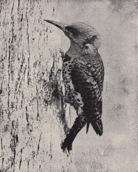
Fig. 139.—The yellow-hammer, Colaptes auratus. (Photograph by W. E. Carlin; permission of G. O. Shields.)
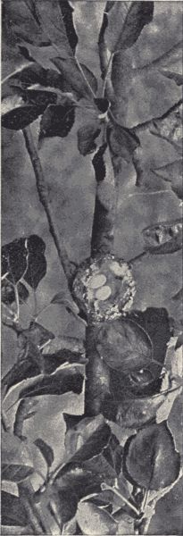
Fig. 140.—Nest and eggs of ruby-throat
humming-bird, Trochilus
colubris, seen from above, in apple-tree.
(Photograph by E. G. Tabor;
permission of Macmillan Co.)
The largest and one of the most interesting woodpeckers is the ivory-billed (Campephilus principalis), twenty inches long, glossy blue-black, with a high head-crest which is scarlet in the male. This bird lives in the heavily wooded swamps of the Southern States. Among the more abundant and widespread, and hence better known, woodpeckers are the yellow-hammers (fig. 139)[Pg 356] or flickers (Colaptes auratus in the East, C. cafer in the West), the red-headed woodpecker (Melanerpes erythrocephalus), with its crimson head and neck and pure-white "vest"; and the black-and-white downy (Dryobates pubescens) and hairy (D. villosus) woodpeckers or "sap-suckers." The California woodpecker (M. formicivorus), a near relative of the red-headed woodpecker, has the curious habit of boring small holes in the bark of oak- or pine-trees and sticking acorns into these holes. Sometimes thousands of acorns are put into the bark of one tree, to which the birds come occasionally to break open some acorns and feed on the grubs inside.
The whippoorwills, chimney-swifts and humming-birds (Macrochires).—All the birds of this order are remarkable for their power of flight. They have long and pointed wings; their feet are small and weak and used only for perching or clinging. All feed on insects, which are caught on the wing by the short-beaked, wide-mouthed swifts and whippoorwills and extracted from flower-cups by the humming-birds with their long and slender bills. The whippoorwill (Antrostomus vociferus) is common in the woods of the East and is readily known by its call. Its two brown-blotched white eggs are laid loose on the ground or on a log or stump. The night-hawk (Chordeiles virginianus), common over the whole country, is seen at twilight flying vigorously about in its search for insects. Its nesting habits are like those of the whippoorwill. The sooty-brown chimney-swifts (Chætura pelagica), popularly confused with the swallows, are the common inhabitants of old chimneys, in which they build their curious saucer-shaped open-work nests. Their eggs are pure white and number four or five. Of the humming-birds but one species, the ruby-throat (Trochilus colubris), is to be found in the Eastern States, but in the western and especially southwestern parts of the country[Pg 357] several other species occur. In all seventeen species have been found in the United States. The nests (fig. 140) of the hummers are very dainty little cups lined with hair or wool or plant-down. The ruby-throat lays two tiny pure-white eggs.
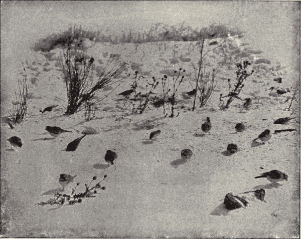
Fig. 141.—Horned larks, Otocoris alpestris, and snowflakes, Plectrophenax nivalis. (Photograph from life by H. W. Menke; permission of Macmillan Co.)
The perchers (Passeres).—Nearly one-half of the birds of North America belong to the great order Passeres, and of all the known birds of the world more than half are included in it. The Passeres or perching birds include the familiar song-birds and a great majority of the birds of the garden, the forest, the roadside, and the field. The feet of these birds always have four toes and are fitted for perching. The syrinx or musical apparatus is, in most, well developed. The nesting and other domestic habits are various, but the young are always hatched in a helpless condition and have to be fed and otherwise cared for by the parents for a longer or shorter time. The North American species of this order are grouped into[Pg 358] eighteen families, as the fly-catcher family (Tyrannidæ), the crow family (Corvidæ), the sparrows and finches (Fringillidæ), the swallows (Hirundinidæ), the warblers (Mniotiltidæ), the wrens (Troglodytidæ), the thrushes, robins and bluebirds (Turdidæ), etc. In this book nothing can be said of the various species which belong to this order. However, as the passerine birds are those which most immediately surround us and which, by their familiar songs and nesting habits, most interest us, the out-door study of birds by beginning students will be devoted chiefly to the members of this order, and many species will soon be got acquainted with. The robin and bluebird will introduce us to the shyer and less familiar song-thrushes; the study of the kingbird or bee-martin[Pg 359] will interest us in some of the other fly-catchers; from the familiar chipping sparrow and tree-sparrow we shall be led to look for their cousins the swamp-sparrows and song-sparrows, and the larger grosbeaks and cross-bills, and so on through the order.
Determining and studying the birds of a locality.—To identify the various species of birds in the locality of the school it will be necessary to have some book giving the descriptions of all or most of the species of the region, with tables and keys for tracing out the different forms. Such manuals or keys are numerous now; the study of birds is one of the most popular lines of nature study, and a host of bird books has been published in the last few years. The best general manual is Coues's "Key to the Birds of North America," which includes not only keys for tracing and descriptions of all the known species of birds on this continent, but also accounts of the distribution, of the nesting and eggs, and of the plumage of the young birds, besides a thorough introduction to the anatomy and physiology of birds, and directions for collecting and preserving them. Jordan's "Manual of Vertebrates" gives keys and short descriptions of the birds found east of the Missouri River; Chapman's "Handbook of the Birds of Eastern North America" is excellent. To be able to use these manuals it is necessary to have the bird's body in hand; and that means usually death for the bird. Recently there have been published several bird-keys which attempt to make it possible to determine species, the commoner ones at any rate, without such close examination. The birds in these books are usually grouped wholly artificially (without any reference to their natural relationships) according to such salient characteristics as color, markings, size, habit of perching, or running, or flying, etc. These characteristics are such as can presumably be made out in the living[Pg 360] bird by aid of an opera-glass or often with the unaided eye. Such books make no pretence to be scientific manuals nor to include any but the more usual and strongly marked species. They are usually limited to the birds of a restricted region. Such books are readily obtainable. There are several popular illustrated "bird-magazines" devoted to accounts of the life and habits of birds. Of these "Bird-lore" is the organ of the Audubon Society for the Protection of Birds.
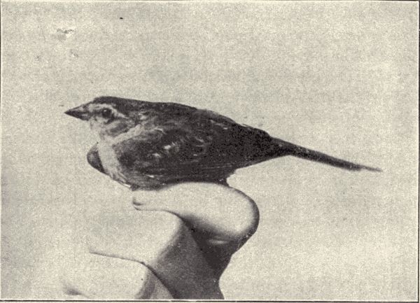
Fig. 142.—Western chipping sparrow, Spizella socialis arizonae. (Photograph from life by Eliz. and Jos. Grinnell.)
In trying to become acquainted with the birds of a locality it must be borne in mind that the bird-fauna of any region varies with the season. Some birds live in a certain region all the year through; these are called residents. Some spend only the summer or breeding season in the locality, coming up from the South in spring and flying back in autumn; these are summer residents. Some spend only the winter in the locality, coming down from[Pg 361] the severer North at the beginning of winter and going back with the coming of spring; these are winter residents. Some are to be found in the locality only in spring and autumn as they are migrating north and south between their tropical winter quarters and their northern summer or breeding home; these are migrants. And finally an occasional representative of certain bird species whose normal habitat does not include the given locality at all will appear now and then blown aside from its regular path of migration or otherwise astray; these are visitants. As to the relative importance, numerically, of these various categories among the birds which may be found in a certain region and thus form its bird-fauna we may illustrate by reference to a definite region. Of the 351 species of birds which have been found in the State of Kansas (a region without distinct natural boundaries and fairly representative of any Mississippi valley region of similar extent), 51 are all-year residents; 125 are summer residents, 36 are winter residents, 104 are migrants, and 35 are rare visitants.
It must also be kept in mind in using bird-keys and descriptions to determine species that the descriptions and keys refer to adult birds and in ordinary plumage. Among numerous birds the young of the year, old enough to fly and as large as the adults, still differ considerably in plumage from the latter; males differ from females, and finally both males and females may change their plumage (hence color and markings) with the season. The seasonal changes of plumage accomplished by molting may be marked or hardly noticeable. "All birds get new suits at least once a year, changing in the fall. Some change in the spring also, either partially or wholly, while others have as many as three changes—perhaps, to a slight extent, a few more.... It is claimed by some that now all new colors are acquired by molt, and by[Pg 362] others that in some instances (young hawks) an infusion or loss, as the case may be, of pigment takes place as the feather forms, and continues so long as it grows."
There is much lack and uncertainty of knowledge concerning the molting and change of plumage by birds, and careful observations by bird-students should be made on the subject.
In connection with learning the different kinds of birds in a locality, together with their names, observations should be made, and notes of them recorded, on their habits and on the relation or adaptation of structure and habit to the life of the bird. Some of the special subjects for such observation are pointed out in the following paragraphs. A suggestive book, treating of the adaptive structure and the life of birds is Baskett's "The Story of the Birds."
Bills and feet.—The interesting adaptation of structure to special use is admirably shown in the varying character of the bills and feet of birds. The various feeding habits and uses of the feet of different birds are readily observed, and the accompanying modification of bills and feet can be readily seen in birds either freshly killed or preserved as "bird-skins." Such skins may be made as directed on p. 467, or may be bought cheaply of taxidermists. A set of such skins, properly named, will be of great help in studying birds, and should be in the high-school collection. In some cases the general structure of feet and bills may be seen in the live birds by the use of an opera-glass. The characters of bills and feet are much used in the classification of birds, so that any knowledge of them gained primarily in the study of adaptations will have a secondary use in classification work.
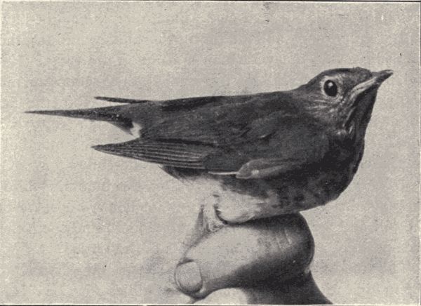
Fig. 143.—Russet-backed thrush, Turdus ustulatus. (Photograph from life by Eliz. and Jos. Grinnell.)
Note the foot of the robin, bluebird, catbird, wrens, warblers and other passerine or perching birds. It has three unwebbed toes in front, and a long hind toe perfectly[Pg 363] opposable to the middle front one. This is the perching foot. Note the so-called zygodactyl foot of the woodpecker, with two toes projecting in front and partly yoked together, and two similarly yoked projecting behind. Note the webbed swimming foot of the aquatic birds; note the different degrees of webbing, from the totipalmate, where all four toes are completely webbed, palmate, where the three front toes only are bound together but the web runs out to the claws, to the semi-palmate, where the web runs out only about half way. Note the lobate foot of the coots and phalaropes. Note the long slender wading legs of the sandpipers, snipe and other shore birds; the short heavy strong leg of the divers; the small weak leg of the swifts and humming-birds, almost always on the wing; the stout heavily nailed foot of the scratchers, as the hens, grouse, and turkeys; and the strong grasping talons, with their sharp long[Pg 364] curving nails, of the hawks and owls and other birds of prey. In all these cases the fitness of the structure of the foot to the special habits of the bird is apparent.
Similarly the shape and structural character of the bill should be noted, as related to its use, this being chiefly concerned of course with the feeding habits. Note the strong hooked and dentate bill of the birds of prey; they tear their prey. Note the long slender sensitive bill of the sandpipers; they probe the wet sand for worms. Note the short weak bill and wide mouth of the night-hawk and whippoorwill and of the swifts and swallows; they catch insects in this wide mouth while on the wing. Note the flat lamellate bill of the ducks; they scoop up mud and water and strain their food from it. Note the firm chisel-like bill of the woodpeckers; they bore into hard wood for insects. Note the peculiarly crossed mandibles of the cross-bills; they tear open pine-cones for seeds. Note the long sharp slender bill of the humming-birds; they get insects from the bottom of flower-cups. Note the bill and foot of any bird you examine, and see if they are specially adapted to the habits of the bird.
The tongues and tails of birds are two other structures the modifications and special uses of which may be readily observed and studied. Note the structure and special use of the tongue and tail of the woodpeckers; note the tongue of the humming-bird; the tail of the grackles.
Flight and songs.—The most casual observation of birds reveals differences in the flight of different kinds, so characteristic and distinctive as to give much aid in determining the identity of birds in nature. Note the flight of the woodpeckers; it identifies them unmistakably in the air. Note the rapid beating of the wings of quail and grouse; also of wild ducks; the slow heavy flapping of the larger hawks and owls and of the crows; and the splendid soaring of the turkey-buzzard and of the gulls.[Pg 365] This soaring has been the subject of much observation and study but is still imperfectly understood. The soaring bird evidently takes advantage of horizontal air-currents, and some observers maintain that upward currents also must be present. The principal hopes for the invention of a successful flying-machine rest on the power of soaring possessed by birds. The speed of flight of some birds is enormous, the passenger-pigeon having been estimated to attain a speed of one hundred miles an hour. The long distances covered in a single continuous flight by certain birds are also extraordinary, as is also the total distance covered by some of the migrants. "It is said that some plovers that nest in Labrador winter in Patagonia, their long wings easily carrying them this great distance."
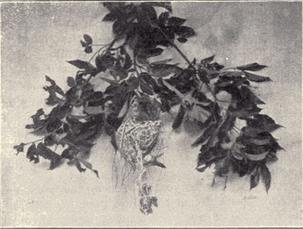
Fig. 144.—Oriole's nest with skeleton of blue jay suspended from it; the blue jay probably came to the nest to eat the eggs, became entangled in the strings composing the nest, and died by hanging. (Photograph by S. J. Hunter.)
Varying even more than the manner and power of flight among different birds are the vocal utterances, the cries and calls and singing. By their calls and songs alone many birds may be identified although they remain unseen. The field-student of birds comes to know them by their songs; knows what birds they are; knows what they are doing or not doing; knows what time in their life-season it is, whether they are mating, or brooding, or preparing to migrate; knows whether they are frightened, or self-confident, whether in distress or happy. Little urging and suggestion are needed to induce the student to attend to the songs. But the naturalist should not only hear and enjoy them, but by observation and the recording of repeated observations, he should come to understand the significance of the calls and songs.
As to how these sounds are made, attention has already been called (see p. 338) to the voice-organ or syrinx. The condition of this organ varies much in birds, as would be expected from the differing character of vocal utterances. Dissections will make these differences apparent.
Nesting and care of young.—Among the birds' most interesting instincts and habits are those domestic ones which include mating, nest-building, and care of the young. Birds' eggs and birds' nests are always attractive objects of search and collection for boys, and most boys have a considerable personal knowledge of the domestic habits of the commoner summer birds of their region. With this interest and unsystematized knowledge as a basis the teacher should be able to get from the class much excellent field-work and personal observation. The first thing to undertake in this study is the gathering of data regarding the character of the nests of different species, their situation, the time of nesting, the participation or non-participation of the male in nest-building,[Pg 367] etc.; also the number of eggs, their size and color markings, the length of incubation, the help or lack of help of the male in brooding, etc. In connection with this gathering of data in the field by note-taking, sketching, and photographing, nests and eggs can be collected (see directions on page 469). Let only one clutch of eggs of each species be taken for the common high-school collection, and if more than one nest is desired take used and deserted nests. When the nestlings are hatched, the bringing of food, the defence of the home, and the teaching of the young to fly should all be observed and noted.
Some attempt should be made to systematize the miscellaneous data obtained. Do all the members of a group have similar nesting habits? Note the early nesting of birds of prey; note the nests of the woodpeckers in holes in trees; note the nesting of the various swallows. Is there any significance in the colors and markings of eggs? Observe the protective coloration obvious in some (see Chap. XXXI). Are there differences in the condition of the newly hatched nestlings? Note the helpless altricial young of the robin; the independent precocial young of the quail.
The strong influence of the mating passion will be made plain by observations on the fighting, love-making, singing, and general behavior of the birds in the mating season. The expression of the mental and emotional traits, the psychic phenomena of birds, are most emphasized at this time, and reveal the possession among animals lower than man of many characteristics which are too commonly ascribed as the exclusive attributes of the human species.
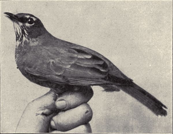
Fig. 145.—Western robin, Merula migratoria propinqua. (Photograph from life by Eliz. and Jos. Grinnell.)
Local distribution and migration.—As explained in Chapter XXXII, the geographical distribution of animals is a subject of much importance, and offers good opportunities in its more local features for student field-work.[Pg 368] The field-study of the birds of a given locality will comprise much observation bearing directly on zoogeography or the distribution of animals. Certain birds will be found to be limited to certain parts of even a small region, the swimmers will be found in ponds and streams and the long-legged shore-birds on the pond- or stream-banks, or in the marshes and wet meadows, although a few like the upland plover, curlews, and godwits are common on the dry upland pastures. Distinguish the ground-birds from the birds of the shrubs and hedge-rows and these again from the strictly forest-birds. Find the special haunts of swallows and kingfishers. Which are the shy birds driven constantly deeper into the wild places or being exterminated by the advance of man; which birds do not[Pg 369] retreat but even find an advantage in man's seizure of the land, obtaining food from his fields and gardens?
Make a map on large scale of the locality of the school, showing on it the topographic features of the region, such as streams, ponds, marshes, hills, woods, springs, wild pastures, etc., also roads and paths, and such landmarks as schoolhouses, county churches, etc. On this map indicate the local distribution of the birds, as determined by the data gradually gathered; mark favorite nesting-places of various species, roosting-places of crows and blackbirds, feeding-places, and bathing- and drinking-places of certain kinds, the exact spots of finding rare visitants, rare nests, etc., etc. The making of such a zoogeographical map will be a source of great interest and profit to the students.
As already mentioned, many of the birds of a locality are "migrants," that is, they breed farther north, but spend the winter in more southern latitudes. These migrants pass through the locality twice each year, going north in the spring and south in the autumn. They are much more likely to be observed during the spring migration than in the fall, as the flight south is usually more hurried. The observation of the migration of birds is very interesting, and much can be done by beginning students. Notes should be made recording the first time each spring a migrating species is seen, the time when it is most abundant and the last time it is seen the same spring. Similar records should be made showing the movements of the birds in the fall. A series of such records covering a few years will show which are the earliest species to appear, which the later, and which the last. Such records of appearance and disappearance should also be kept for the summer residents, those birds that come from the South in the spring, breed in the locality, and then depart for the South again in the[Pg 370] autumn. Notes on the kinds of days, as stormy, clear, cold, warm, etc., on which the migration seems to be most active; on the greater prevalence of migratory flights by day or by night; on the height from the earth at which the migrants fly, etc., are all worth while. The Division of Biological Survey, U. S. Department of Agriculture, keeps records of notes on migration sent in by voluntary observers and furnishes blanks to be filled out by each observer. A suggestive book about migration, and one giving the records for many species at many points in the Mississippi valley is Cooke's "Bird Migration in the Mississippi Valley." Migration is discussed in most bird-books.
Feeding habits, economics, and protection of birds.—The feeding habits of birds are not only interesting, but their determination decides the economic relation of birds to man, that is, whether a particular bird species is harmful or beneficial to man. Casual observation shows that birds eat worms, grains, seeds, fruits, insects. A single species often is both fruit-eating and insect-eating. Do fruits or do insects compose the chief food-supply of the species? To determine this more than casual observation is necessary. The birds must be watched when feeding at different seasons. The most effective way of determining the kind of food which the bird takes is to examine the stomachs of many individuals taken at various times and localities. Much work of this kind has been done, especially by the investigators connected with the Division of Biological Survey of the U. S. Department of Agriculture, and pamphlets giving the results of these investigations can be had from the Division. It has been distinctly shown that a great majority of birds are chiefly beneficial to man by eating noxious insects and the seeds of weeds. Many birds commonly reputed to be harmful, and for that reason shot by farmers and fruit-growers, have been[Pg 371] proved to do much more good than harm. Some few birds have been proved to be, on the whole, harmful. An investigation of the food habits of the crow, a bird of ill-repute among farmers, based on an examination of 909 stomachs shows that about 29 per cent of the food for the year consists of grain, of which corn constitutes something more than 21 per cent, the greatest quantity being eaten in the three winter months. All of this must be either waste grain picked up in fields and roads, or corn stolen from cribs and shocks. May, the month of sprouting corn,[Pg 372] shows a slight increase over the other spring and summer months. On the other hand the loss of grain is offset by the destruction of insects. These constitute more than 23 per cent of the crow's yearly diet, and the larger part of them are noxious. The remainder of the crow's food consists of wild fruit, seeds and various animal substances which may on the whole be considered neutral.
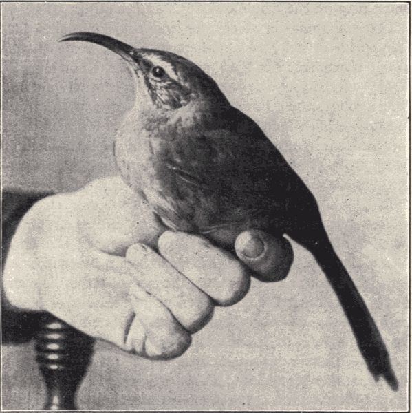
Fig. 146.—Sickle-billed thrasher, Harporhynchus redevivus. (Photograph from life by Eliz. and Jos. Grinnell.)
The slaughter of birds for millinery purposes has become so fearful and apparent in recent years that a strong movement for their protection has been inaugurated. Rapacious egg-collecting, legislation against birds wrongly thought to be harmful to grains and fruit, and the selfish wholesale killing of birds by professional and amateur hunters, help in the work of destruction. Apart from the brutality of such slaughter, and the extermination of the most beautiful and enjoyable of our animal companions, this destruction[18] works strongly against our material interests. Birds are the natural enemies of insect pests, and the destroying of the birds means the rapid increase and spread, and the enhanced destructive power of the pests. It is asserted by investigators that during the past fifteen years the number of our common song-birds has been reduced to one-fourth. At the present rate, says one author, extermination of many species will occur during the lives of most of us. Already the passenger-pigeon and Carolina paroquet, only a few years ago abundant, are practically exterminated. Protect the birds!
Technical Note.—It is best to catch specimens alive in a good trap. A live trap well baited and placed in some old granary should furnish plenty for class use. White mice can often be obtained at "bird-stores." When mice are not procurable, use rats. A rat is perhaps preferable on account of its size, but all essential structures can readily be made out in the mouse. Specimens should be killed by chloroform as described for the toad, p. 5.
Structure (fig. 147).—Compare the external characters of the mouse with those of the toad and sparrow. The mouse, unlike the other vertebrates so far studied, is thickly covered with hair all over its body except on the tip of the nose and the soles of the feet. Where are the nostrils placed? What are the large leaf-like expansions called pinnæ situated just back of the eyes? Pull open the mouth and note the large incisor teeth on the upper and lower jaws. Cut one corner of the mouth back and observe the large flat-topped molar teeth on both jaws. How does the attachment of the large fleshy tongue differ from the condition in the toad? The toad's tongue is for snapping up insects, whereas in the mouse this organ serves to move food about in the mouth. On the tongue are numerous small taste-papillæ. Notice the long hairs, "feelers," on each side of the nose. Note the similarity between the front paws and our own hands; each has four fingers with a small rudimentary thumb on the inner[Pg 374] side of the paw. How does the hind foot of the mouse differ from the foot of man? Posteriorly the body is terminated by a long tail. At the root of the tail is a small aperture, the anus, and just below, or ventral to it, is the opening from the kidneys and reproductive organs.
Technical Note.—Place the mouse on its back in a dissecting-pan and cut through the skin from anus to the lower jaw. Extend the legs, pin down each foot and pin out the cut edges of the skin. Now carefully cut forward through the body-wall from the anal region and on through the breast-bones and ribs. Pin each side out.
Near the hindmost pair of ribs note a sheet of muscles, the diaphragm, which extends across the body-cavity, dividing it into an anterior portion, the thoracic cavity, and a posterior, the abdominal cavity. What are the most conspicuous organs in the thoracic cavity? Leading anteriorly to the mouth-cavity is a long tube, the trachea, composed of a series of cartilaginous parts of rings placed end to end. Note at its anterior end the glottis and epiglottis. Insert a blowpipe into the glottis and inflate the lungs, which will fill all the otherwise unfilled space in the thoracic cavity. The abdominal cavity contains the viscera suspended in a fold of the lining membrane, as in the other vertebrates studied. Note lying against the diaphragm a large, red, glandular structure, the liver. Separate the two large lobes of the liver and expose the opalescent gall-bladder. By passing a canula into this and ligaturing, the cystic duct may be injected. Beneath the liver is a large loop-shaped expansion of the alimentary canal, the stomach. Arising from the right end of the stomach is the narrow duodenum, which gradually merges into the very much convoluted small intestine, or ileum, which is followed by the large intestine, or colon, the last part of which is a straight tube, the rectum. The small intestine occupies most of the space in the peritoneal[Pg 375] cavity. Within the loop of the pylorus will be found an irregular pinkish mass of tissue, the pancreas. Beneath the stomach on the left side of the body lies a very dark glandular mass not much unlike the liver but altogether detached from it. This structure is the spleen, a ductless gland.
Note dorsally of the trachea a long tube passing through the diaphragm and connecting the mouth with the stomach. What is this tube? Note the Eustachian tubes extending from the mouth to the ears. The median part of the roof of the mouth is the palate, hard in front, soft behind. A pair of small bodies at the sides of the soft palate near its hinder end are the tonsils. At the posterior angle of the lower jaw are glandular bodies, the sub-maxillary glands, which lead by a short duct anteriorly to open on the floor of the mouth. On the sides of the neck just below the ears are pink or yellowish bodies, the parotid glands, opening anteriorly in the sides of the mouth-cavity. These two sets of glands are collectively known as the salivary glands, the function of which is to secrete the saliva. Push apart the sub-maxillary glands and note below them overlying the trachea on either side two dark-red lobes connected by a band of tissue. These constitute the thyroid gland, another of the so-called ductless glands. Within the thoracic cavity anterior to the heart note a mass of pinkish tissue, the thymus gland. Observe the large masseter muscles, which cover the jaws. What is their function? On either side of the neck lies a large blood-vessel, the external jugular vein, which collects blood from the head and carries it down to the heart. Note the large pectoral muscles which cover the breast and extend out into the arms, and which are so strong and highly developed in the sparrow. The head is supported by large muscles which run down the back of the neck to the ribs. Others are attached to the ribs, which they[Pg 376] raise and lower. These movements, together with the contraction of the diaphragm, cause the expansion and contraction of the thoracic cavity whereby the lungs are regularly filled and emptied. Note that the abdomen is covered by a double layer of muscular tissue, the outer part made up of the external oblique muscles, the inner by the internal oblique muscles.
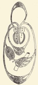
Fig. 148.—Diagram of
the circulation of the
blood in a mammal; a,
auricles; l, lung; lv,
liver; p, portal vein
bringing blood from the
intestine; v, ventricles;
the arrows show the direction
of the current;
the shaded vessels
carry venous blood, the
others arterial blood.
(From Kingsley.)
Examine the heart. How many auricles has it? The ventricles in the mouse, as in the bird, are entirely separated, forming two complete compartments, a right and a left ventricle. The blood flowing from the veins of the body is collected in the right auricle, thence it passes into the right ventricle, whence it is conveyed to the lungs; returning it flows through the left auricle into the left ventricle, whence it is forced through the arteries of the body. For a study of the circulatory system in mammals (fig. 148), a rat or a rabbit should be injected by the teacher and an advanced text-book, as Parker's "Zootomy" or Marshall and Hurst's "Practical Zoology," used as a guide. A sheep's heart is very good to cut open for a class demonstration.
Make a drawing of the organs observed thus far in the dissection.
The kidneys in the mouse are situated in the dorsal region next to the backbone. They consist of two bean-shaped smooth glands. From them a pair of ducts, the ureters, can be traced down to a median thin-walled[Pg 377] muscular sac, the bladder. The bladder opens to the exterior of the body by means of a short tube, the urethra. Cut open a kidney longitudinally and examine the cut surfaces.
The two egg-glands of the female mouse lie in the median portion of the abdominal cavity, somewhat below the kidneys, and from the vicinity of each runs an egg-tube. These tubes meet below the bladder, and open to the exterior of the body through the aperture noted below the anus. In the posterior parts of these tubes lie until birth the developing embryos.
Technical Note.—For a study of the nervous system place the specimen ventral side down and cut through the skull with the bone-cutters or heavy scissors, exposing the brain and spinal cord.
Note the large brain (fig. 149), composed of small optic lobes, large cerebrum, cerebellum, and medulla oblongata, followed by the long spinal cord. Note the nerves arising from the brain and spinal cord.
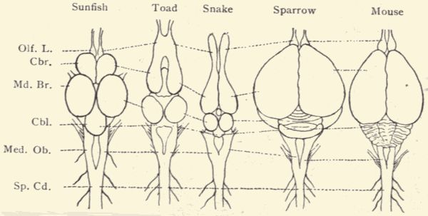
Fig. 149.—Diagram of brains of vertebrates; Olf. L., olfactory lobes; Cbr., cerebrum; Md. Br., midbrain (optic lobes); Cbl., cerebellum; Med. Ob., medulla oblongata; Sp. Cd., spinal cord. (From specimens.)
For a careful dissection of the mammalian nervous system a larger mammal, such as a cat or dog or rabbit, should be used. For guide use a text-book such as, for the dog, Howell's "Dissection of the Dog"; for the cat, Reighard and Jennings' "Anatomy of the Cat"; and for the rabbit, Parker's "Zootomy" or Marshall and Hurst's[Pg 378] "Practical Zoology." Make a good preparation of the brain and preserve it for future use in some fluid like Fischer's fluid (see page 453).
Technical Note.—Prepare a well-cleaned skeleton by boiling a specimen in a soap solution and thoroughly cleansing it (see p. 452).
Note the very compact skeleton of the mouse. Note the closely sutured skull. How many cervical or neck vertebræ are there? The ribs are attached to the thoracic vertebræ. How many pairs of ribs? The bony thorax supports the shoulder-girdle and bones of the fore legs. The thorax is followed by a series of ribless vertebræ, the lumbar vertebræ, which in the posterior region of the body fuse with the pelvic girdle supporting the hind limbs. The body vertebræ are succeeded by the very much smaller caudal vertebræ. Compare the skeleton of the mouse with that of the bird; also with that of the toad. For directions for a detailed study of the skeleton see in Parker's "Zootomy" an account of the skeleton of the rabbit, pp. 263-286.
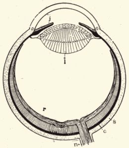
Fig. 150.—Diagram of vertebrate
eye; c, choroid; i, iris; l, lens; n,
optic nerve; r, retina; s, sclerotic.
(From Kingsley.)
Technical Note.—For the study of the eye (fig. 150) the teacher should obtain the eye of some large mammal, as the ox or sheep, with which to make a class demonstration. The eye of a rabbit or cat can of course be used. For an account of the vertebrate eye see Parker and Haswell's "Text-book of Zoology," Vol. II. pp. 103-107. For a study of the ear use a bird or mammal, and see pp. 107-110 of the same book.
Life-history and habits.—The house-mouse is not a native of North America, but was introduced into this country from Europe, to which, in turn, it came from Asia,[Pg 379] its original habitat. The mouse came to this country in the vessels of early explorers. Similarly the brown and black rats, now so abundant all over North America, and members of the same genus as the mouse, were introduced from Europe. Accompanying man in his travels the mouse has spread from Asia until it is now to be found over the whole world.
The habits of mice are well known; their fondness for living in our homes and outbuildings makes them familiar acquaintances. Their food is varied; they seem to thrive best, however, on a vegetable diet. Grains and nuts are favorite foods. The house-cat is their greatest enemy, but man takes advantage of their instinct to go into holes by constructing traps with funnel or tunnel entrances which, baited with cheese or other favorite food, are fatally attractive. In climbing, mice are aided by the tail. Their strong hind legs enable them to stand erect, and even to take several steps in this posture. They can swim readily, although naturally they rarely take to water. Their special senses are keen, the senses of hearing and taste being unusually well developed. Their "singing," which has been the subject of much discussion, seems to be actually a voluntary and normal performance which, however, hardly deserves to be called singing, but rather a slightly varied peeping or whistling.
The mouse is a prolific mammal, producing from four to six times a year broods of from four to eight young. The mouse makes a cosy nest of straw, bits of paper, feathers, wool or other soft materials, and in this the young are born. The newly born mice are very small and are blind and helpless. They are odd little creatures, being naked and almost transparent. They grow rapidly, being covered with hair in a week, although not opening their eyes for about two weeks. A day or two after their eyes are open they begin to leave the nest, and hunt for food for themselves.
The mammals constitute the highest group of animals, including man, the monkeys and apes, the quadrupeds, the bird-like bats and fish-like seals and whales; in all about 2500 species. They are found everywhere except on a few small South Sea islands. Only a few species, however, have a world-wide distribution. The name Mammalia is derived from the mammary or milk glands with which the females are provided and by the secretion of which the young of this class, born free in all but a few of the lowest forms, are nourished for some time after birth. In size mammals range from the tiny pigmy-shrew and harvest mouse, which can climb a stem of wheat, to the great sulphur-bottom whale of the Pacific Ocean, which attains a length of a hundred feet and a weight of many tons. Mammals differ from fishes and batrachians and agree with reptiles and birds in never having external gills; they differ from reptiles and agree with birds in being warm-blooded and in having a heart with two distinct ventricles and a complete double circulation; finally, they differ from both reptiles and birds in having the skin more or less clothed with hair, the lungs freely suspended in a thoracic cavity separated from the abdominal by a muscular partition, the diaphragm, and in the possession by the females of mammary glands. In economic uses to man mammals are the most important of all animals. They furnish the greater portion of the animal food of many human races, likewise a large amount of their clothing. Horses, asses, oxen, camels, reindeer, elephants, and llamas are beasts of burden and draught; swine, sheep, cattle, and goats furnish flesh, and the two latter milk for food; the wool of sheep, the furs of the carnivores, and the leather of cattle, horses, and others are used for clothing, while the bones and horns of various mammals serve various purposes.
Body form and structure.—The mammalian body varies greatly. Its variety of form and general organization is explained by the facts that, although most of the species live on the surface of the earth, some are burrowers in the ground, some flyers in the air, and some swimmers in the water. Mammals never have more than two pairs of limbs; in most cases both pairs are well developed and adapted for terrestrial progression. In the aerial bats the fore limbs are modified into organs of flight; among the aquatic seals, sea-lions, walruses, and whales both sets are modified to be swimming flippers or paddles. In many of these aquatic forms the hind limbs are greatly reduced or even completely wanting.
Most mammals are externally clothed with hair, which is a peculiarly modified epidermal process. Each hair, usually cylindrical, is composed of two parts, a central pith containing air, and an outer more solid cortex; each hair rises from a short papilla sunk at the bottom of a follicle lying in the true skin. In some mammals the hairs assume the form of spines or "quills," as in the porcupine. The hairy coat is virtually wanting in whales and is very sparse in certain other forms, the elephant, for example, which has its skin greatly thickened. The claws of beasts of prey, the hooves of the hoofed mammals, and the outer horny sheaths of the hollow-horned ruminants are all epidermal structures.
The bones of mammals are firmer than those of other vertebrates, containing a larger proportion of salts of lime. Among the different forms the spinal column varies largely in the number of vertebræ, this variation being chiefly due to differences in length of tail. Apart from the caudal vertebræ their usual number is about thirty. The mammalian skull is very firm and rigid, all the bones composing it, excepting the lower jaw, the tiny auditory ossicles, and the slender bones of the hyoid arch, being[Pg 382] immovably articulated together. The correspondence between the bones of the two sets of limbs is very apparent. The number of digits varies in different mammals, and also in the fore and hind limbs of a single species. Among the Ungulates the reduction in the number of digits is especially noticeable; the forefoot of a pig has four digits, that of the cow two, and that of the horse one. The two short "splint" bones in the horse are remnants of lost digits. The teeth are important structures in mammals, being used not only for tearing and masticating food, but as weapons of offence and defence. A tooth consists of an inner soft pulp (in old teeth the pulp may become converted into bone-like material) surrounded by hard white dentine or ivory, which is covered by a thin layer of enamel, the hardest tissue known in the animal body. A hard cement sometimes covers as a thin layer the outer surface of the root, and may also cover the enamel of the crown. The teeth in most forms are of three groups: (a) the incisors, with sharp cutting edges and simple roots, situated in the centre of the jaw; (b) the canines, often conical and sharp-pointed, next to the incisors; (c) next the molars, broad and flat-topped for grinding, and divided into premolars and true molars. There is great variety in the character and arrangement of these structures in mammals, their variations being much used in classification. The number and arrangement of the teeth is expressed by a dental formula, as, for example, in the case of man
2—2 1—1 2—2 3—3
i ——, c ——, p ——, m —— = 32.
2—2 1—1 2—2 3—3
The mouth is bounded by fleshy lips. On the floor of
the mouth is the tongue, which bears the taste-buds or
papillæ, the organs of taste. The œsophagus is always
a simple straight tube, but the stomach varies greatly,[Pg 383]
[Pg 384]
being usually simple, but sometimes, as in the ruminants
and whales, divided into several distinct chambers. The
intestine in vegetarian mammals is very long, being in a
cow twenty times the length of the body. In the carnivores
it is comparatively short—in a tiger, for example,
but two or three times the length of the body.
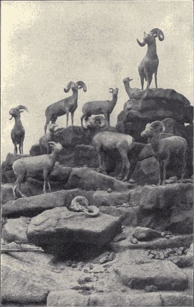
Fig. 151.—A group of Rocky Mountain sheep, or "big horns," Ovis canadensis, including males, females and young. (Photograph by E. Willis from specimens mounted by Prof. L. L. Dyche, University of Kansas.)
The blood of mammals is warm, having a temperature of from 35° C. to 40° C. (95° F. to 104° F.). It is red in color, owing to the reddish-yellow, circular, non-nucleated blood-corpuscles. The circulation is double, the heart being composed of two distinct auricles and two distinct ventricles. Air is taken in through the nostrils or mouth and carried through the windpipe (trachea) and a pair of bronchi to the lungs, where it gives up its oxygen to the blood, from which it takes up carbonic-acid gas in turn. At the upper end of the trachea is the larynx or voice-box, consisting of several cartilages attaching by one end to the vocal cords and by the other to muscles. By the alteration of the relative position of these cartilages the cords can be tightened or relaxed, brought together or moved apart, as required to modulate the tone and volume of the voice.
The kidneys of mammals are more compact and definite in form than those of other vertebrates. In all mammals except the Monotremes they discharge their product through the paired ureters into a bladder, whence the urine passes from the body by a single median urethra. Mammary glands, secreting the milk by which the young are nourished during the first period of their existence after birth, are present in both sexes in all mammals, though usually functional in the female only.
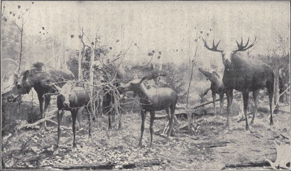
Fig. 152.—A group of moose, Alce americana, showing male, female, and young. (Photograph by E. Willis from specimens mounted by Prof. L. L. Dyche, University of Kansas.)
The nervous system and the organs of special sense
reach their highest development in the mammals. In
them the brain is distinguished by its large size, and by
the special preponderance of the forebrain or cerebral[Pg 385]
[Pg 386]
hemispheres over the mid- and hind-brain. Man's brain
is many times larger than that of all other known mammals
of equal bulk of body, and three times as large as
that of the largest-brained ape. In man and the higher
mammals the surface of the forebrain is thrown into many
convolutions; among the lowest the surface is smooth.
Of the organs of special sense, those of touch consist of
free nerve-endings or minute tactile corpuscles in the skin.
The tactile sense is especially acute in certain regions, as
the lips and end of the snout in animals like hogs, the
fingers in man, and the under surface of the tail in certain
monkeys. All the other sense-organs are situated on the
head. The organs of taste are certain so-called taste-buds
located in the mucous membrane covering certain
papillæ on the surface of the tongue. The organ of
smell, absent only in certain whales, consists of a ramification
of the olfactory nerves over a moist mucous membrane
in the nose. The ears of mammals are more highly
developed than those of other vertebrates both in respect
to the greater complexity of the inner part and the size
of the outer part. A large outer ear for collecting the
sound-waves is present in all but a few mammals. A
tympanic membrane separates it from the middle ear in
which is a chain of three tiny bones leading from the
tympanum to the inner ear, composed of the three semicircular
canals and the spiral cochlea. The eyes (fig. 150)
have the structure characteristic of the vertebrate eye, consisting
of a movable eyeball composed of parts through
which the rays of light are admitted, regulated, and concentrated
upon the sensitive expansion, retina, of the optic
nerve lining the posterior part of the ball. The eye is protected
by two movable lids. In almost all mammals below
the Primates there is a third lid, the nictitating membrane.
In some burrowing rodents and others the eye is quite
vestigial and even concealed beneath the skin.
Development and life-history.—All mammals except the Monotremes give birth to free young. The two genera of Monotremes produce their young from eggs hatched outside the body; Tachyglossus lays one egg which it carries in an external pouch, while Ornithorhynchus deposits two eggs in its burrow. The embryo of other mammals develops in the lower portion of the egg-tube, to the walls of which it is intimately connected by a membrane called the placenta. (In the kangaroos and opossums, Marsupialia, there is no placenta.) Through this placenta blood-vessels extend from the body of the mother to the embryo, the young developing mammal thus deriving its nourishment directly from the parent.
The duration of gestation (embryonic or prenatal development in the mother's body) varies from three weeks with the mouse, eight weeks with the cat, nine months with the stag, to twenty months with the elephant. Like the birds, the young of some mammals, the carnivores for example, are helpless at birth, while those of others, as the hoofed mammals, are very soon able to run about. But all are nourished for a longer or shorter time by the milk secreted by the mammary gland of the mother.
Habits, instinct, and reason.—Despite the wonderful examples of instinct and intelligence shown by many insects and by the other vertebrates, especially the birds, it is among mammals that we find the highest development of these qualities and of reason. In the wary and patient hunting for prey by the carnivora, in the gregarious and altruistic habits of the herding hoofed mammals, in the highly developed and affectionate care of the young shown by most mammals, and in the loyal friendship and self-sacrifice of dogs and horses in their relations to man, we see the culmination among animals of the development of the functions of the nervous system. In the characteristics[Pg 388] of intelligence and reason man of course stands immensely superior to all other animals, but both intelligence and reason are too often shown by many of the other mammals not to make us aware that man's mental powers differ only in degree, not in kind, from those of other animals.
Pure instinct is hereditary, and purely instinctive actions are common to all the individuals of a species. Those actions which the individual could not learn by teaching, imitation, or experience are instinctive. The accurate pecking at food by chicks just hatched from an incubator is purely instinctive. Purely instinctive also is the laying of eggs by a butterfly on a certain species of plant which may have to be sought for over wide acres, so that the caterpillars when hatched shall find themselves on their own special food-plant. Yet the butterfly never ate of this plant and will never see its young. Such elaborate instincts as these have been developed from the simplest manifestations of sensation and nervous function, just as the complex structures of the body have been developed from simple structures (see Chapter XXIX).
The feeding and domestic habits and the whole general behavior of animals are extremely interesting subjects of observation and study. And such observation intelligently pursued will be of much value. The point to be kept ever in mind is that all animal habits are connected with certain conditions of life; that in every case there is an answer to the question "why." This answer may not be found; in many cases it is extremely difficult to get at, but often it is simple and obvious and can be found by the veriest beginner.
Classification.—The mammals of North America represent eight orders. Three additional mammalian orders, namely, the Monotremata, including the extraordinary duck-bills (Ornithorhynchus) and a species of Tachyglossus[Pg 389] in Australia and Tasmania; the Edentata, including the sloths, armadillos, and ant-eaters found in tropical regions; and the Sirenia, including the marine manatees and dugongs, are not represented (except by a single manatee) in North America. In the following paragraphs some of the more familiar mammals representing each of the eight orders represented in North America are referred to.
The opossums (Marsupialia).—The opossum (Didelphys virginiana) is the only North American representative of the order Marsupialia, the other members of which are limited exclusively to Australia and certain neighboring islands. The kangaroos are the best known of the foreign marsupials. After birth the young are transferred to an external pouch, the marsupium, on the ventral surface of the mother, in which they are carried about and fed. The opossum lives in trees, is about the size of a common cat, and has a dirty-yellowish woolly fur. Its tail is long and scaly, like a rat's. Its food consists chiefly of insects, although small reptiles, birds, and bird's eggs are eaten. When ready to bear young the opossum makes a nest of dried grass in the hollow of a tree, and produces about thirteen very small (half an inch long) helpless creatures. These are then placed by the mother in her pouch. Here they remain until two months or more after birth. Probably all the North American opossums found from New York to California and especially common in the Southern States belong to a single species, but there is much variety among the individuals.
The rodents or gnawers (Glires).—The rabbits, porcupines, gophers, chipmunks, beavers, squirrels, and rats and mice compose the largest order among the mammals. They are called the rodents or gnawers (Glires) because of their well-known gnawing powers and proclivities.[Pg 390] The special arrangement and character of the teeth are characteristic of this order. There are no canines, a toothless space being left between the incisors and molars on each side. There are only two incisor teeth in each jaw (rarely four in the upper jaw), and these teeth grow continuously and are kept sharp and of uniform length by the gnawing on hard substances and the constant rubbing on each other. The food of rodents is chiefly vegetable.
Of the hares and rabbits the cottontail (Lepus nuttalii) and the common jack-rabbit (L. campestris) are the best known. The cottontail is found all over the United States, but shows some variation in the different regions. There are several species of jack-rabbits, all limited to the plains and mountain regions west of the Mississippi River. The food of rabbits is strictly vegetable, consisting of succulent roots, branches, or leaves. Rabbits are very prolific and yearly rear from three to six broods of from three to six young each. There are two North American species of porcupines, an Eastern one, Erethizon dorsatus, and a Western one, E. epixanthus. The quills in both these species are short, being only an inch or two in length, and are barbed. In some foreign porcupines they are a foot long. They are loosely attached in the skin and may be readily pulled out, but they cannot be shot out by the porcupine, as is popularly told. The little guinea-pigs (Cavia), kept as pets, are South American animals related to the porcupines.
The pocket gophers, of which there are several species mostly inhabiting the central plains, are rodents found only in North America. They all live underground, making extensive galleries and feeding chiefly on bulbous roots. The mice and rats constitute a large family of which the house-mice and rats, the various field-mice, the wood-rat (Neotoma pennsylvanica) and the muskrat (Fiber zibethicus) are familiar representatives. The common[Pg 391] brown rat (Mus decumanus) was introduced into this country from Europe about 1775, and has now nearly wholly supplanted the black rat (M. rattus), also a European species, introduced about 1544. The beaver (Castor canadensis) is the largest rodent. It seems to be doomed to extermination through the relentless hunting of it for its fur. The woodchuck or ground-hog (Arctomys monax) is another familiar rodent larger than most members of the order. The chipmunks and ground-squirrels are commonly known rodents found all over the country. They are the terrestrial members of the squirrel family, the best known arboreal members of which are the red squirrel (Sciurus hudsonicus), the fox-squirrel (S. ludovicianus), and the gray or black squirrel (S. carolinensis). The little flying squirrel (Sciuropterus volans) is abundant in the Eastern States.
The shrews and moles (Insectivora).—The shrews and moles are all small carnivorous animals, which, because of their size, confine their attacks chiefly to insects. The shrews are small and mouse-like; certain kinds of them lead a semi-aquatic life. There are nearly a score of species in North America. Of the moles, of which there are but few species, the common mole (Scalops aquaticus) is well known, while the star-nosed mole (Condylura cristata) is recognizable by the peculiar rosette of about twenty cartilaginous rays at the tip of its snout. Moles live underground and have the fore feet wide and shovel-like for digging. The European hedgehogs are members of this order.
The bats (Chiroptera).—The bats (fig. 153), order Chiroptera, differ from all other mammals in having the fore limbs modified for flight by the elongation of the forearms and especially of four of the fingers, all of which are connected by a thin leathery membrane which includes also the hind feet and usually the tail. Bats are chiefly nocturnal,[Pg 392] hanging head downward by their hind claws in caves, hollow trees, or dark rooms through the day. They feed chiefly on insects, although some foreign kinds live on fruits. There are a dozen or more species of bats in North America, the most abundant kinds in the Eastern States being the little brown bat (Myotis subulatus), about three inches long with small fox-like face, high slender ears, and a uniform dull olive-brown color, and the red bat (Lasiurus borealis), nearly four inches long, covered with long, silky, reddish-brown fur, mostly white at tips of the hairs.
The dolphins, porpoises, and whales (Cete).—The dolphins, porpoises, and whales (Cete) compose an order of more or less fish-like aquatic mammals, among which[Pg 393] are the largest of living animals. In all the posterior limbs are wanting, and the fore limbs are developed as broad flattened paddles without distinct fingers or nails. The tail ends in a broad horizontal fin or paddle. The Cete are all predaceous, fish, pelagic crustaceans, and especially squids and cuttlefishes forming their principal food. Most of the species are gregarious, the individuals swimming together in "schools." The dolphins and porpoises compose a family (Delphinidæ) including the smaller and many of the most active and voracious of the Cete. The whales compose two families, the sperm-whales (Physeteridæ) with numerous teeth (in the lower jaw only) and the whalebone whales (Balænidæ) without teeth, their place being taken in the upper jaw by an array of parallel plates with fringed edges known as "whalebone." The great sperm-whales or cachalots (Physeter macrocephalus) found in southern oceans reach a length (males) of eighty feet, of which the head forms nearly one-third. Of the whalebone whales, the sulphur-bottom (Balænoptera sulfurea) of the Pacific Ocean, reaching a length of nearly one hundred feet, is the largest, and hence the largest of all living animals. The common large whale of the Eastern coast and North Atlantic is the right whale (Balæna glacialis); a near relative is the great bowhead (B. mysticetus) of the Arctic seas, the most valuable of all whales to man. Whales are hunted for their whalebone and the oil yielded by their fat or blubber. The story of whale-fishing is an extremely interesting one, the great size and strength of the "game" making the "fishing" a hazardous business.
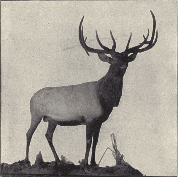
Fig. 154.—Male elk or wapiti, Cervus canadensis. (Photograph by E. Willis from specimen mounted by Prof. L. L. Dyche, University of Kansas.)]
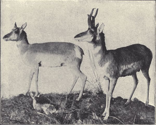
Fig. 155.—Antelope, male, female, and young, Antilocapra americana. (Photograph by E. Willis from specimens mounted by Prof. L. L. Dyche, University of Kansas.)
The hoofed mammals (Ungulata).—The order Ungulata includes some of the most familiar mammal forms. Most of the domestic animals, as the horse, cow, hog, sheep, and goat, belong to this order, as well as the familiar deer, antelope, and buffalo of our own land and[Pg 394] the elephant, rhinoceros, hippopotamus, giraffe, camel, zebra, etc., familiar in zoological gardens and menageries. The order is a large one, its members being characterized by the presence of from one to four hooves, which are the enlarged and thickened claws of the toes. The Ungulates are all herbivorous, and have their molar teeth fitted for grinding, the canines being absent or small. The order is divided into the Perissodactyla or odd-toed forms, like the horse, zebra, tapir, and rhinocerus, and the Artiodactyla or even-toed forms, like the oxen, sheep, deer,[Pg 395] camels, pigs, and hippopotami. The Artiodactyls comprise two groups, the Ruminants and Non-ruminants. All of the native Ungulata of our Northern States belong to the Ruminants, so called because of their habit of chewing a cud. A ruminant first presses its food into a ball, swallows it into a particular one of the divisions of its four-chambered stomach, and later regurgitates it into[Pg 396] the mouth, thoroughly masticates it, and swallows it again, but into another stomach-chamber. From this it passes through the other two into the intestine.
The deer family (Cervidæ) comprises the familiar Virginia or red deer (Odocoileus americanus) of the Eastern and Central States and the white-tailed, black-tailed, and mule deers of the West, the great-antlered elk or wapiti (Cervus canadensis) (fig. 154), the great moose (Alce americana) (fig. 152), largest of the deer family, and the American reindeer or caribou (Rangifer caribou). All species of the Cervidæ have solid horns, more or less branched, which are shed annually. Only the males (except with the reindeer) have horns. The antelope (Antilocapra americana) (fig. 155) common on the Western plains also sheds its horns, which, however, are not solid and do not break off at the base as in the deer, but are composed of an inner bony core and an outer horny sheath, the outer sheath only being shed. The family Bovidæ includes the once abundant buffalo or bison (Bison bison) (frontispiece), the big-horn or Rocky Mountain sheep (Ovis canadensis) (fig. 151), and the strange pure-white Rocky Mountain goat (Oreamnos montanus). The buffalo was once abundant on the Western plains, travelling in enormous herds. But so relentlessly has this fine animal been hunted for its skin and flesh that it is now practically exterminated (fig. 156). A small herd is still to be found in Yellowstone Park, and a few individuals live in parks and zoological gardens. In all of the Bovidæ the horns are simple, hollow, and permanent, each enclosing a bony core.
The carnivorous mammals (Feræ).—The order Feræ includes all those mammals usually called the carnivora, such as the lions, tigers, cats, wolves, dogs, bears, panthers, foxes, weasels, seals, etc. All of them feed chiefly on animal substance and are predatory, pursuing[Pg 397] and killing their prey. They are mostly fur-covered and many are hunted for their skin. They have never less than four toes, which are provided with strong claws that are frequently more or less retractile. The canine teeth are usually large, curved, and pointed.
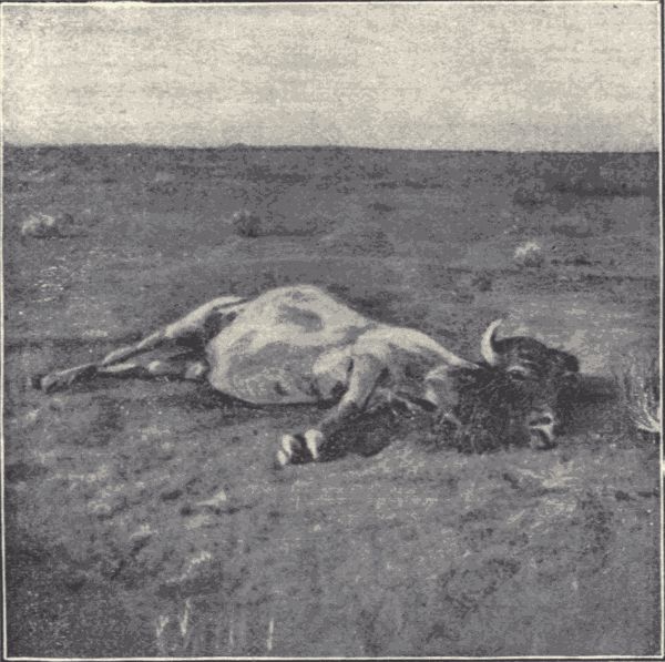
Fig. 156.—A buffalo, Bison bison, killed for its skin and tongue, on the plains of Western Kansas thirty years ago. (Photograph by J. Lee Knight.)
While most of the Feræ live on land, some are strictly aquatic. The true seals, fur-seals, sea-lions, and walruses comprise the aquatic forms, all being inhabitants of the ocean. The true seals, of which the common harbor seal (Phoca vitulina) is our most familiar representative, have the limbs so thoroughly modified for swimming that they are useless on land. The fur-seals, sea-lions, and walruses use the hind legs to scramble about on the rocks or[Pg 398] beaches of the shore. The fur-seals (fig. 157) live gregariously in great rookeries on the Pribilof or Fur Seal Islands, and the Commander Islands in Bering Sea.
The bears are represented in our country by the widespread brown, black, or cinnamon bear (Ursus americanus) and the huge grizzly bear (U. horribilis) of the West. The great polar bear (Thalarctos maritimus) lives in arctic regions. The otters, skunks, badgers, wolverines, sables, minks, and weasels compose the family Mustelidæ, which includes most of the valuable fur-bearing animals. Some of the members of this family lead a semi-aquatic or even strictly aquatic life and have webbed feet. The wolves, foxes, and dogs belong to the family Canidæ. The coyote (Canis latrans), the gray wolf (C. nubilus), and the red fox (Vulpes pennsylvanicus) are the most familiar representatives of this family, in addition to the dog (C. familiaris), which is closely allied to the wolf. "Most carnivorous of the carnivora, formed to devour, with every offensive weapon specialized to its utmost, the Felidæ, whether large or small, are, relatively to their size, the fiercest, strongest, and most terrible of beasts." The Felidæ or cat family includes the lions, tigers, hyenas, leopards, jaguars, panthers, wildcats, and lynxes. In this country the most formidable of the Felidæ is the American panther or puma (Felis concolor). It reaches a length from nose to root of tail of over four feet. Its tail is long. The wildcat (Lynx rufus) is much smaller and has a short tail.
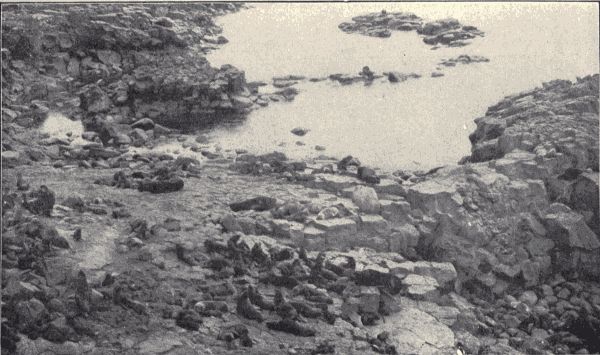
Fig. 157.—The Lukanin rookery of fur seals, Callorhinus alascanus, on St. Paul Island, Pribilof Group, Bering Sea. (Photograph from life by the Fur Seal Commission.)
The man-like mammals (Primates).—The Primates,
the highest order of mammals, includes the lemurs,
monkeys, baboons, apes, and men. Man (Homo sapiens)
is the only native representative of this order in our
country. All the races and kinds of men known, although
really showing much variety in appearance and body
structure, are commonly included in one species. The[Pg 399]
[Pg 400]
chief structural characteristics which distinguish man from
the other members of this order are the great development
of his brain and the non-opposability of his great toe.
Despite the similarity in general structure between him
and the anthropoid apes of the Old World, in particular
the chimpanzee and orang-outang, the disparity in size of
brain is enormous.
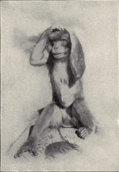
Fig. 158.—"Bob Jordan," a monkey of the genus Cercopithecus. (Photograph from life by D. S. Jordan.)
The lowest Primates are the lemurs found in Madagascar, in which island they include about one-half of all the mammalian species found there. The brain is much[Pg 401] less developed in the lemurs than in any of the other monkeys. The monkeys and apes may be divided into two groups, the lower, platyrrhine monkeys, found in the New World, and the higher, catarrhine forms, limited to the Old World. The platyrrhine monkeys have wide noses in which the nostrils are separated by a broad septum and with the openings directed laterally. These monkeys are mostly smaller and weaker than the Old World forms and are always long-tailed, the tail being frequently prehensile. They include the howling, squirrel, spider, and capuchin monkeys common in the forests of tropical South America. The catarrhine monkeys have the nose-septum narrow and the openings of the nostrils directed forwards, and the tail is wanting in numerous members of the group. They include the baboons, gorillas, orang-outangs, and chimpanzees. These apes have a dentition approaching that of man, and in all ways are the animals which most nearly resemble man in physical character.
Technical Note.—Multiplication, or increase by geometric ratio, among animals can be illustrated by noting the many eggs laid by a single female moth or beetle or fly or mosquito or any other common insect (or almost any other non-mammalian animal). The production of many live young by each female rose aphid can be readily seen; the number of young in a litter of kittens or pups or rabbits is a good illustration. From this geometric increase it is obvious that there must be a great crowding of animals and a struggle among them for existence. This struggle and the downfall of the many and success of the victorious few can be observed by rearing in a small jar of water all the young of a single brood of water-tigers (larva of Dyticus) or other aquatic predaceous insect. The strongest young will live by killing and eating the weaker of their own kind. In a spider's egg-sac the young after hatching do not immediately leave the sac, but remain in it for several days. During this time they live on each other, the strongest feeding on the weaker. Thus out of many spiderlings hatched in each sac comparatively few issue. This can be readily observed. Open several egg-sacs and count the eggs in them. Let the spiderlings hatch and issue from some other egg-sacs belonging to the same species of spider. The number of issuing spiderlings will always be much less than that of the eggs. The actual working of natural selection and the forming of new species can of course be seen only in results, and not in process. The great variety of adaptation, the fitness of adaptive structures, can be readily illustrated among the commonest animals. Animals showing certain striking and unusual adaptations will perhaps make the matter more obvious. To all teachers will occur numerous opportunities of illustrating, by reference[Pg 404] to actual processes or to obvious results, the principles of this chapter.
The multiplication and crowding of animals.—In the reproduction or multiplication of animals the production of young proceeds in geometric ratio, that is, it is truly a multiplication. Any species of animal, if its multiplication proceeded unchecked, would sooner or later be sufficiently numerous to populate exclusively the whole world. The elephant is reckoned the slowest breeder of all known animals. It begins breeding when thirty years old and goes on breeding until ninety years old, bringing forth six young in the interval, and surviving until a hundred years old. Thus after about eight hundred years there would be, if all the individuals lived to their normal age limit, 19,000,000 elephants alive descended from the first pair. A few years more of unchecked multiplication of the elephant and every foot of land on the earth would be covered by them. But the rate of multiplication of other animals varies from a little to very much greater than that of the elephant. It has been shown that at the normal rate in increase in English sparrows, if none were to die save of old age, it would take but twenty years to give one sparrow to every square inch in the State of Indiana. The rate of increase of an animal, each pair producing ten pairs annually and each animal living ten years, is shown in the following table:
| Years. | Pairs produced. | Pairs alive at end of year. |
| 1 | 10 | 11 |
| 2 | 110 | 121 |
| 3 | 1,210 | 1,331 |
| 4 | 13,310 | 14,641 |
| 5 | 146,410 | 161,051 |
| 10 | ...... | 25,937,424,600 |
| 20 | ...... | 700,000,000,000,000,000,000 |
Some animals produce vast numbers of eggs or young; for example, the herring, 20,000; a certain eel, several millions; and the oyster from 500,000 to 16,000,000. Supposing we start with one oyster and let it produce one million of eggs. Let each egg produce an oyster which in turn produces[19] one million of eggs, and let these go on increasing at the same rate. In the second generation there would be one million million of oysters, and in the fourth, i.e. the great great grandchildren of the first oyster, there would be one million million million million of oysters. The shells of these oysters would just about make a mass the size of the earth.
But it is obvious that all the new individuals of any animal produced do not live their normal duration of life. All animals produce far more young than can survive. As a matter of fact, which we may verify by observation, the number of individuals of animals in a state of nature is, in general, about stationary. There are about as many squirrels in the forest one year as another, about as many butterflies in the field, about as many frogs in the pond. Some species increase in numbers, as for example, the rabbit in Australia, which was introduced there in 1860 and in fifteen years had become so abundant as to be a great pest. Other species decrease, as the buffaloes, which once roamed our great plains in enormous herds and are now represented by a total of a few hundred individuals, and the passenger-pigeon, whose migrating flocks ten years ago darkened the air for hours in parts of the Mississippi valley, where now it is a rare bird. But the hand of man is the agent which has helped to increase or to check the multiplication of these animals. In nature such quick changes rarely occur.
The struggle for existence.—The numbers of animals are stationary because of the tremendous mortality occasioned by the constant preying on eggs and young and adults by other animals, because of strenuous and destructive climatic and meteorological conditions, and because there is not space and food for all born, not even, indeed, for all of a single species, let alone all of the hundreds of thousands of species which now inhabit the earth. There is thus constantly going on among animals a fearful struggle for existence. In the case of any individual this struggle is threefold: (1) with the other individuals of his own species for food and space; (2) with the individuals of other species, which prey on him, or serve as his prey, or for food and space; and (3) finally with the conditions of life, as with the cold of winter, the heat of summer, or drouth and flood. Sometimes one of these struggles is the severer, sometimes another. With the communal animals the struggle among individuals is lessened—they help each other; but when the struggle with the conditions of life are easiest, as in the tropics or in the ocean, the struggle among individuals becomes intensified. Each strives to feed itself, to save its own life, to produce and safeguard its young. But in spite of all their efforts only a few individuals out of the hosts produced live to maturity. The great majority are destroyed in the egg or in adolescence.
Variation and natural selection.—What individuals survive of the many which are born? Those best fitted for life; those which are a little stronger, a little swifter, a little hardier, a little less readily perceived by their enemies, than the others. They are the winners in the struggle for existence; they are the survivors. And this survival of the fittest, as it is called, is practically a process of selection by Nature. Nature selects the fittest to live and to perpetuate the species. Their progeny again[Pg 407] undergo the struggle and the selecting process, and again the fittest live. And so on until adjustment or harmonizing of animals' bodies and habits with the conditions of life, with their environment, comes to be extremely fine and nearly perfect.
It is evident, of course, that such a natural selection or survival of the fittest and consequent adaptation to environment presupposes differences among the individuals of a species. And this is an observed fact. No two individuals, although of the same brood, are exactly alike at birth; there always exist slight variations in structure and performance of functions. And these slight variations are the differences which determine the fate of the individual. One individual is a little larger or stronger or swifter or hardier than its mates. The existence of this variation we know from our observation of the young kittens or puppies of a brood. So it is with all animals. Thus natural selection depends upon two factors, namely, the excess in the production of new individuals and the consequent struggle for existence among them, and the existence of variations which give certain individuals slight advantages in this struggle.
Adaptation and adjustment to surroundings.—The action of natural selection obviously must, and does, result in a fine adaptation and adjustment of the structure and habits of animals to their surroundings. If a certain species or group of individuals cannot adapt itself to its environment, it will be crowded out by others that can. A slight advantageous variation comes in time by the continuously selective process to be a well-developed adaptation.
The diverse forms and habits possessed by animals are chiefly adaptations to their special conditions of life. The talons and beak of the eagle, the fishing-pouch of the pelican, the piercing chisel-like bill of the woodpecker,[Pg 408] and the sensitive probing-bill of the snipe are adaptations connected with the special feeding habits of these birds. The quills of the porcupine, the poison-fangs of the rattlesnake, the sting of the yellow-jacket, and the antlers of the deer are adaptations for self-defence. The fins and gills of fishes, the shovel-like fore feet of the mole, the wings of birds and insects and bats, the toe-pads of the tree-toad, the leaping-legs of the grasshopper, all these are adaptations concerned with the special life-surroundings of these animals.
Adaptations may relate to habits and behavior as well as to structure. Plainly adaptive are such habits as the migration of birds and some other animals, most of the habits connected with food-getting, and especially striking and interesting those connected with the production and care of the young, including nest-making and home-building.
Species-forming.—It is evident that through the cumulative action of natural selection, animals of a structural type considerably (even unlimitedly) different from any original type may in time be produced by the gradual modification of the original type under new conditions. If, for example, a few individuals of a mainland species should come to be thrown as waifs of wave and storm upon an island, and if these should be able to maintain themselves there and produce young, increasing so as to occupy the new territory, there would be produced in time a new type of individual conforming or adapted to the conditions obtaining in the island, these conditions being, of course, almost certainly different from those of the mainland. Thus as an offshoot or derivation from the original type still existing on the mainland we should have the new island-inhabiting type. Now when these island individuals come to differ so much, structurally and physiologically, from the mainland type that they cannot,[Pg 409] even if opportunity offers, successfully mate or interbreed with mainland individuals the island type constitutes a new species. That is, our distinction between species rests not only on structural differences, but on the impossibility of interbreeding (at least for the production of fertile young). Such a combination of the action of natural selection and the condition of isolation (as illustrated by the case of island animals), is probably the most potent factor in the production of new species of animals (and plants).
For accounts of the struggle for existence, variations, adaptations, natural selection and species-forming see Darwin's "Origin of Species," Wallace's "Island Life," and Romanes' "Darwin and After Darwin," I.
Artificial selection.—When a selection among the individuals of a species, that is, the choosing and preserving of individuals which show a certain trait or traits and the destroying of those individuals not possessing this trait, is done by man, it is called artificial selection. To artificial selection we chiefly owe all the many races or varieties of our domesticated animals and plants. For example, from the ancestral jungle fowl have been developed by artificial selection (and by cross-breeding) all our kinds of domestic fowl, as Brahmas, black Spanish, bantams, game-cocks, etc.; from the wild rock-dove have been developed our various fancy pigeons, as carriers, pouters, fantails, etc.
For an account of artificial selection see Darwin's "Plants and Animals under Domestication," and Romanes' "Darwin and After Darwin," I.
Social life and gregariousness.—Technical Note.—Students should refer to examples of gregariousness from their own observations of animals. The roosting together of crows and of blackbirds; the gathering of swallows preparatory to migration; the flocking of geese and ducks, with leaders, in their migratory flights, all can be readily observed. From observation or general reading students will be more or less familiar with prairie-dog villages, beaver-dams and marshes, the one-time great herds of bison, etc.
The struggle for existence is always operative; but in some cases one or more phases of it may be ameliorated. For example, the amelioration of the struggle among individuals of one species obtains in a lesser or greater degree in the case of those animals which exhibit a social life, of which mutual aid and mutual dependence are the basis. The honey-bee and the ants are familiar examples of animals which show a high degree of social life. They live, indeed, a truly communal life, where the fate of the individual is bound up in the fate of the community. But there are many animals which show a much lower degree of mutual aid and a far less coherent society. The simplest form of social life exists among those animals in which many individuals of one species keep together, forming a great band or herd. In this case there is not nearly so much mutual aid or mutual dependence as in that of the honey-bee, and the safety of the individual is not wholly bound up in the fate of the herd. Such[Pg 411] animals are said to be gregarious in habit, and this gregariousness is undoubtedly advantageous to the individuals of the band. The great herds of reindeer in the North, and of the bison or buffalo which once ranged over the Western American plains are examples of a gregariousness in which mutual protection from enemies, like wolves, seems to be the principal advantage gained. The bands of wolves which hunted the buffalo show the advantage of mutual help in aggression as well as in protection. Prairie-dogs live in great villages or communities which spread over many acres. By shrill cries they tell each other of the approach of enemies, and they seem to visit each other and to enjoy each other's society a great deal, although that they are thus afforded much actual active help is not apparent. The beavers furnish a well-known and very interesting example of mutual help; they exhibit a communal life although a simple one. They live in villages or communities, all helping to build the dam across the stream which is necessary to form the marsh or pool in which the nests or houses are built.
Communal life.—Technical Note.—See technical notes, pp. 212 et seq, for directions for work in connection with the study of the communal life of ants, bees, and wasps.
When many individuals of a species live together in a community in which the different kinds of work are divided more or less distinctly among the different members and where each individual works primarily for the whole and not for himself; where there is, in other words, a thorough mutual help and mutual dependence among the members of the community accompanied by a division of labor, the life of the species is truly communal. Those animals which show the most elaborate and specialized communal life are the termites or white ants, the social bees and wasps, and the true ants. Of these the ants and honey-bees[Pg 412] stand first. As already explained (see pp. 220 et seq), there are among these communal insects several different kinds of individuals in each species. With most animals there are two kinds only, males and females, which may or may not show differences in color, form, etc., so that they are readily distinguishable. Among all the communal insects, however, there are always three kinds of individuals, males, females, and workers, these last being infertile individuals. With the social wasps and social bees the workers are all infertile females and are smaller than the fertile forms; with the termites there are besides the fertile males and females, which are winged, workers which are wingless, and also peculiar wingless individuals called soldiers which have very large jaws and whose business it is to fight off attacking enemies of the community. Among the ants the workers are also wingless, while the males and females are winged. The worker ants in many species are of two kinds, so-called worker majors and worker minors, differing markedly in size. All the ant workers are good soldiers, but with some the fighting is done almost wholly by certain especially large-headed and large-jawed ones which may be called soldier-workers.
Thus among all strictly communal animals there is a specialization or differentiation of individuals accompanying the division of labor. Special individuals have a certain part of the work of the community to do, and they are specially modified in structure to do this work. This structural modification may make them incapable of performing certain other labor or work which is necessary for their living and which must be done for them, therefore, by others. Thus the mutual interdependence of the individuals composing a colony is very real. The worker honey-bees cannot perpetuate the species; honey-bees would die out were it not for the males and females. But[Pg 413] the males and females have given up the functions of food-getting and of caring for their young; did not the workers do these things for them, the community would die out quite as soon.
The advantages of communal or social life, of co-operation and mutual aid are real. Those animals that have adopted such a life are among the most successful of all in the struggle for existence. The termite worker is one of the most defenseless and for those animals that prey on insects one of the most toothsome insects, and yet the termite is one of the most abundant and successfully living insect kinds in all the tropics. Ants are everywhere and are everywhere successful. The honey-bee is a popular type of successful life. The artificial protection afforded it by man may aid it in its struggle for existence, but it gains this protection because of certain features of its communal life, and in nature the honey-bee takes care of itself well. Co-operation and mutual aid are among the most important factors which help in the struggle for existence.
Commensalism.—Technical Note.—Examine ants' nests to find myrmecophilous insects. If on the seashore search for hermit-crabs with sea-anemones on shell. If inland, try to have some preserved specimens showing the crabs and sea-anemones.
The phases of living together and mutual help just discussed concerned in each instance a single species of animal. All the members of a pack of wolves or of a honey-bee community belong to a single species. But there are numerous instances known of the mutually advantageous association of individuals of two different species. Such an association is called commensalism or symbiosis.
The hermit-crabs live, as has been learned (p. 154), in the shells of molluscs, most of the body of the crab being concealed within the shell, only the head and the grasping[Pg 414] and walking legs protruding. In some species of hermit-crabs there is always to be found on the shell near the opening a sea-anemone. "This sea-anemone is carried from place to place by the crab, and in this way is much aided in obtaining food. On the other hand, the crab is protected from its enemies by the well-armed and dangerous tentacles of its companion. On the tentacles there are many thousand long slender stinging threads, and the fish that would eat the hermit-crab must first deal with the stinging anemone." If the sea-anemone be torn away from the shell the crab will wander about seeking another anemone. When he finds one, he struggles to loosen it from the rock to which it is attached, and does not rest until he has torn it loose and placed it on his shell.
In the case of the hermit-crab and the sea-anemone there is no doubt of the mutual advantage derived from their communal life. But this mutual advantage is not so obvious in some cases of commensalism, where indeed most or all of the advantage often seems to lie with one of the animals, while the other derives little or none, but on the other hand suffers no injury. For example, "small fish of the genus Nomeus may often be found accompanying the beautiful Portuguese man-of-war (Physalia) as it sails slowly about on the ocean's surface. These little fish lurk underneath the float among the various hanging thread-like parts of the man-of-war which are provided with stinging cells. They are protected from their enemies by their proximity to these stinging threads, but of what advantage to the man-of-war their presence is is not understood." Similarly in the nests of the various species of ants and termites many different kinds of other insects have been found. "Some of these are harmful to their hosts, in that they feed on the food-stores gathered by the industrious and provident ant, but[Pg 415] others appear to feed only on refuse or useless substances in the nest. Some may be of help to their hosts by acting as scavengers. Over one thousand species of these myrmecophilous (ant-loving) and termitophilous (termite-loving) insects have been recorded by collectors as living habitually in the nests of ants and termites."
Parasitism.—Technical Note.—As examples of temporary external parasites find and examine fleas and ticks on dogs and cats, red mites on house-flies and grasshoppers (at the bases of the wings), etc. As examples of permanent external parasites find bird-lice on pigeons or domestic fowls or on other birds. Note the absence of wings and the peculiarly modified body shape of these parasites. Examine a bird-louse under the microscope; note the absence of compound eyes (it has simple eyes) and absence of wings; note bits of feathers, its food, in stomach, showing through the body. Find, as examples of internal parasites, intestinal worms or flukes. Examine trichinized pork to see Trichinæ in muscles. Examine preserved specimens of tapeworms. Collect pupæ of some common butterfly or moth and keep them in the schoolroom until either the butterflies or ichneumon flies issue. Some will surely be parasitized, and yield ichneumon flies (parasites) instead of a butterfly. As examples of degeneration by quiescence examine barnacles (found on outer rocks of seashore at low tide; easily obtained as preserved specimens by inland schools) and the females of scale-insects. These insects may be found on oleanders (the black scale, Lecanium oleæ) or fruit-trees (the San Jose scale, Aspidiotus perniciosus). Note the great degeneration of the adult female of the San Jose scale; it has no eyes, antennæ, wings, or legs. The young may be found crawling about at certain times of the year; they have eyes, antennæ and legs.
In addition to the various ways of living together among animals, already described, namely, the social and communal life of individuals of a single species and the commensal and symbiotic life of individuals of different species, there is another and very common kind of association among animals. This is the association of parasite and host; the association between two sorts of animals whereby one, the parasite, lives on or in the other, the host, and at the expense of the host. In this association the parasite gains advantages great or small, sometimes even obtaining all the necessities of life, while the host gains nothing,[Pg 416] but suffers corresponding disadvantage, often even the loss of life itself. Parasitism is a phenomenon common in all the large groups of animals, though the parasites themselves are mostly invertebrates. There are parasitic Protozoa, worms, crustaceans, insects, and molluscs, and a few vertebrates.
Some parasites, like the fleas and lice, live on the surface of the body of the host. These are called external parasites. Others, as the tapeworms, live exclusively inside the body; such are called internal parasites. Again, some, as the bird-lice, which are external parasites feeding on the feathers of birds, spend their whole lifetime on the host; they are called permanent parasites. Others, as a flea, which leaps on or off its host as caprice directs, or like certain parasites which as young live free and active lives, finally attaching themselves to some host and remaining fixed there for the rest of their lives, are called temporary parasites. Such a grouping is purely arbitrary and exists simply for the sake of convenience. It is not rigid, nor does it class parasites in their proper natural groups.
When various parasites are examined it will be noted that practically in all cases the body of a parasite is simpler in structure than the body of other animals closely related to it; that is, species which live parasitically, obtaining their food from and being carried about by a host, have simpler bodies than related forms that live free active lives, competing for food with other animals about them. This simplicity is not primitive, but results from the loss or atrophy of the structures which the special mode of life of the parasite renders useless. Many parasites are attached firmly by hooks or suckers to their host, and do not move about independently of it. They have no need of the power of locomotion, and accordingly are usually without wings, legs, or other locomotory organs.[Pg 417] Because they have no need of locomotion they have no need of organs of orientation, those special sense organs like the eyes, ears, and feelers which serve to guide and direct the moving animal; and most fixed parasites will be found to have no eyes, or any of those organs accessory to locomotion, and which serve for the detection of food or of enemies. Because these important organs, which depend for their successful activity on a well-organized nervous system, are lacking, the nervous system of parasites is usually very simple. Again, because the parasite usually feeds on the already digested food or the blood of its host, most parasites have a very simple alimentary canal, or even none at all. Finally, as the fixed parasite leads a wholly sedentary and inactive life, the breaking down and rebuilding of tissue in its body goes on very slowly and in minimum degree, so that there is little need of highly developed respiratory and circulatory systems; and most fixed and internal parasites have these systems of organs decidedly simplified. Altogether the body of a fixed permanent parasite is so simplified and so wanting in all those special structures which characterize the active, complex animals that it often presents a very different appearance from those forms with which we know it to be nearly related. This simplicity due to loss or reduction of parts is called degeneration. Such simplicity of body-structure due to degeneration is, however, essentially different in its origin from the simplicity of the lower simpler animals. In them the simplicity of body is primitive; they are generalized animals; the simplicity of degeneration is acquired; it is really an adaptation, or specialization.
An excellent example of body degeneration due to the adoption of a parasitic habit is that of Sacculina (fig. 159), a crustacean parasitic on other crustaceans, namely, crabs. The young Sacculina is an active, free-swimming larva[Pg 418] essentially like a young prawn or crab. After a short period of independent existence it attaches itself to the abdomen of a crab, and lives as a parasite. It completes its development under the influence of this parasitic life, and when adult bears absolutely no resemblance to such a typical crustacean as a crab or crayfish. Its body external to the host crab is simply a pulsating tumor-like sac, with no mouth-parts, no legs, and internally hardly any well-developed organs except those of reproduction. Degeneration here is carried very far.
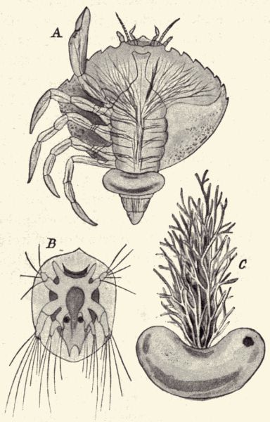
Fig. 159.—Sacculina, a parasitic crustacean; A, attached to a crab, the root-like processes of the parasite penetrating the body of the host; B, the active larval condition; C, the adult removed from its host. (After Haeckel.)
Various parasites have been referred to in Part II under their proper branch and class. The worms include an unusually large number of them, such as the tapeworms, trichinæ and other intestinal forms, all of which live as internal parasites in the alimentary canal or in other organs of higher animals, especially the vertebrates. Many crustaceans are parasitic, usually living, like the fish-lice, as fixed external parasites on fishes, other crustaceans, etc., but with a free and active larval stage. Among the insects, on the contrary, many of the parasitic forms (as the ichneumon flies) are free and active in the adult stage, but live as internal grubs or maggots in the larval stage. The ichneumon flies (of the order Hymenoptera) are four-winged, slender-bodied insects which lay their eggs either on or in (by means of a sharp piercing ovipositor) some caterpillar or beetle grub, into the body of which the young grub-like ichneumon larvæ burrow on hatching. The parasites feed on the body-tissues of the host, not attacking, however, such organs as the heart or nervous system, which would produce the immediate death of the host. The caterpillar lives with the ichneumon grubs within it usually until nearly time for its pupation. Often, indeed, it pupates with the parasite still in its body. But it never comes to maturity. The larval ichneumons pupate either within the body of its host, or in a tiny silken cocoons outside of its body (fig. 160). From the cocoons the winged adult ichneumons issue; and after mating the females find another caterpillar on whose body to lay their eggs.
Degeneration can be produced by other causes than parasitism. It is evident that if for any other reason an animal should adopt an inactive fixed life it would degenerate. The barnacles (see fig. 37) are excellent examples of degeneration through quiescence. They are crustaceans related most nearly to the crabs and shrimps. The[Pg 420] young barnacle just from the egg is a six-legged, free-swimming larva (nauplius) with a single eye, greatly like a young prawn or crab. It develops during its independent life two compound eyes and two large antennæ. But soon it attaches itself to some stone or shell, or pile or ship's bottom, giving up its power of locomotion, and its further development is a degeneration. It loses its compound eyes and antennæ, and acquires a protecting shell. Its swimming feet become modified into grasping organs, and it loses most of its outward resemblance to the typical members of its class. The Tunicata or ascidians compose a whole group of animals which are fixed in their adult condition and have thus become degenerate. They have been likened to a "mere rooted bag with a double neck." In their young stage they are free-swimming, active, tadpole-like or fish-like larvæ, possessing organs much like those of the adult simplest fish or fish-like animals. Their larval structure reveals, however, the relationships of the ascidians to the vertebrates, a relationship which is not at all apparent in the degenerate adults. Certain insects live sedentary or fixed lives. All the members of one large family, the Coccidæ, or scale-insects (figs. 62 and 63), have females which as adults are wingless and in some cases have no legs, eyes, or antennæ, while the males are all winged and have legs and the[Pg 421] special sense organs. The males lead a free active life, but the females have nearly or quite given up the power of locomotion, attaching themselves by means of their sucking beak to some plant, where they obtain a sufficient food-supply (plant-sap) and lay their eggs. In both males and females the larvæ are little active crawling six-legged creatures with legs, eyes, and antennæ.
We are accustomed perhaps to think of degeneration
as necessarily implying a disadvantage in life. It is true
that a blind, footless, degenerate animal could not cope
with the active, keen-sighted, highly organized non-degenerate
in free competition. But free competition is
exactly what the degenerate animal has nothing to do
with. Certainly the Sacculina and the scale-insects live
well; they are admirably adapted to the kind of life they
lead. A parasite enjoys certain obvious advantages in life,
and even extreme degeneration is no drawback (except as
we shall see later), but gives it a body which demands less
food and care. As long as the host is successful in eluding
its enemies and avoiding accident and injury the parasite
is safe. Its life is easy as long as the host lives.
But the disadvantages of parasitism and degeneration are
nevertheless obvious. The fate of the parasite is bound
up with the fate of the host. "When the enemy of the
host crab prevails, the Sacculina goes down without a
chance to struggle in its own defence. But far more important
than the disadvantage in such particular or individual
cases is the fact that the parasite cannot adapt itself
in any considerable degree to new conditions. It has
become so modified, so specialized to adapt itself to the
very special conditions under which it now lives, it has
gone so far in giving up organs and functions, that if
present conditions change and new ones come to exist
the parasite cannot adapt itself to them. The independent
free-living animal holds itself, one may say, able and[Pg 422]
[Pg 423]
ready to adapt itself to any new conditions of life. The
parasite has risked everything for the sake of a sure and
easy life under the present existing conditions. Change
of conditions means its extinction."
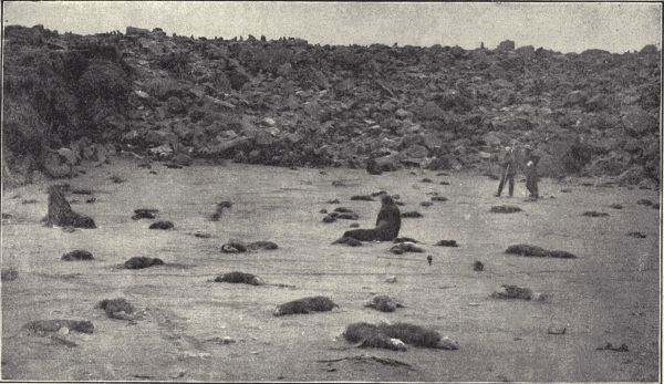
Fig. 161.—Young fur seals, Callorhinus ursinus, of the Tolstoi rookery, St. Paul Island, Bering Sea, killed by a parasitic intestinal worm, Uncinaria sp. (Photograph by the Fur Seal Commission.)
For an elementary account of commensalism and parasitism see Jordan and Kellogg's "Animal Life," pp. 172-200. The account here given is based on the author's previously written account in "Animal Life." See also Van Beneden's "Animal Parasites and Messmates."
Technical Note.—For an appreciation of the reality of protective resemblances observations must be made in the field. Examples are easily found. Locusts, katydids, green caterpillars, lizards, crouching rabbits, and brooding birds are readily observed instances of general protective resemblance. For examples of variable resemblance examine specimens of a single locust species taken from different localities; the individuals of the various species of the genus Trimerotropis show much variation to harmonize with their surroundings. Collect a number of larvæ (caterpillars) of one of the swallow-tail butterflies (Papilio), and when ready to pupate put them separately into pasteboard boxes lined inside with differently colored paper. The chrysalids will show in their coloration the influence of the different colors of the lining paper, their immediate environment. As examples of special protective resemblance observe inch- or span-worms (larvæ of Geometrid moths). The walking-stick is not uncommon; many spiders that inhabit flower-cups show striking protective color patterns; and the Graptas or comma-butterflies which resemble dead leaves may be examined.
To illustrate warning colors, find, if possible, the larvæ (caterpillars) of the common milkweed or monarch butterfly (Anosia plexippus), and offer them to birds, at the same time offering other caterpillars, and note the results. For terrifying or threatening appearance find specimens of the large green tobacco- or tomato-worm (larva of the five-spotted sphinx-moth, Phlegethontius carolina), or other sphingid larvæ.
The butterflies illustrating the striking example of mimicry, described on p. 432, can be found in most parts of the country. Syrphid and other flies which mimic bees and wasps can readily be found on flowers.
Each student should search for himself for examples of protective resemblance.
Use of color.—The prevalence of color and the oftentimes striking and intricate coloration patterns of animals[Pg 425] demand some explanation. As naturalists are accustomed to find the frequently bizarre and seemingly inexplicable shapes and general structure of animals readily explained by the principle of adaptation, that is, special modification of body-structure to fit special conditions of life, so they look to use as the chief explanation of color and markings. Some uses are obvious; bright colors and striking patterns may serve to attract mates or to avail as recognition marks by which individuals of a kind may readily recognize each other. The white color of arctic animals probably serves to help keep them warm by preventing radiation of heat from the body; on the other hand dark color may also help to keep animals warm by absorbing heat. "But by far the most widespread use of color is for another purpose, that of assisting the animal in escaping from its enemies or in capturing its prey."
It is common knowledge that the young and old, too, of many kinds of ground-inhabiting animals, when startled by an enemy will not run, but crouching close to the ground remain immovable, trusting to remain unperceived. But a blue or crimson rabbit, however still it might keep, would be easily seen by its enemy and killed. Rabbits, however, which are good examples of animals having this habit of lying close, are neither blue nor green nor red, but are colored very much like the ground on which they crouch. This harmonious coloration is as necessary to the success of this habit as is the keeping still. A grasshopper flying or leaping in the air is conspicuous; when it alights how inconspicuous it is! Unless one has followed it closely in its flight and has kept the eye fixed on it after alighting it is usually impossible to distinguish it from its surroundings. And this is greatly to the advantage of the grasshopper in its efforts to escape its enemies, that is, in its struggle for existence. On the other hand a green katydid would be very conspicuous[Pg 426] in a dusty road. But dusty roads are precisely where katydids do not rest. They alight among the green leaves of a tree or shrub. The animals that live in deserts are almost all obscurely mottled with gray and brownish and sand-color so as to harmonize in color with their habitual environment. The arctic hares and foxes and grouse which live in regions of perpetual snow are pure white instead of red or brown or gray like their cousins of temperate and warm regions.
These cases of an animal's color and markings harmonizing with the usual environment are called instances of protective resemblance; that is, they are resemblances for a purpose, that purpose being to render the animal indistinguishable from its surroundings and thus to aid it in escaping its enemies. Such protective resemblances are obviously of great value to animals, and, like other advantageous modifications, have been produced by the action of natural selection. Those individuals of a species most conspicuous and hence most readily perceived by enemies are the first (under ordinary circumstances) to be captured and eaten. The less conspicuous live and produce young like themselves. Of these young the least conspicuous are again saved and so over and over again through thousands of generations until this natural selecting of the protectively colored results in the production of the wonderfully specialized examples of resemblance to which attention is called in the following paragraphs.
General, variable, and special protective resemblance.—In the brooks most fishes are dark olive or greenish above and white below. To the birds and other enemies which look down on them they are colored like the bottom. To their fish-enemies which look up from below they are like the white light above them in color and their forms are not clearly seen. The green tree-frogs and tree-snakes which live habitually among green foliage;[Pg 427] the mottled gray and tawny lizards and birds and small mammals of the plains and deserts, and the white hares and foxes and owls and ptarmigan of the snowy arctic regions—all show a general protective resemblance.
Sometimes an animal changes color when its surroundings change. Certain hares and grouse of northern latitudes are white in winter when the snow covers all the ground, but in summer when much of the snow melts, revealing the brown and gray rocks and withered leaves, they put on a grayish and brownish coat of hair or feathers. A small insect called the toad-bug (Galgulus) lives abundantly on the banks of a pond on the campus of Stanford University. The shores of this pond are covered in some places with bits of bluish rock, in others with bits of reddish rock, and in still others with sand. Specimens of the toad-bug collected from the blue rocks are bluish or leaden in color, those from the red rocks are reddish, and those from the sand are sand-colored. Changes of color to suit the surroundings can be quickly[Pg 428] made by some animals. The chameleons of the tropics change momentarily from green to brown, blackish, or golden. There is a little fish (Oligocottus snyderi) common in the tide-pools of the Bay of Monterey in California whose color changes quickly to harmonize with the rocks it happens to rest above. Such changing coloration to suit the surroundings may be called variable protective resemblance.
Very striking are those cases of protective resemblance
in which the animal resembles in color and shape, sometimes
in extraordinary detail, some particular object or
part of its usual environment. This may be called special
protective resemblance. The larvæ of the Geometrid
moths called inch-worms or span-worms are twig-like in
appearance, and have the habit, when disturbed, of standing
out stiffly from the twig or branch on which they rest,
so as to resemble in attitude as well as color and markings
a short or broken twig. To increase this simulation
the body of the larva often has a few irregular spots or
humps resembling the scars left by fallen leaves, and it
also lacks the middle prop-legs of the body common to
other lepidopterous larvæ, which would tend to destroy
the illusion so successfully carried out by it. The common
twig-insect or walking-stick (fig. 162) with its wingless,
greatly elongate, brown or greenish body and legs is when
at rest quite indistinguishable from the twigs on which it
lies. Another excellent example of special protective
resemblance is furnished by the famous green-leaf insect
(Phyllium) of the tropics, which has broad leaf-like wings
and body of a bright green color with markings which
imitate the leaf-veins, and small irregular yellowish spots
which simulate decaying or stained or fungus-covered
spots in the leaf. Most striking of all, however, is the
large dead-leaf butterfly Kallima (fig. 163) of the East
Indian region. The upper sides of the wing are dark[Pg 429]
[Pg 430]
with purplish and orange markings not at all resembling
a dead leaf. But the butterflies when at rest hold their
wings together over the back, so that only the under sides
of them are exposed. These are exactly the color of a
dry dead leaf with markings mimicking midrib and
oblique veins, and, most remarkable of all, what are
apparently two holes like those made in leaves by insects,
but in the butterfly imitated by two small circular spots
free from scales and hence clear and transparent. When
Kallima alights it holds the wings in such position that the
combination of all four produces with remarkable fidelity
the simulation of a dead leaf still attached to the twig by
a short pedicel or leaf-stalk (imitated by a short "tail"
on the hind wings). The head and legs of the butterfly
are concealed beneath the wings.
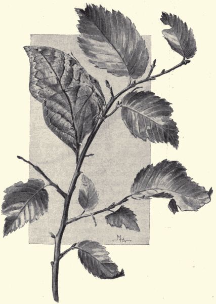
Fig. 163.—The dead-leaf butterfly, Kallima sp., a remarkable case of special protective resemblance. (From specimen.)
Warning colors, terrifying appearances, and mimicry.—While many animals are so colored as to harmonize with their habitual or usual environment, others on the contrary are very brightly colored and marked in such bizarre and striking pattern as to be conspicuous. There is no attempt at concealment; it is obvious that conspicuousness is the object sought or at least produced by the coloration. Animals like these, we shall find, are in almost all cases specially protected by special weapons of defence such as stings or poison-fangs, or by the secretion of an acrid, ill-tasting fluid in the body. Many caterpillars have been found, by observation in nature and by experiment, to be distasteful to insectivorous birds. Now it is obvious that it would be advantageous to these caterpillars to be readily recognized by birds. After a few trials the bird learns by experience to let these distasteful larvæ alone; their conspicuous markings serve as warning colors. The black-and-yellow-banded caterpillar of the common milkweed or monarch butterfly (Anosia plexippus) is a good example of such protection by a combination[Pg 431] of distastefulness and warning coloration. The little lady-bird beetles are mostly distasteful to birds; they are brightly and conspicuously marked. Certain little Nicaraguan frogs have a bright livery of red and blue, in strong contrast to the dull concealing colors of other frogs in their region. By offering these little blue and red frogs to hens and ducks the naturalist Belt found that they are distasteful to the birds.
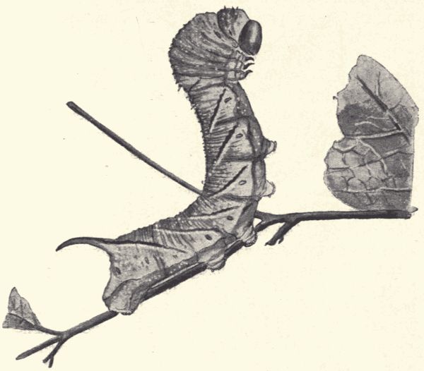
Fig. 164.—The larva of the pen-marked sphinx-moth, Sphinx chersis, showing terrifying attitude. (After Comstock.)
Certain animals which are without special means of defence and are not distasteful are yet so marked or shaped, and so behave as to present a threatening or terrifying appearance. The large green caterpillars of the sphinx-moths, the tomato- and tobacco-worms, are[Pg 432] familiar examples, each larva having a sharp horn on the back of the next to last body-segment (fig. 164). When disturbed the caterpillar assumes a threatening attitude, and the horn seems to be an effective weapon of defence. As a matter of fact it is not at all a weapon of defence, being weak, not provided with poison, and altogether harmless.
But it would plainly be to the advantage of a defenceless animal, one without poison-fangs or sting and without an ill-tasting substance in its body, to be so marked and shaped as to mimic some other specially defended or inedible animal sufficiently to be mistaken for it and thus to escape attack. Such cases have been noted, especially among insects. This kind of protective resemblance may be called mimicry. A most striking case is that presented by the familiar monarch and viceroy butterflies (fig. 165). The monarch (Anosia plexippus) is perhaps the most abundant and widespread butterfly of our country. It is a fact well known to entomologists that it is distasteful to birds and is let alone by them. It is conspicuous, being large and chiefly red-brown in color. The viceroy (Basilarchia archippus), also red-brown and patterned almost exactly like the monarch, is not, as its appearance would seem to indicate, a very near relation of the latter, but on the contrary it belongs to a genus of butterflies all of which, except the viceroy and one other, are black and white in color and of different pattern from the monarch. The viceroy is not distasteful to birds, but by its extraordinary simulation or mimicking of the monarch it is not distinguished from it and so is not molested. In the tropics there have been discovered numerous examples of mimicry among insects. The members of two large families of butterflies (Danaidæ and Heliconidæ) are distasteful to birds and are mimicked by members of other butterfly families (especially the Pieridæ).
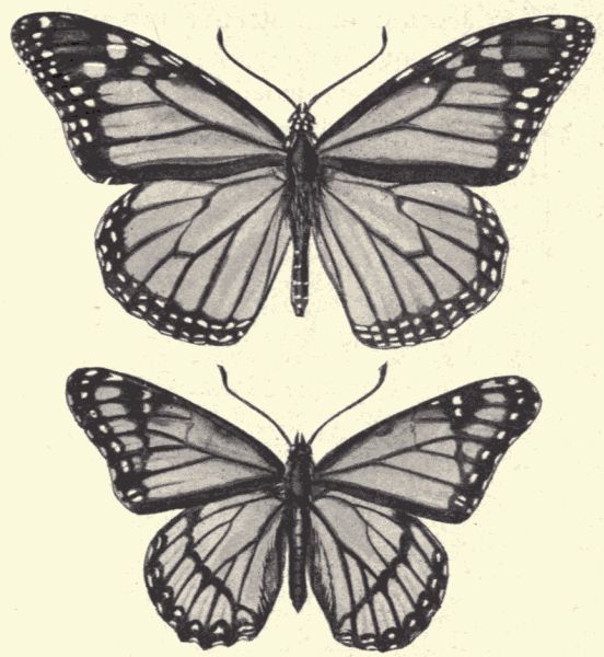
Fig. 165.—The monarch butterfly, Anosia plexippus (above), distasteful to birds, and the viceroy, Basilarchia archippus (below), which mimics it. (From specimens.)
Alluring coloration.—A few animals show what is called alluring coloration; that is, they display a color pattern so arranged as to resemble or mimic a flower or other lure, and thus entice to them other animals, their natural prey. Certain Brazilian fly-catching birds have a brilliantly colored crest which can be displayed in the shape of a flower-cup. The insects attracted by the false flower furnish the bird with food. In the tribe of fishes called the "anglers" or "fishing frogs," the front rays of the dorsal fin are prolonged in the shape of long slender filaments, the foremost and longest of which has a flattened and divided extremity. The angler conceals itself in the mud or in the cavities of a coral reef, and waves the filament back and forth. Small fish are attracted[Pg 434] by the lure, mistaking it for worms writhing about. When they approach they are engulfed in the mouth of the angler, which in some species is of enormous size. One of these angler species is known to fishermen as the "all-mouth."
For a fuller account of protective resemblances and mimicry see Jordan and Kellogg's "Animal Life," pp. 201-223. For still more extended accounts see Poulton's "Colours of Animals," and Beddard's "Animal Coloration."
Technical Note.—The larger aspects or phenomena of the distribution of animals over the earth on land and in sea cannot be studied personally in the field by the student, but many local features of distribution can be so observed and studied. The restriction of certain kinds of animals to certain kinds of habitat, the presence and character and effectiveness of barriers, some of the modes of distribution, the presence and successful life of introduced foreign species such as the black and brown rats, the English sparrow, the German and Asiatic cockroaches, the gradual change of range or distribution of certain kinds of animals through the influence of a change in environment (caused by man in cutting off forests, cultivating heretofore wild pastures, etc.) all offer favorable and profitable opportunities for personal observation.
An excellent and feasible piece of field-work in distribution is the making of a zoological survey of the locality in which the school is situated. A map of the locality should be made on a generous scale, which should include all prominent physical features of the region, such as streams, ponds, hills, woodlands, marshes, etc., and on this map should be indicated the places where the various animals of the local fauna occur. Some of the animal species will have a limited range, and the limits of this range should be shown. This map and faunal list can be added to and perfected by successive classes. For fuller discussions of the geographical distribution of animals see Jordan and Kellogg's "Animal Life," Beddard's "Zoogeography," Heilprin's "The Distribution of Animals," and Wallace's "Geographical Distribution."
Geographical distribution.—It is a matter of common knowledge with all of us that there are no wild lions or camels or kangaroos or monkeys or ostriches or nightingales in North America. Ostriches are found only in Africa and South America, kangaroos only in Australia,[Pg 436] lions only in Asia and Africa. On the other hand there are no opossums in Europe or grizzly bears or rattlesnakes anywhere else in the world than in this country. That is, certain kinds of animals have a certain limited range of occurrence or distribution. It is obvious, too, that certain animals live only on land, while others live only in water, and of these latter some are restricted to the ocean, while others live only in fresh water. All of the facts regarding the dispersion or diffusion of animals on land and in water make up the science of the geographical distribution of animals, or, as it is sometimes called, zoogeography. Under this subject are included not only the facts of the present actual distribution or occurrence of animals over the world, but the facts concerning the reasons for this distribution, the modes of travel and dispersion, the character and influence of barriers to the spread, and the results, in the adaptation of old forms and the production of new forms, of the phenomena of distribution.
Just as maps are made to show graphically the facts of political geography, which concerns the position and extent of the various powers and States which claim the allegiance of the people, and the facts of physical geography, which concerns the physical character of the earth's surface, so maps are made to show the geographical distribution of animals. Because of the great numbers of animal species no one map can show the distribution of all species, but a series of maps of the world or of a continent or of a State or county or more limited region could be made (and many such have been made) showing the distribution of selected species. In a map of a limited locality, say of a few square miles, the occurrence and distribution of most of the commoner and more familiar animals can be shown, and each high school should possess such a map (see technical note at beginning of this chapter).
Laws of distribution.—The laws governing the distribution of animals over the earth's surface have been recently[20] expressed in a simple statement as follows: Every species of animal is found in every part of the earth unless (a) its individuals have been unable to reach this region on account of barriers of some sort; or (b) having reached it, the species is unable to maintain itself, through lack of capacity for adaptation, through severity of competition with other forms, or through destructive conditions of environment; or (c) having entered and maintained itself it has become so altered in the process of adaptation as to become a species distinct from the original type.
Modes of migration and distribution.—That animals should be continually trying to extend their range is obvious from what we know of their rapid increase by multiplication. In a region which can provide food for but one thousand wolves, there is a production each year of several times one thousand. These new wolves must struggle among themselves for food, or migrate, if possible, to new regions as yet not inhabited by wolves. The wolf's mode of migration or distribution is walking or running, and so with other mammals except the bats and aquatic forms. Birds and bats can fly, and can thus migrate more swiftly, farther, and over barriers which would stop mammals. Most insects can fly. Worms can only crawl and very slowly at that. Fishes can swim, but if they are in a landlocked sheet of water, they cannot go beyond its confines. Marine animals can migrate from ocean to ocean, and land animals from continent to continent unless checked by barriers (see next paragraph).
But besides such voluntary and independent modes of distribution long journeyings may be made involuntarily, or by a passive migration as it may be called. Parasites,[Pg 438] for example, are carried by their hosts in all their travels; the tiny Tardigrada and Rotifera, which can be desiccated and yet restored to active life by coming again into water, are carried in the dried mud on the feet of birds or other animals. On floating objects in rivers or in ocean currents land-animals may be carried long distances. Man, the greatest traveller of all, is responsible for the widened distribution of many animals. Thus have come to us in ships from Europe the black and brown rats, the English sparrow, the Hessian fly, the commonest cockroaches of our houses and many other forms. And these animals have been carried involuntarily all over the United States in railway-cars and wagons.
Barriers to distribution.—As is indicated in the paragraph on the modes of migration, a considerable sheet of water is obviously a barrier to the further travelling of a walking or crawling land-animal, although no barrier to a winged form. Similarly a strip of land is a barrier to a strictly aquatic animal as a fish. Or a high fall in the stream may serve as an insuperable barrier, making it impossible for any fish below the fall to reach the upper part of the stream. Numerous cases of this kind are known in the Rocky Mountains and Sierra Nevada, where a stream may be well supplied with trout below a fall, and quite bare of these fish above the fall. In the Yosemite Valley in California trout live in the Merced River below the great Vernal and Nevada falls, but above these falls the Merced contains no trout. To fresh-water swimming animals salt water may be a barrier; thus some kinds of fresh-water fishes are limited to one of two near-by streams although the mouths of these streams empty near each other into the ocean. The amphibious batrachians, at home in fresh water and on land, are killed by contact with sea-water. Earthworms also are killed by salt water. Thus the narrowest ocean strait is[Pg 439] as effective a barrier to these animals as a whole sea. High mountain ranges and broad deserts are barriers to many land-animals, partly because of the physical obstacles, partly because of the differences in temperature and in food-supply.
Temperature and climate (as distinct from temperature) and the ocean are the three great barriers when we consider the animal kingdom as a whole, and look for the causes which determine the chief zoogeographical divisions of the earth's surface. Most of the tropical animals cannot endure frost, hence the isothermal line of frost is a line across which few tropical animals venture. Most arctic animals are enfeebled by heat, and the isothermal line which marks off the region in which frost occurs the year round is another great zoogeographical boundary. But while these lines are limits for localized species, some animals, as birds, especially, keep within a relatively uniform temperature by migrations across these lines. It should be borne in mind that the gradual decrease in temperature met with in going north or south from the tropics is also met in the ascent of high mountains. The summits of lofty peaks, even in the tropics, are truly arctic in character; they are snow-covered, and the animals and plants on them are truly arctic. Thus in the ascent of a single mountain a whole series of life-zones from tropical to arctic can be traversed.
Climate, as distinct from temperature, establishes limits of distribution. The animals of Eastern North America accustomed to a humid atmosphere cannot live in the dry plains and deserts of the West. Closely associated with climate is the nature of the plant-growth covering the land; here are forests and luxuriant meadows, there are sparse tough grasses of the dry plateau. The limits of a special kind of plant-growth often are the limits of distribution of certain animals.
The third great barrier, the ocean, is perhaps the most obvious of all in its influence. It is only in rare cases that any land-animal can independently cross a great ocean. Thus the land-animals of Australia differ from those of all other countries, and those of Africa and South America have developed almost independently of one another. The ocean is, as already mentioned, also a barrier for fresh-water aquatic animals, and even marine fishes which live normally in shallow waters along the shore rarely venture across the great depths of mid-ocean.
The obstacles or barriers met with determine the limits of a species. Each species broadens its range as far as it can. It attempts unwittingly, through natural processes of increase, to overcome the obstacles of ocean or river, of mountain or plain, of woodland or prairie or desert, of cold or heat, of lack of food or abundance of enemies—whatever the barriers may be. The degree of hindrance offered by any barrier differs with the nature of the animal trying to pass it. That which forms an impassable obstacle to one species may be a great aid to the spread of another. "The river which blocks the monkey or the cat is the highway of the fish and turtle. The waterfall which limits the ascent of the trout is the chosen home of the ouzel."
Faunæ and zoogeographic areas.—The term fauna is applied to the animals of any region considered collectively. Thus the fauna of Illinois includes the entire list of animals found naturally in that State. The fauna of a schoolyard comprises all the animals found living naturally in the yard. The fauna of a pond includes all the animal inhabitants of the pond. (Flora is used similarly of all the plants in a given region.) The relation of one fauna to another depends on the character and effectiveness of the barriers between, and the physical character of the two regions. Thus the fauna of Illinois differs but little[Pg 441] from that of Indiana or Iowa, because there are no barriers between the States, and they are alike physically. On the other hand the fauna of California differs much from that of the Eastern States because of the great barriers (the desert and the Sierra Nevada Mountains) which lie between it and these States, and because of the great differences in the physical and climatic conditions of the two regions.
The land-surface of the earth has been divided by zoogeographers into seven great realms of animal life, based on the distributional characters shown by these various regions. These realms are separated by barriers of which the chief are the presence of the sea and the occurrence of frost. These realms are named, from their geographical region, the Arctic, the North Temperate, the South American, the Indo-African, the Madagascar, the Patagonian, and the Australian. Of these the Australian alone is sharply defined. Most of the others are surrounded by a broad fringe of debatable ground that forms a transition to some other zone.
Habitat and species.—The habitat of a species of animal is the region in which it is found in a state of nature. It is currently believed that the habitat of any animal is the whole of that region for which it is best adapted. But this is not necessarily true. In fact in most cases it is not true. The trout naturally debarred from the rivers in Yellowstone Park by the waterfalls could live there well if the barrier could be passed. In the case of one stream it has been passed and the trout flourish above the fall. The success of the black and brown rats and the English sparrow in America, of the rabbit in Australia, of bumblebees and house-flies in New Zealand, all of which animals had a natural habitat not including these regions, but by artificial means have been carried over the barriers and into the new territory, prove[Pg 442] that "habitat" is not necessarily coincident with "only fit region." Shad, striped bass, and catfish from the Potomac River have been introduced into and now thrive in the Sacramento River in California. In fact the whole work of the introduction and diffusion of valuable food-animals in territory not naturally included in the habitat of the species is based on our knowledge that the habitat of a species is often determined by physical barriers rather than by exclusive fitness of environment. Within the natural habitat the environment is fit for the species' existence, outside of it the environment may be fit.
But there occur numerous instances where a species successful in leaving its original habitat is unsuccessful in attempting to maintain itself on new ground. Man has introduced various animals from one country to another. The English sparrow (naturally debarred from this country by the ocean barrier), brought to America from Europe, has covered its new territory rapidly and maintains itself with brilliant success. But the nightingale, the starling and skylark which have been repeatedly introduced and set free are unable to maintain themselves here.
Species-extinguishing and species-forming.—Accompanying the constant slow migrating of species into new habitats and the constant slow changing of environment and conditions everywhere is to be seen a constant modification of the fauna of any region due to the inability of some species to maintain their ground, the predominating increase of others, and the modifying or adaptive changing of others into new forms. In 1544 the black rat of Europe was introduced into America and it soon crowded out the native rats, being in its turn crowded out by the European brown rat (the present common rat in buildings), introduced about 1775. Here we have the original native species unable to maintain itself in competition with introduced forms.
With a change of environing conditions, certain species are unable to maintain themselves. With the destruction of the forests going on in parts of our country the great host of wood-creatures, the bears, squirrels, the wood-birds and insects, can no longer maintain themselves, and grow rare and disappear. Man often also influences the status of a species by checking its increase either by actual slaughter, as with the bison and passenger-pigeon, or by making adverse changes in its environment, as by destroying forests, or putting the plains under cultivation.
In the discussion of "species-forming" (see p. 408) it was shown that adaptation may lead to the altering of species, and to the formation of new ones (under the influence of natural selection). With the gradual change of conditions, or with the facing of new conditions because of an unusual migration to or invasion of new territory, those individuals of the species exposed to the new conditions must adapt themselves in structure and habit in order to meet successfully the new demands. By the cumulative action of natural selection these adaptive changes are emphasized; and this emphasis may come to be so pronounced that the part of the species represented in this newly acquired territory, if isolated from the original stock, is so altered as to be quite distinct in appearance from the old. If these changed individuals are also physiologically distinct from the old stock, i.e. are unable to mate with them, a new species is established. As already mentioned, the peopling of islands from mainlands is an excellent and readily observable example of the phenomena referred to in the third law of distribution.
Equipment of pupils.—Each pupil should have a laboratory note-book of about 8 × 10 inches, opening at the end, in which both drawings and notes can be made. The paper should be unruled and of good quality (not too soft). Each pupil should have also instruments of his own as follows: scalpel, pair of small scissors, spring forceps, pair of dissecting-needles, small glass pipette, and paper of ribbon-pins for pinning out specimens. The cost of this outfit need not exceed $1.00. The laboratory should furnish him with a dissecting-dish and a dissecting-microscope, or at least a lens.
Laboratory drawings and notes.—Each pupil should make the drawings called for in the directions for the laboratory exercises. These drawings should be in outline, and put in by pencil; the lines may be inked over if preferred. Shading should be used sparingly, if at all. Each drawing and all the organs and animal parts represented in it should be fully named. See the anatomical plates in this book for example. With such complete "labelling," little note-taking need be done in connection with the dissections.
Notes should be made of any observations which cannot be represented in the drawings; for example, on the[Pg 448] behavior of the living animals. All notes referring to matters of life-history should be dated.
Field-observations and notes.—Scattered through this book will be found numerous suggestions for student field-work, for the observation of the life-history and habits and conditions of animals in nature. As explained in the Preface, the initiation and direction of such work must be left to the teacher. But its importance both because of its instructiveness and its interest is great. Pupils should not only be incited to make individual observations whenever and wherever they can, but the teacher should make little field-excursions with the class or with parts of it at various times, to ponds or streams or woods, and "show things" to all. The life-history and feeding-habits of insects, the web-making of spiders, the flight, songs, nesting, and care of young of birds, the haunts of fishes, the development of frogs, toads, and salamanders, the home-building and feeding-habits of squirrels, mice, and other familiar mammals are all (as has been called attention to at proper places in the book) specially fit subjects for field-observation.
Each pupil should keep a field note-book, recording from day to day, under exact date, any observations he may make. Let the most trivial things be noted; when referred to later in connection with other notes they may not seem so trivial. The field note-book should be smaller than the laboratory note- and drawing-book, small enough to be carried in the pocket. Notes should be made on the spot of observation; do not wait to get home. Sketches, even rough ones, may be advantageously put into the book. Students with photographic cameras can do some very interesting and valuable field-work in making photographs of animals, their nests and favorite haunts. Such photographic work is very effectively used now in the illustration of books about animals and[Pg 449] plants (see the reproductions of photographs in this book). If the class is making a collection the collecting notes or data made in the field-books of the different pupil collectors should all be transferred to a common "Notes on Collections" book kept by the whole class.
Equipment of laboratory.—The equipment of the laboratory or classroom will, of necessity, depend upon the opportunities afforded the teacher by the school officers to provide such facilities as instruments, books, and charts. If dissections are to be seriously and properly made, however, some equipment is indispensable. Flat-topped tables, not over 30 inches high, a few compound microscopes (one is much better than none), as many simple lenses, or, far better, simple dissecting-microscopes, as there are students, dissecting-dishes, a pair of bone-clippers, one injecting-syringe, a bunch of bristles, water, a few simple reagents and some inexpensive glassware, as slides, cover-glasses, watch-crystals, and fruit- or battery-jars for live cages and aquaria, make up a sufficient equipment for good work. Much can be done with less, and perhaps a little more with some additional facilities.
The dissecting-pans should be of galvanized iron or tin, oblong, about 6 × 8 inches by 2 inches deep, with slightly flaring sides. If an iron wire be run around the margin, and the margin bent back over it, it will strengthen the dish, and make a broader and smoother edge for the hands to rest on. Diagonally across the dish, about one-fourth inch from the bottom, should run a thick wire. A layer of paraffin one-half inch thick[Pg 451] should cover the bottom. It should be poured in melted, when the diagonal wire will be imbedded in it and will hold it in place. Acids must not be put into the pan.
The reagents necessary are alcohol of 95 per cent and 85 per cent, and formalin of 4 per cent (the formaldehyde sold by druggists is 40 per cent and should be diluted ten times with water), these for preserving material for dissection; chloroform for killing specimens; glycerin for making temporary microscopic mounts, and 20 per cent nitric acid for preparing specimens for study of the nervous system. In addition there will be needed the few other materials mentioned in the following paragraphs as necessary in the preparation of injecting-fluids, the staining of fresh tissue and preserving by special methods.
A list of reference books desirable in the laboratory is appended as a separate paragraph (see p. 454).
Collecting and preparing material for use in the laboratory.—As directions have been given in the "technical notes" scattered through the book for the collecting and preparing of all the various kinds of animals chosen as subjects of the laboratory exercises, it will only be necessary to give here directions for making certain special mixtures and for the special preparation of specimens by injection, etc. Specimens to be used for dissection should be kept in alcohol of 85 per cent or in formalin of 4 per cent. Alcohol is better for the earthworm, but for the other examples formalin is either better or as good, and as it is much cheaper it may well be chosen for the general preservative.
Methyl green, a stain used for coloring fresh tissues. Dissolve the methyl green powder in water, using about as much powder as the water will take up. Add a few drops of acetic acid.
Injecting-masses.—Injections are best made with preparations of French gelatine, but white glue will answer[Pg 452] most purposes. For fine injection use a combination of the following: 1 part of a solution of gelatine, 1 part to 4 parts of water; 1 part of a saturated solution of lead acetate in water, and 1 part of a saturated solution of potassium bichromate in water. A mixture of these when hot gives a beautiful yellow injection-mass which, filtered, will pass through the finest capillaries. For different colorings use dry paints, which come in ultramarine blue, vermilion, and green. The gelatine should be thoroughly soaked before the coloring-matter is added. A mistake is generally made in using the injection-mass too thick. One part by weight of gelatine to six or even more parts of water is a good proportion. The gelatine as well as glue-masses should be made in a water-bath, which consists of one dish placed within another outer one containing warm water. The mass should be injected warm, not hot, after which the injected specimen is to be placed in cold water until the injecting-mass has set. Glue (the ordinary white kind) can be used for most injections just as the gelatine was used, but should not be so much diluted. All injection-masses should be filtered through a cloth before using.
Preparing skeletons.—In general, skeletons are best cleaned by boiling. After most of the flesh has been cut away the skeleton should be boiled in a soap solution until the remaining parts of the muscles are thoroughly softened. The soap solution is made of 2,000 c.c. of water, preferably distilled, 12 grams of saltpetre, and 75 grams of hard soap (white). Heat these until dissolved, then add 150 c.c. of strong ammonia. This stock solution is mixed with four or five parts of water, when the mixture is ready for use. The bones after boiling are rinsed in cold water, brushed and picked clean, then left to dry on a clean surface.
Preserving anatomical preparations.—Many specimens[Pg 453] worth keeping will be found, and for them a solution known as Fischer's formula is suggested as good, especially for brains. Fischer's formula is made up as follows: 2,000 c.c. of water, 50 c.c. of formalin, 100 grams of sodium chloride, and 15 grams of zinc chloride. These are mixed together until thoroughly dissolved. Open preparations well before placing them in the liquid and use about twenty times the volume of the object to be preserved.
To keep fresh dissections.—For materials which are dissected fresh and must be kept over for several days in a fresh condition add a few drops of carbolic acid to the water which covers them. Carbolized water (2 per cent in water) will preserve a great many tissues for a long time. Hearts will remain for years in a supple condition in this solution.
Obtaining marine animals, microscopic preparations, etc.—For schools not on the seashore the marine animals such as starfishes, etc., which are to be dissected or examined as examples of the branches to which they belong must be obtained as preserved specimens from dealers in such supplies. Among such dealers on the Atlantic coast are the Marine Biological Laboratory, Woods Holl, Mass.; F. W. Walmsley, Academy of Natural Sciences, Philadelphia, Pa.; and H. H. and C. S. Brimley, Raleigh, N. C.; on the Pacific coast the Supply Department, Hopkins Seaside Laboratory, Stanford University, California. Ward's Natural Science Establishment, Rochester, N. Y., supplies almost any biological specimens asked for. This establishment furnishes already made dissections and sets illustrating life-history and metamorphosis. The few permanent microscopic preparations which are mentioned in the book as desirable to have can be made by the teacher if he has had any training in microscopical technic. If not, they may be bought[Pg 454] cheaply of such dealers in natural history supplies as the Bausch & Lomb Optical Co., Rochester, N. Y.; the Kny-Scheerer Co., 17 Park Place, New York City; Queen & Co., 1010 Chestnut Street, Philadelphia, Pa., and numerous others. From these dealers also can be bought all of the laboratory supplies, such as lenses, slides, cover-glasses, dissecting-scalpels, scissors and needles, etc., mentioned in this book.
Reference books.—Throughout the preceding chapters exact references have been made to various books, as many of which as possible should be in the school-library. Some of these references have been made with special regard to the teacher, but most with special regard to the pupil. All of the books referred to are included in the following list. For the convenience of the prospective buyer, the names of the publishers and prices of the books are appended. In buying books, it is of course not necessary to order from the various publishers. A list of the books desired may be handed to any book-dealer, who will order them and who should in most cases be able to get them for a little less than publisher's list prices.
Baskett, J. N. The Story of the Birds. 1899, D. Appleton & Co. $0.65.
Beddard, Frank. Animal Coloration. 1892, Macmillan Co. $3.50.
---- Zoogeography. 1895, Macmillan Co. $1.60.
Bendire, Chas. Directions for Collecting, Preparing, and Preserving Birds' Eggs and Nests. Distributed by U. S. National Museum.
Bird Lore, an Illustrated Journal about Birds. Macmillan Co. $1.00 a year.
Cambridge Natural History, Vols. V (Peripatus), $4.00, VI (Insects), $3.50. Macmillan Co.
Chapman, Frank. Handbook of the Birds of Eastern North America. 1899. D. Appleton & Co. $3.00.
Comstock, J. H. Manual for the Study of Insects. 1897, Comstock Publishing Co. $3.75.
---- Insect Life. 1901, D. Appleton & Co. $1.50.
---- and Kellogg, V. L. Elements of Insect Anatomy. 1901, Comstock Publishing Co. $1.00.
Cooke, W. W. Bird Migration in the Mississippi Valley. Distributed by the Division of Biological Survey, U. S. Dept. Agric.
Cowan, T. W. Natural History of the Honey-bee. 1890, London: Houlston. 1s. 6d.
Coues, Elliott. Key to North American Birds. 1890, Estes and Lauriat. $7.50.
Darwin, Chas. The Formation of Vegetable Mold through the action of Worms. D. Appleton & Co. $1.50.
---- Origin of Species. 1896, Caldwell. $0.75.
---- The Structure and Distribution of Coral Reefs. D. Appleton & Co. $2.00.
---- Plants and Animals under Domestication. D. Appleton & Co.
Davie, Oliver. Methods in the Art of Taxidermy. 1894, Oliver Davie & Co., Columbus, O. $10 net.
Gage, S. H. Life History of the Toad. Teacher's Leaflets No. 9, April, 1898, prepared by College of Agriculture, Cornell University, Ithaca, N. Y.
Heilprin, A. The Distribution of Animals. 1886, D. Appleton & Co. $2.00.
Hodge, C. F. The Common Toad. Nature Study Leaflet, Biology Series No. 1. 1898, published by C. H. Hodge, Worcester, Mass.
Holland, W. J. The Butterfly Book. 1899, Doubleday and McClure Co. $3.00.
Hornaday, W. T. Taxidermy and Zoological Collecting. 1897, Chas. Scribner's Sons. $2.50 net.
Howell, W. H. Dissection of the Dog. 1889, Henry Holt & Co. $1.00.
Huxley, T. H. The Crayfish: an introduction to the Study of Zoology. D. Appleton & Co. $1.75.
Jordan, D. S. Manual of Vertebrate Animals of the Northern United States, 8th ed. 1899. A. C. McClurg & Co. $2.50.
---- and Evermann, B. W. Fishes of North and Middle America, 4 vols. 1898-1900, Distributed by U. S. National Museum.
---- and Kellogg, V. L. Animal Life. 1900, D. Appleton & Co. $1.20.
Lubbock, John. Ants, Bees, and Wasps. 1882. D. Appleton & Co. $2.00.
Marshall, H. M., and Hurst, C. H. Practical Biology, 5th ed. G. P. Putnam's Sons. $3.50.
Martin, H. W., and Moale, W. A. Handbook of Vertebrate Dissection, 3 parts. 1881, Macmillan Co.
Part 1. How to dissect a Chelonian (red-bellied slider terrapin);
Part 2. How to dissect a bird (pigeon);
Part 3. How to dissect a rodent (rat).
McCook, Henry. American Spiders and their Spinning Work, 3 vols. 1889-1893, H. C. McCook, Phila., Pa. $30.00.
Miall, L. C. The Natural History of Aquatic Insects. 1895, Macmillan Co. $1.75.
Parker, T. J. A Course of Instruction in Zootomy. 1884, Macmillan Co. $2.25.
---- Lessons in Elementary Biology. 1897, Macmillan Co. $2.65.
---- and Haswell, W. A. Textbook of Zoology, 2 vols. 1897, Macmillan Co. $9.00.
Peckham, George W. and E. J. On the Instincts and Habits of the Solitary Wasps. 1898, sold by Des Forges & Co., Milwaukee, Wis. $2.00.
Potts, E. Fresh-water Sponges. 1887, Phil. Acad. of Sciences.
Poulton, E. B. The Colors of Animals. 1890, D. Appleton & Co. $1.75.
Reighard, J. E., and Jennings, H. S. The Anatomy of the Cat. 1901, Henry Holt & Co. $4.00.
Ridgway, R. Directions for Collecting Birds. Distributed by U. S. National Museum.
Riverside Natural History, 6 vols. Houghton, Mifflin & Co. $30.00.
Romanes, Geo. Darwin and After Darwin, I. 1895-97, Open Court Publishing Co.
Scudder, S. H. The Life of a Butterfly. 1893, Henry Holt & Co. $1.00.
Van Beneden, E. Animal Parasites and Messmates. 1876, D. Appleton & Co. $1.50.
Wallace, A. R. The Geographical Distribution of Animals. 1876, Harper & Bros. $10.00.
Wallace, A. R. Island Life. 1881, Harper & Bros. $4.00.
Much good work in observing the behavior and life-history of some kinds of animals can be done by keeping them alive in the schoolroom under conditions simulating those to which they are exposed in nature. The growth and development of frogs and toads from egg to adult, as well as their feeding habits and general behavior, can all be observed in the schoolroom as explained in Chapter XII. Harmless snakes are easily kept in glass-covered boxes; snails and slugs are contented dwellers indoors; certain fish live well in small aquaria, and many other familiar forms can be kept alive under observation for a longer or shorter time. But from the ease with which they are obtained and cared for, the inexpensiveness of their live-cages, and the interesting character of their life-history and general habits, insects are, of all animals, the ones which specially commend themselves for the schoolroom menagerie. In the technical notes in the chapter (XXI) devoted to insects are numerous suggestions regarding the obtaining and care of certain kinds of insects which may be reared and studied to advantage in the schoolroom. In the following paragraphs are given directions for making the necessary live-cages and aquaria for these insects.
Live-cages and aquaria.—Prof. J. H. Comstock has so well described the making of simple and inexpensive[Pg 458] cages and aquaria in his book, "Insect Life," that, with his permission, his account is quoted here.
Live-cages.—"A good home-made cage can be built by fitting a pane of glass into one side of an empty soap-box. A board, three or four inches wide, should be fastened below the glass so as to admit of a layer of soil being placed in the lower part of the cage, and the glass can be made to slide, so as to serve as a door (fig. 166). The glass should fit closely when shut, to prevent the escape of the insects.
"In rearing caterpillars and other leaf-eating larvæ, branches of the food-plant should be stuck into bottles or cans which are filled with sand saturated with water. By keeping the sand wet the plants can be kept fresh longer than in water alone, and the danger of the larvæ being drowned is avoided by the use of sand.
"Many larvæ when full-grown enter the ground to pass the pupal state; on this account a layer of loose soil should be kept in the bottom of a breeding-cage. This soil should not be allowed to become dry, neither should it be soaked with water. If the soil is too dry the pupæ will not mature, or if they do so the wings will not expand fully; if the soil is too damp the pupæ are liable to be drowned or to be killed by mold.
"It is often necessary to keep pupæ over winter, for a large proportion of insects pass the winter in the pupal state. Hibernating pupæ may be left in the breeding-cages or removed and packed in moss in small boxes. Great care should be taken to keep moist the soil in the[Pg 459] breeding-cages, or the moss if that be used. The cages or boxes containing the pupæ should be stored in a cool cellar, or in an unheated room, or in a large box placed out of doors where the sun cannot strike it. Low temperature is not so much to be feared as great and frequent changes of temperature.
"Hibernating pupæ can be kept in a warm room if care be taken to keep them moist, but under such treatment the mature insects are apt to emerge in midwinter.
"An excellent breeding-cage is represented by fig. 167. It is made by combining a flower-pot and a lantern-globe. When practicable, the food-plant of the insects to be bred is planted in the flower-pot; in other cases a bottle or tin can filled with wet sand is sunk into the soil in the flower-pot, and the stems of the plant are stuck into this wet sand. The top of the lantern-globe is covered with Swiss muslin. These breeding-cages are inexpensive, and especially so when the pots and globes are bought in considerable quantities. A modification of this style of breeding-cage that is used by the writer differs only in that large glass cylinders take the place of the lantern-globes. These cylinders were made especially for us by a manufacturer of glass, and cost from six to eight dollars per dozen, according to size, when made in lots of fifty.
"When the transformation of small insects or of a small number of larger ones are to be studied, a convenient[Pg 460] cage can be made by combining a large lamp-chimney with a small flower-pot.
"The root-cage.—For the study of insects that infest the roots of plants, the writer has devised a special form of breeding-cage known as the root-cage. In its simplest form this cage consists of a frame holding two plates of glass in a vertical position and only a short distance apart. The space between the plates of glass is filled with soil in which seeds are planted or small plants set. The width of the space between the plates of glass depends on the width of two strips of wood placed between them, one at each end, and should be only wide enough to allow the insects under observation to move freely through the soil. If it is too wide the insects will be able to conceal themselves. Immediately outside of each glass there is a piece of blackened zinc which slips into grooves in the ends of the cage, and which can be easily removed when it is desired to observe the insects in the soil.
"Aquaria.—For the breeding of aquatic insects aquaria are needed. As the ordinary rectangular aquaria are expensive and are liable to leak we use glass vessels instead.
"Small aquaria can be made of jelly-tumblers, glass finger-bowls, and glass fruit-cans, and larger aquaria can be obtained of dealers. A good substitute for these is what is known as a battery-jar (fig. 168). There are several sizes of these, which can be obtained of most dealers in scientific apparatus.
"To prepare an aquarium, place in the jar a layer of sand; plant some water-plants in this sand, cover the sand with a layer of gravel or small stones, and then add the required amount of water carefully, so as not to disturb the plants or to roil the water unduly. The growing plants will keep the water in good condition for aquatic animal life, and render changing of the water unnecessary,[Pg 461] if the animals in it live naturally in quiet water. Among the more available plants for use in aquaria are the following:
"Waterweed, Elodea canadensis.
"Bladderwort, Utricularia (several species).
"Water-starwort, Callitriche (several species).
"Watercress, Nasturtium officinale.
"Stoneworts, Chara and Nitella (several species of each).
"Frog-spittle or water-silk, Spirogyra.
"A small quantity of duckweed, Lemna, placed on the surface of the water adds to the beauty of an aquarium.
"When it is necessary to add water to an aquarium on account of loss by evaporation, rain water should be used to prevent an undue accumulation of the mineral-water held in solution in other water."
Making collections.—Much is to be learned about animals by "collecting" them. But the collecting[Pg 462] should be done chiefly with the idea of learning about the animals rather than with the notion of getting as many specimens as possible. To collect, it is necessary to find the animals alive; one learns thus their haunts, their local distribution, and something of their habits, while by continued work one comes to know how many and what different kinds or species of each group being collected occur in the region collected over. Collecting requires the sacrifice of life, however, and this will always be kept well in mind by the humane teacher and pupil. Where one set of specimens will do, no more should be collected. The author believes that high-school work in this line should be almost exclusively limited to the building up of a common school collection. Let a single set of specimens be brought together by the combined efforts of all the members of the class, and let it be well housed and cared for permanently. Each succeeding class will add to it; it may come in time to be a really representative exhibition of the local fauna.
The high-school collection should include not only adult specimens of the various kinds of animals, forming a systematic collection, as it is called, but also all kinds of specimens which illustrate the structure and habits of the animals in question and which will constitute a so-called biological collection. Specimens of the eggs and all immature stages; dissections preserved in alcohol or formalin showing the external and internal anatomy; nests, cocoons, and all specimens showing the work and industries of the various animals; in short, any specimen of the animal itself in embryonic or postembryonic condition, or any parts of the animal, or anything illustrating what the animal does or how it lives, all these should be collected as assiduously as the adult individuals. Each specimen in the collection should be labelled with the name of the animal, the date, and locality, and the name[Pg 463] of the collector, with any particular information which will make it more instructive. If such special data are too voluminous for a label, they should be written in a general note-book called "Notes on Collections" (kept in the schoolroom with the collection), the specimen and corresponding data being given a common number so that their association may be recognized. In the following paragraphs are given brief directions for catching, pinning up, and caring for insects, for making skins of birds and mammals, and for the alcoholic preservation of other kinds of animals.
Fig. 169.—Insect killing-bottle; cyanide
of potassium at bottom, covered with
plaster of Paris. (From Jenkins and
Kellogg.)
Insects.—For catching insects there are needed a net, a killing-bottle, a few small vials of alcohol, and a few small boxes to carry home live specimens, cocoons, galls, etc. For preparing and preserving the insects there are needed insect-pins, cork- or pith-lined drawers or boxes, and small wide-mouthed bottles of alcohol.
The net, about 2 feet deep, tapering and rounded at its lower end, is made of cheesecloth or bobinet (not mosquito-netting, which is too frail), attached to a ring, one foot in diameter, of No. 3 galvanized iron wire, which in turn is fitted into a light wooden or cane handle about three and a half feet long.
The killing-bottle (fig. 169) is prepared by putting a few small lumps (about a teaspoonful) of cyanide of potassium[Pg 464] into the bottom of a wide-mouthed bottle holding about four ounces, and covering this cyanide with wet plaster of Paris. When the plaster sets it will hold the cyanide in place, and allow the fumes given off by its gradual volatilization to fill the bottle. Insects dropped into it will be killed in from two or three to ten minutes. Keep a little tissue paper in the bottle to soak up moisture and to prevent the specimens from rubbing. Also keep the bottle well corked. Label it "Poison," and do not breathe the fumes (hydrocyanic gas). Insects may be left in it over night without injury to them.
Butterflies or dragon-flies too large to drop into the killing-bottle may be killed by dropping a little chloroform or benzine on a piece of cotton, to be placed in a tight box with them. Larvæ (caterpillars, grubs, etc.) and pupæ (chrysalids) should be dropped into the vials of alcohol.
In collecting, visit flowers, sweep the net back and forth over the small flowers and grasses of meadows and pastures, look under stones, break up old logs and stumps, poke about decaying matter, jar and shake small trees and shrubs, and visit ponds and streams. Many insects can be collected in summer at night about electric lights, or a lamp by an open window.
When the insects are brought home or to the schoolroom they must be "pinned up." Buy insect-pins, long, slender, small-headed, sharp-pointed pins, of a dealer in naturalists' supplies (see p. 453). These pins cost ten cents a hundred. Order Klaeger pins, No. 3, or Carlsbaeder pins, No. 5. These are the most useful sizes. For larger pins order Klaeger No. 5 (Carlsbaeder No. 8); for smaller order Klaeger No. 1 (Carlsbaeder No. 2). Pin each insect straight down through the thorax (fig. 170) (except beetles, which pin through the right wing-cover near the middle of the body). On each pin below the[Pg 465] insect place a small label with date and locality of capture. Insects too small to be pinned may be gummed on to small slips of cardboard, which should be then pinned up. Keep the insects in drawers or boxes lined on the bottom with a thin layer of cork, or pith of some kind. (Corn-pith can be used; also in the West, the pith of the flowering stalk of the century plant.) The cheapest insect-boxes and very good ones, too, are cigar-boxes. But unless well looked after they let in tiny live insects which feed on the dead specimens. For a permanent collection, therefore, it will be necessary to have made some tight boxes or drawers. Glass-topped ones are best, so that the specimens may be examined without opening them. A "moth-ball" (naphthaline) fastened in one corner of the box will help keep out the marauding insects.
Butterflies, dragon-flies, and other larger and beautiful-winged insects should be "spread," that is, should be allowed to dry with wings expanded. To do this spreading- or setting-boards (figs. 171 and 172) are necessary. Such a board consists of two strips of wood fastened a short distance apart so as to leave between them a groove for the body of the insect, and upon which the wings are held in position until the insect is dry. A narrow strip of pith or cork should be fastened to the lower side of the two[Pg 466] strips of wood, closing the groove below. Into this cork is thrust the pin on which the insect is mounted. Another strip of wood is fastened to the lower sides of the cleats to which the two strips are nailed. This serves as a bottom and protects the points of the pins which project through the piece of cork. The wings are held down, after having been outspread with the hinder margins of the fore wings about at right angles to the body, by strips of paper pinned down over them.
"Soft specimens" such as insect larvæ, myriapods, and spiders should be preserved in bottles of alcohol (85 per cent). Nests, galls, stems, and leaves partly eaten by insects, and other dry specimens can be kept in small pasteboard boxes.
For a good and full account of insect-collecting and preserving, with directions for making insect-cases, etc., see Comstock's "Insect Life," pp. 284-314.
Birds.—In collecting birds, shooting is chiefly to be relied on. Use dust-shot (the smallest shot made) in small loads. For shooting small birds it is extremely desirable to have an auxiliary barrel of much smaller bore than the usual shotgun which can be fitted into one of the regular gun-barrels. In such[Pg 467] an auxiliary barrel use 32-calibre shells loaded with dust-shot instead of bullets. Plug up the throat and vent of shot birds with cotton, and thrust each bird head downward into a cornucopia of paper. This will keep the feathers unsoiled and smooth.
Birds should be skinned soon after bringing home, after they have become relaxed, but before evidences of decomposition are manifest. The tools and materials necessary to make skins are scalpel, strong sharp-pointed scissors, bone-cutters, forceps, corn-meal, a mixture of two parts white arsenic and one part powdered alum, cotton, and metric-system measure. Before skinning, the bird should be measured. With a metric-system measure carefully take the alar extent, i.e. spread from tip to tip of outstretched wings; length of wing, i.e. length from wrist-joint to tip; length of bill in straight line from base (on dorsal aspect) to tip; length of tarsus, and length of middle toe and claw.
To skin the bird, cut from anus to point of breast-bone through the skin only. Work skin away on each side to legs; push each leg up, cut off at knee-joint, skin down to next joint, remove all flesh from bone, and pull leg back into place; loosen skin at base of tail, cut through vertebral column at last joint, being careful not to cut[Pg 468] through bases of tail-feathers; work skin forward, turning it inside out, loosening it carefully all around, without stretching, to wings; cut off wings at elbow-joint, skin down to next joint and remove flesh from wing-bones; push skin forward to base of skull, and if skull is not too large (it is in ducks, woodpeckers, and some other birds), on over it to ears and eyes; be very careful in loosening the membrane of ears and in cutting nictitating membrane of eyes; do not cut into eyeball; remove eyeballs without breaking; cut off base of skull, and scoop out brain; remove flesh from skull, and "poison" the skin by dusting it thoroughly with the powdered arsenic and alum mixture. Turn skin right side out, and clean off fresh blood-stains by soaking them up with corn-meal; wash off dried blood with water, and dry with corn-meal. Corn-meal may be used during skinning to soak up blood and grease.
There remains to stuff the skin. Fill orbits of eyes with cotton (this can be advantageously done before skin is reversed); thrust into neck a moderately compact, elastic, smooth roll of cotton about thickness of the natural neck; make a loose oval ball of size and general shape of bird's body and put into body-cavity with anterior end under the posterior end of neck-roll; pull two edges of abdominal incision together over the cotton, fasten, if necessary, with a single stitch of thread, smooth feathers, fold wings in natural position, wrap skin, not tightly, in thin sheet of cotton (opportunity for delicate handling here) and put away in a drawer or box to dry. Before putting away tie label to leg, giving date and locality of capture, sex and measurements of bird, and name of collector. Before bird is put into permanent collection it should be labelled with its common and scientific name.
The mounting of birds in lifelike shape and attitude is hard to do successfully; and a collection of mounted birds[Pg 469] demands much more room and more expensive cabinets than one of skins. For instructions for the mounting of birds see Davie's "Methods in the Art of Taxidermy," pp. 39-57; or Hornaday's "Taxidermy and Zoological Collecting." For a more detailed account of making bird-skins, see also these books, or Ridgway's "Directions for Collecting Birds."
In collecting birds' nests cut off the branch or branches on which the nest is placed a few inches above and below the nest, leaving it in its natural position. Ground-nests should have the section of the sod on which they are placed taken up and preserved with them. If the inner lining of the nest consists of feathers or fur put in a "moth-ball" (naphthaline).
To preserve birds' eggs they should be emptied through a single small hole on one side by blowing. Prick a hole with a needle and enlarge with an egg-drill (obtain of dealers in naturalists' supplies, see p. 453.) Blow with a simple bent blowpipe with point smaller than the hole. After removing contents clean by blowing in a little water, and blowing it out again. After cleaning, place the egg, hole downward, on a layer of corn-meal to dry. Label each egg by writing on it near the hole a number. Use a soft pencil for writing. This number should refer to a record (book) under similar number, or to an "egg-blank," containing the following data: name of bird, number of eggs in set, date and locality, name of collector, and any special information about the eggs or nest which the collector may think advisable. The eggs may be kept in drawers or boxes lined with cotton, and divided into little compartments.
For detailed directions for collecting and preserving birds' eggs and nests, see Bendire's "Directions for Collecting, Preparing, and Preserving Birds' Eggs and[Pg 470] Nests" or Davie's "Methods in the Art of Taxidermy," pp. 74-78. [21] Mammals.—Any mammal intended for a scientific specimen should be measured in the flesh, before skinning, and as soon after death as practicable, when the muscles are still flexible. (This is particularly true of larger species, such as foxes, wildcats, etc.) The measurements are taken in millimetres, a rule or steel tape being used. (1) Total length: stretch the animal on its back along the rule or tape and measure from the tip of the nose (head extended as far as possible) to the tip of the fleshy part of tail (not to end of hairs). (2) Tail: bend tail at right angles from body backward and place end of ruler in the angle, holding the tail taut against the ruler. Measure only to tip of flesh (make this measurement with a pair of dividers). (3) Hind foot: place sole of foot flat on ruler and measure from heel to tip of longest toe-nail (in certain small mammals it is necessary to use dividers for accuracy). The measurements should be entered on the label, along with such necessary data as sex, locality, date, and collector's name.
Skin a mammal as soon after death as possible. Lay mammal on back and with scissors or scalpel open the skin along belly from about midway between fore and hind legs to vent, taking care not to cut muscles of abdomen. Skin down on either side of the body by working the skin from flesh with fingers till hind legs appear. Use corn-meal to stanch blood or moisture. With left hand grasp a leg and work the knee from without into the opening just made; cut the bone at the knee, skin leg to heel and clean meat off the bone (leaving it attached of course to foot). In animals larger than squirrels skin down to tips[Pg 471] of toes. Do the same with other leg. Skin around base of tail till the skin is free all around so that a grip can be secured on body; then with thumb and forefinger hold the skin tight at base of tail and slowly pull out the tail. In small mammals this can be done readily, but in foxes it is often necessary to split the skin up along the under side and dissect it off the tail-bones. After the tail is free skin down the body, using the fingers (except in large mammals) till the fore legs are reached; treat the fore legs in the same manner as hind legs, thrusting elbow out of the skin much as a person would do in taking off a coat; cut bone at elbow; clean fore-arm bone. Skin over neck to base of ears. With scalpel cut through ears close to skull. With scalpel dissect off skin over the head (taking care not to injure eyelids) down to tip of nose, severing its cartilage and hence freeing skin from body. Sew mouth by passing needle through under lip and then across through two sides of the upper lip; draw taut and tie thread. Poison skin thoroughly. Turn skin right side out. Next sever the skull carefully from body, just where the last neck-vertebra joins the back of the skull. It is necessary to keep the skull, because characters of bone and teeth are much used in classification. Remove superfluous meat from the skull and take out brain with a little spoon made of a piece of wire with loop at end. Tag the skull with a number corresponding to that on skin, and hang up to dry. A finished specimen skull is made by boiling it a short time and picking the meat off with forceps, further cleaning it with an old tooth-brush, when it is placed in the sun to bleach. Care must be taken always not to injure bones or dislodge teeth.
Mammals are stuffed with cotton or tow; the latter is used in species from a gray squirrel up. Large mammals stuffed with cotton do not dry readily, and often spoil. Being much thicker-skinned than birds, mammals require[Pg 472] more care in drying and ordinarily require a much longer period. Soft hay may be substituted for tow; never use feathers or hair. Roll a longish wad of cotton about the size of body and insert with forceps, taking care to form the head nearly as in life. Split the back end of the cotton and stuff each hind leg with the two branches thus formed. Roll a piece of cotton around end of forceps and stuff fore legs. Place a stout straight piece of wire in the tail, wrapping it slightly to give the tail the plump appearance of life. (If the cotton cannot be reeled on to the wire evenly, leave it off entirely.) Make the wire long enough to extend half way up belly. Sew up slit in belly. Lay mammal on belly and pin out on a board by legs, with the fore legs close beside head, and hind legs parallel behind, soles downward. Be sure the label is tied securely on right hind leg.
For directions for preparing and mounting skeletons of birds, mammals, and other vertebrates, see the books of Davie and Hornaday already referred to.
Fishes, batrachians, reptiles, and other animals.—The most convenient and usual way of preserving the other vertebrates (not birds or mammals) is to put the whole body into 85 per cent alcohol or 4 per cent formalin. Batrachians should be kept in alcohol not exceeding 60 per cent strength. Several incisions should always be made in the body, at least one of which should penetrate the abdominal cavity. Anatomical preparations are similarly preserved. By keeping the specimens in glass jars they may be examined without removal. Fishes should not be kept in formalin more than a few months, as they absorb water, swell, and grow fragile.
Of the invertebrates all, except the insects, are preserved in alcohol or formalin. The shells of molluscs can be preserved dry, of course, in drawers or boxes divided into small compartments.
☞Illustrations are indicated by an asterisk
Acanthia lectularia, *188.
Acarina, 230.
Acmara spectorum, *248.
Actinozoa, 97, 102.
Adaptation, 407.
Adder, spreading, 321.
Ægialitis vocifera, 349.
Agalenidæ, 235.
Agkistrodon piscivorous, 323.
Aix sponsa, 347.
Albatross, 346.
Alca impennis, 345.
Alce americana, *385, 396.
Alligator, 326.
Alligator mississippensis, 326.
Alternation of generations, 96.
Amblophtes rupestris, 282.
Amblystoma, 297, 298.
Amblystoma maculatum, 299.
Ameiurus, 282.
Amœba, *32;
structure and life of, 31.
Amphioxus, 278.
Anaconda, 324.
Anas boschas, 347.
Anatomy defined, 3.
Anguilla, 284.
Anguillula, 140.
Anolis principalis, 319.
Anosia plexippus, anatomy of larva of, 177;
external structure of, 171, *172;
life of, 175;
mimicked by Basilarchia archippus, *433.
Anseres, 347.
Ant, little black, *224;
little brown, 223.
Antelope, *395, 396.
Antenna of carrion beetle, *184.
Antilocapra americana, *395, 396.
Antrostomus vociferus, 356.
Ant, 212, 218, 223.
Anura, 299.
Ape, 401.
Aphidiæ, 200.
Apis florea, comb of, *222.
Apis mellifica, *218.
Appearance, terrifying, 430.
Aquarium, 457.
Aquarium, battery-jar, *461.
Aquila chrysætos, 342.
Arachnida, 144, 229.
Arctomys monax, 391.
Ardea herodias, 358.
Ardea virescens, 347.
Argiope sp., *236.
Argonaut, 257.
Argonauta argo, 257.
Ariolimax californica, *252.
Arthogastra, 230.
Arthropoda, 144.
Ascidian, 259, *261.
Aspidiotus aurantii, *198.
Asterias sp., structure and life of, 108.
Asterias, *109;
cross-section of, *112.
Asterias ocracia, *122.
Asterina mineata, *122.
Asteroidea, 120, 121.
Attidæ, 235.
Auk, great, 345.
Aves, 327.
Aythya vallisneria, 347.
Ayu, 283.
Back-swimmer, 197, 199.
Balanus, *153.
Balæna glacialis, 393.
Balæna mysticetus, 393.
[Pg 474]Barbadoes earth, 82.
Barnacle, *153, 155;
sessile, 155;
stalked, 155.
Barn-owl, 353.
Bartramia longicauda, 349.
Bascanium constrictor, 321.
Bass, 282.
Bat, hoary, *392.
Batrachia, 291.
Batrachians, 291;
body form and structure of, 292;
classification of, 295;
life-history and habits of, 295;
structure of, 292.
Bat, 391.
Bead-snake, 322.
Bear, 398.
Beaver, 391.
Bed-bug, *188.
Bee, 212;
solitary, 216.
Beetle, great water-scavenger, 163;
external structure, *164;
internal structure, *167;
antenna of carrion, *184;
Colorado potato, 209.
Beetle, 206;
carrion, 209;
whirligig, 206.
Bell-animalcule, structure and life of, 75.
Bills of birds, 362.
Bipinnaria, 119.
Bird, frigate, 346;
man-of-war, 346;
outline of body showing external regions, *330;
ruby-throat humming, nest and eggs of, *357.
Bird-louse, *194.
Birds, 327;
bills and feet of, 362;
body form and structure of, 336;
care of young, 366;
classification of, 340;
collecting, 466;
determining, 359;
development and life-history of, 339;
feeding habits of, 370;
flight and songs of, 364;
migration of, 367;
molting of, 361;
nesting of, 366;
protection of, 370.
Bird-skins, making, 466.
Bison bison, 396, *397.
Bittern, 348.
Blacksnake, 321.
Blissus leucopterus, 198.
Blood, circulation of, in mammal, *376.
Blood of toad, structure of, 40.
Blow-fly, 201, 202;
section through compound eye of, *185.
"Bob Jordan" (monkey), *400.
Bobwhite, 350.
Bombus, 216.
Bombyx mori, anatomy of larva of, *178.
Bonasa umbellus, 350.
Books, reference, 454.
Borer, peach-tree, *210.
Botaurus lentiginosus, 348.
Box tortoise, 315.
Brachynotus nudus, *153.
Brains of vertebrates, *378.
Branch, defined, 73.
Branta canadensis, 347.
Breeding cage, 458, 459.
Brittle-stars, 120, 121, 122.
Bubo virginianus, 353.
Buffalo, 396, *397.
Bufo lentiginosus, 301;
dissection of, 5.
Bullfrog, 299.
Bumblebee, 216.
Bunodes californica, 103.
Buteo, 353.
Butterfly, external structure of, 171, *172;
life of, 175;
monarch, anatomy of larva of, 177;
dead leaf, *429;
mimicked by viceroy, *433.
Butterflies, 205;
setting-board for, *466, 467.
Buzzard, turkey, 352.
Cachalot, 393.
Cage, lamp-chimney and flower-pot breeding, *459;
soap-box breeding, *458.
Cake-urchin, 124.
Calcarea, 91.
Calliphora vomitoria, 202;
section through compound eye of, *185.
Callorhinus alascanus, *399.
Callorhinus ursinus, parasitized, *422.
Cambarus sp., dissection of, 18;
life of, 146.
Camphephilus principalis, 355.
Cancer productus, *153.
Canis familiaris, 398.
Canis latrans, 398.
Canis nubilus, 398.
Canvas-back, 347.
Carcharodon, 280.
Caribou, 396.
Cassowary, 343.
[Pg 475]Castor canadensis, 391.
Caterpillar, apple tent, 208;
forest tent, 209.
Catfish, 282.
Cathartes aura, 352.
Cavia, 390.
Cell, defined, 37.
Cell differentiation, degrees of, 54.
Cell products, 38.
Cell wall, 38.
Centiped, *228, 229;
skein, *228.
Centipeds, 226.
Centrocercus urophasianus, 350.
Centrurus sp., *236.
Cephalpoda, 246.
Cercopithicus, *400.
Cervus canadensis, *394, 395.
Ceryle alcyon, 354.
Cete, 393.
Cetorhinus, 270, 280.
Chætura pelagica, 356.
Chain-snake, 320.
Chalk, 81.
Chameleon, green, 318.
Chelonia, 313, 314.
Chelonia mydas, 315.
Chelydra serpentina, 314.
Chen hyperborea, 247.
Chicken-hawk, 353.
Chimney-swift, 356.
Chinch bug, 198.
Chipmunk, 391.
Chiroptera, 391.
Chitin, 145, 158.
Chlorostomum funebrale, *248.
Chordata, 259;
classification of, 260.
Chordeiles virginianus, 356.
Chromatophore, 256.
Chub, 282.
Chrysemys, 314.
Cicada, 199;
seventeen-year, *200;
septendecim, *200, 197.
Circulation of blood in mammal, *376.
Circus hudsonius, 352.
Cistudo carolina, 314.
Clams, 246;
hard shell, 247;
soft-shell, 247.
Class, defined, 73.
Classification, basis and signification of, 65;
defined, 3;
example of, 68.
Clisiocampa americana, larvæ, *208.
Clisiocampa disstria, caterpillars, *209;
life-history of, 207.
Clupea harengus, 284.
Cobra-da-capello, 324.
Coccidæ, 198.
Coccyges, 354.
Coccyzus, 354.
Cock, chapparal, 354.
Cockroach, 192.
Codfish, 284.
Cœcilians, 302.
Cœlenterata, 92;
classification of, 96;
development and life-history of, 95;
form of, 93;
skeleton of, 95;
structure of, 94.
Colaptes auratus, *355.
Colaptes cafer, 355.
Coleoptera, 206.
Colinus virginianus, 350.
Collections, making, 461.
Color, use of, 424.
Colors, warning, 430.
Colubridæ, 319.
Columba fasciata, 351.
Columba livia, 351.
Columbæ, 351.
Colymbus auritus, 343.
Comb-building of honey-bee, *221.
Comb of East Indian honey-bee, *222.
Commensalism, 155, 413.
Communal life, 411.
Condor, California, 352.
Condylura cristata, 391.
Conjugation, 35, 60.
Conotrachelus cratægi, *212, 213.
Conotrachelus nenuphar, *214.
Constrictor, boa, 324.
Conurus carolinensis, 353.
Coot, American, 349.
Copperhead, 322.
Coral, 95;
branching, *104;
red, 106.
Coral islands, 104, 106.
Coral polyps, 104.
Coral reefs, 106.
Corals, 92, 102, 104.
Coregonus, 283.
Corisa, 197.
Corisa sp., *199.
Cormorant, 346.
Cornea of eye of horse-fly, *186.
Cottontail, 390.
Coyote, 398.
Crab, 151, 152;
soft-shelled, 154.
Crabs, *153.
Crane, sand-hill, 348;
whooping, 348.
Crayfish, dissection of, 18, *18, 22;
[Pg 476]life of, 146.
Cricket, house, *193.
Cricket, 192.
Crinoid, *126.
Crinoidea, 121, 125.
Crocodile, 326.
Crocodilea, 313, 325.
Crocodilus americanus, 326.
Crotalus, 322.
Crustacea, 144, 146;
form and structure of, 147.
Cryptobranchus, 298.
Ctenophora, 97, 107.
Cuckoos, 354.
Cucumaria, 124.
Cucumber-beetles, 209.
Culex sp., 204, *205.
Curculio plum, *214.
Curculio quince, *212, 213.
Curlew, long-billed, 350.
Cuttlefishes, 255.
Cyclas, 247.
Cyclophis æstivus, 320.
Cyclops, 148, *149.
Cyclostomata, 278.
Cytoplasm, 38.
Dabchick, 343.
Dactylus sp., *249.
Damp bug, *151.
Darters, 282.
Dasyatis, 281.
Decapoda, 151.
Decapods, 256.
Deer, 396.
Degeneration, 417.
Dendrostomium cronjhelmi, *134.
Development, defined, 3;
embryonic, defined, 62;
post-embryonic, defined, 62;
simplest, 59.
Diapheromera femorata, *427.
Diaspis rosæ, *198.
Dictynidæ, 235.
Didelphis virginiana, 390.
Diemystylus torosus, *299.
Diemystylus viridescens, 297.
Dimorphism, 96.
Diptera, 201.
Distribution, barriers to, 437;
geographical,435;
laws of, 436;
local, of birds, 367;
modes of, 437.
Diver, great northern, 343.
Dolphins, 393.
Doris tuberculata, *254.
Draco, 319.
Dragon-flies, 294.
Dragon-fly, *196.
Dragon, flying, 318.
Drawings, 447.
Dryobates pubescens, 355.
Dryobates villosus, 355.
Duck, ruddy, 347.
Dyticus sp., 210.
Dytiscidæ, 207.
Eagle, bald, 352;
golden, 352.
Ear of locust, *187.
Earthworm, anatomy of, *126;
alimentary canal of, *126;
cross-section of, *131;
reproductive organs of, *130;
structure and life of, 127.
Earthworms, 136.
Echinoderm, development of, 119;
structure of, 117;
shape of, 116.
Echinodermata, 108;
classification of, 120.
Echinodoris sp., *254.
Echinoidea, 121, 122, 123.
Eciton, 225.
Ecology, animal, 403.
Ectopistes migratorius, 351.
Eel, 284.
Eft, green, 297;
western brown, *299.
Eggs of birds, collecting, 469.
Eider, 347.
Elasmobranchii, 279.
Elassoma, 271.
Elk, *394, 396.
Epeiridæ, 236.
Ephemerida, 194.
Epialtus productus, *153.
Equipment of laboratory, 450.
Equipment of pupil, 447.
Erethizon dorsatus, 391.
Erethizon epixanthus, 391.
Eretmochelys imbricata, 215.
Erismatura rubida, 347.
Eumeces skeltonianus, *316.
Eupomotic gibbosuc, dissection of, (facing) *263;
life of, 270;
structure of, 263.
Exocætus, 285.
Eye, cornea of compound, of horse-fly, *186;
section through compound, of blow-fly, *185.
Eye of vertebrate, *378.
Falco sparverius, 353.
[Pg 477]Family, defined, 72.
Fauna, 440.
Feather-stars, 121, 125, *126.
Feet of birds, 362.
Felis concolor, 398.
Feræ, 397.
Fever, yellow, and mosquitoes, 205.
Fiber zibethicus, 391.
Fire-flies, 209.
Fishes, 263;
body form and structure of, 271;
classification of, 277;
development and life-history of, 276;
habits and adaptations of, 285.
Fish-hatcheries, 288.
Flat-worms, 137.
Flea, house, *204.
Flickers, 355.
Flies, 201;
chalcid, 214;
ichneumon, 212.
Flight of birds, 366.
Flying fishes, 285.
Food-fishes, 288.
Food of birds, 370.
Foraminifera, 80.
Fox, 398.
Fregata aquila, 346.
Frogs, 299.
Fulica americana, 349.
Fulmars, 345.
Function, defined, 14.
Functions, essential, 15.
Fur-seals, 398, *399.
Fur-seals, parasitized, *422.
Gadus callarias, 284.
Galley-worm, *227.
Gall-flies, 214.
Gallinæ, 350.
Gastropoda, 246.
Gavia imber, 347.
Gavial, 326.
Generation, spontaneous, 58.
Genmules, 85.
Genus, defined, 70.
Geococcyx californianus, 354.
Gephyrean, *134.
Girdler, currant stem, *215.
Glass-snake, 317.
Glires, 391.
Goat, Rocky Mt., 397.
Gonionema vertens, *101.
Goose, Canada, 347.
Gophers, pocket, 391.
Gordius, 140.
Grantia, *47.
Grantia sp., 85.
Grayling, 284.
Grebe,
horned, 343;
pied-billed, 343.
Green, methyl, 351.
Greensnake, 320.
Gregariousness, 410.
Grouse, ruffed, 350.
Grus americana, 348.
Grus mexicana, 348.
Guillemot, 345.
Guinea-pig, 391.
Guinea-worm, 140.
Gull, great black-backed, 345.
Gulls, 345.
Gymnophiona, 302.
Gyrinidæ, 206.
Habitat, 441.
Hag-fishes, 279.
Hair-worms, 140.
Haliætus leucocephalus, 352.
Halictus, 216.
Harporhynchus redivivus, *371.
Hatteria, 312.
Hawk, marsh, 352.
Helmet shells, 255.
Heloderma horridum, *317.
Hemiptera, 197.
Hermit-crab, 154, *153.
Herodiones, 347.
Heron,
great blue, 348;
green, 347.
Herring, 284.
Heteredon platirhinos, 321.
Hippocampus hippocampus, *285.
Holothuroidea, 121, 124.
Homo sapiens, 398.
Honey-bee, *218;
brood-cells of, *219;
building comb, *221;
comb of East Indian, *222;
cross-section of body of pupa of, *191.
Honey-bees, 212.
Honey-dew, food of ants, 223.
Hornets, 217.
Horse-fly, cornea of eye of, *186.
House-fly, 202.
Humming-birds, 356.
Hydra, *47;
structure and life of, 46.
Hydrozoa, 96, 97.
Hydrophilidæ, 207.
Hydrophilus sp.,
external structure of, *164;
internal structure of, *167.
Hygrotrechus, 198, *199.
[Pg 478]Hyla pickeringii, 300.
Hyla versicolor, 300.
Hymenoptera, 212.
Hyptiotes sp., and web, *238.
Iguana, 318.
Imago, 190.
Injecting-masses, 451.
Insect, pinned, *465;
twig, *427;
wingless, *181.
Insecta, 157.
Insectivora, 391.
Insects, classification of, 191;
collecting, 463;
communal, 215;
development and life-history of, 188;
form and structure of, 181;
killing-bottle for, *463;
social, 215.
Invertebrate, defined, 30.
Islands, coral, 104, 106.
Isopod, *151.
Isopoda, 150.
Jack rabbit, 390.
Janus integer, *215.
Jellyfish, *101.
Jellyfishes, 92;
colonial, 97.
Joint-snake, 318.
Julus, *327.
June-beetle, 212.
June beetles, 206.
Kallima, *429.
Kangaroo, 389.
Katydids, 192.
Kelp-crab, *152.
Kill-deer, 349.
Killing-bottle for insects, *463.
Kingfisher, belted, 354.
Laboratory, equipment of, 450.
Lachnosterna, 212.
Lady-birds, 209.
Lagopus, 350.
Lake-lamprey, 279.
Lampetra wilderi, 279.
Lamprey, *278;
brook, 279.
Lampropeltis boylii, *321.
Lampropeltis getulus, 320.
Lancelet, 278.
Larks, horned, *358.
Larus marinus, 345.
Larva, 189;
of Monarch butterfly, anatomy of, 177;
parasitized, *420.
Lasiurus borealis, 392.
Lasiurus cinereus, *392.
Lasius flavus, 223.
Leeches, 136.
Lemurs, 401.
Leucania unipuncta, *211.
Lepidocyrtus americanus, *181.
Lepidoptera, 205.
Leptocardii, 277.
Leptoplana californica, *138.
Lepus campestris, 390.
Lepus nuttali, 390.
Life-history, defined, 62.
Life-processes, essential, 15.
Limicolæ, 349.
Limpets, 255, *248.
Littorina scutulata, *248.
Live cages, 457.
Liver of toad, structure of, 41.
Lizard, *309.
Lizards, 316.
Lobster, 151, 152.
Locust, differential, 156;
ear of, *187;
red-legged, 156, *157;
Rocky Mt., 156;
structure and life of, 156;
two-striped, 156.
Locusts, 192.
Loligo, 257.
Longipennes, 345.
Loon, 343.
Lumbricus sp., alimentary canal of, *131;
cross-section of, *132;
structure and life of, 127.
Lung-fish, 285.
Lycena, scales of wings of, *206.
Lycosidæ, 235.
Lynx rufus, 398.
Macrocheira, 154.
Madrepora cervicornis, *105.
Malaclemmys palustris, 314.
Malaria and mosquitoes, 205.
Mallard, 347.
Mammal, circulation of blood in, *376.
Mammalia, 373.
Mammals, 373;
body form and structure of, 381;
classification of, 389;
development and life-history of, 388;
habits, instinct and reason of, 388;
making skins of, 470.
Man, 398.
Man-of-war, Portuguese, 98, *97.
Marsupialia, 389.
Martesia xylophaga, *251.
[Pg 479]Massasauga, 32.
May-flies, 194.
May-fly, nymph of, *197.
Medusa, *101.
Megalobatrachus, 298.
Megascops asio, *352, 353.
Melanerpes erythrocephalus, 355.
Melanerpes formicivorus, 356.
Melanoplus sp., ear of, *187;
structure and life-history of, 157.
Melanoplus vibittatus, 157.
Melanoplus differentialis, 157.
Melanoplus femur-rubrum, 157, *158.
Melanoplus spretus, 157.
Meleagrina margaritifera, 250.
Merula migratoria propinqua, *368.
Metamorphosis, complete, 171, 188, 189;
incomplete, 171, 189.
Metazoa, defined, 43.
Mice, 391.
Micropterus dolomien, 282.
Micropterus salmoides, 282.
Migration of birds, 367.
Mimicry, 430.
Millipeds, 226.
Mite, cheese, *230.
Mites, 229.
Modifications of structure and function, 29.
Moles, 391.
Mollusca, 239.
Molluscs, 239;
classification of, 246;
development of, 246;
form and structure of, 245.
Molting, 361;
of birds, 361.
Monitor, 318.
Monkey, *400.
Momomerium minutum, *224.
Monster, Gila, *317.
Moose, *385, 396.
Morphology, defined, 3.
Mosquito, 202, *203.
Mosquitoes, 201;
and malaria, 205;
and yellow fever, 205.
Moth, forest tent-caterpillar, life-history of, *207.
Moths, 205.
Mourning dove, 351.
Mouse, life-history and habits of, 379;
structure of, 373.
Mud-eel, 297.
Mud-hen, 349.
Mud-puppies, 297.
Mud-turtle, 313.
Multiplication of one-celled animals, 59;
of many-celled animals, 61.
Murres, *344.
Muscles of toad, structure of, 41.
Mus decumanus, 391.
Mus musculus, structure of, 373.
Mus rattus, 391.
Musk-rat, 391.
Mussel, fresh-water, life-history and habits of, 243;
structure of, 239.
Mya arenaria, 247.
Myotis subulatus, 392.
Myriapoda, 144, 226.
Mytilus californianus, *248.
Myxine, 279.
Names, scientific, 68.
Narcobatis, 281.
Natrix sipedon, 320.
Nautilus, 255;
pearly, 258.
Nautilus pompilius, 258.
Necturus, 297, 298.
Nemathelminthes, 140.
Neotoma pennsylvanica, 391.
Nereid, *134.
Nereis sp., *134.
Nesting of birds, 366.
Nest of oriole, *365.
Nettion carolinense, 347.
Night-hawk, 356.
Night-heron, 348.
Nirmus præstans, *194.
Non-calcarea, 91.
Notes, 447, 448.
Notochord, 259.
Notonecta, 197, 199.
Nucleus, 38.
Nudibranchs, 252, *254.
Numenius longirostris, 350.
Nyctea nyctea, 353.
Nycticorax, 348.
Octopi, 255.
Octopods, 256.
Odocoileus americanus, 396.
Odonata, 194.
Oligochætæ, 136.
Olor, 347.
Ommatostrephes californica, *257.
Oncorhynchus tschawtscha, 283.
One-celled animals, multiplication of, 57.
Ooze, foraminifera, 81;
radiolaria, 81.
[Pg 480]Opheosaurus ventralis, 317.
Ophiuroidea, 121, 122.
Opossum, 389.
Order, defined, 72.
Oreamnos montanus, 397.
Organ, defined, 14.
Orthoptera, 192;
sound-making of, 193.
Orb-web of Epeiridæ, 236.
Ostrea virginiana, 248.
Ostriches, 341, *342.
Otocoris alpestris, *358.
Ovis canadensis, *383, 396.
Owl, burrowing, 353;
great gray, 353;
great horned, 353;
snowy, 353.
Oyster, 248.
Oyster-crab, 154.
Oyster-drills, 255.
Oysters, 246;
"seed" of, 249;
"spat" of, 249.
Pagurus samuelis, *153.
Paludicolæ, 348.
Panther, 398.
Paramœcium, *35;
multiplication of, 60;
structure and life of, 34.
Parasitism, 415.
Paroquet, Carolina, 353.
Parrots, 353.
Passer domesticus, dissection of, (facing) *327;
life-history and habits of, 335;
structure of, 327.
Passeres, 357.
Pearl-oyster, *249.
Pelecanus californicus, 346.
Pelecanus erythrorhynchus, 346.
Pelecanus fuscus, 346.
Pelecypoda, 246.
Pelican, brown, 346;
white, 346.
Pentacrinus sp., *126.
Pentacta frondosa, *125.
Peripatus eiseni, *226.
Perla sp., *182.
Petrels, 345.
Petromyzon marinus, *278.
Phalacrocorax, 346.
Pheasants, 350.
Phoca vitulina, 397.
Phœbe, black, nest and eggs of, *340.
Pholas sp., *250.
Phrynosoma, 318.
Phylloxera, grape, 198, 201.
Phylloxera vastatrix, 198, 201.
Phylum, defined, 73.
Physalia sp., *97.
Physeter macrocephalus, 393.
Physiology, defined, 3.
Pici, 354.
Pickerel-frog, 300.
Pigeon, band-tailed, 351;
passenger, 351.
Pinnotheres, 154.
Pipe-fish, 285.
Pisces, 263.
Pituophis bellona, *323.
Planaria sp., *138.
Planarian, fresh-water, *138;
marine, *138.
Planarians, 137.
Plant-lice, 197, 200.
Planula, 96
Platyhelminthes, 137.
Plectrophenax nivalis, *358.
Plethodon, 297.
Plover, field, 349.
Pluteus, 119.
Podilymbus podiceps, 343.
Poison-fangs of rattlesnake, *324.
Pollicipes polymenus, *153.
Polymorphism, 96.
Polynœ brevisetosa, *134.
Polyps, 92, 97.
Pomoxis annularis, 282.
Pomoxis separoides, 282.
Pond-snails, 252.
Porcupine, 390.
Porcupine-fish, 285.
Porifera, 84.
Porpoises, 393.
Porzana carolina, 349.
Prairie-chicken, 350.
Prawns, 152.
Preparations, preserving anatomical, 452.
Primates, 398.
Pristis pectinatis, 281.
Protophyta, 82.
Protoplasm, described, 39.
Protopterus, 288.
Protozoa, defined, 43, 75;
form of, 78;
marine, 80.
Pseudemys, 313.
Pseudogryphus californianus, 352.
Psittaci, 353.
Ptarmigan, 356.
Puff-adder, 325.
Puffins, 345.
Pulex irritans, *204.
[Pg 481]Pulmonata, 253.
Puma, 398.
Pumpkin seed, life of, 270;
structure of, 263.
Pupa, 189;
cross-section of body of, honey-bee, *191.
Pupation, 189.
Purpura saxicola, *248.
Pygopodes, 343.
Python, 324.
Quail, 350.
Querquedula discors, 347.
Rabbits, 390.
Radiolaria, 80.
Rail, Carolina, 349.
Raja erinacea, 280, *281.
Raja lævis, 281.
Rana catesbiana, 299.
Rana palustris, 300.
Rana sylvatica, 300.
Rangifer caribou, 396.
Raptores, 351.
Ratitæ, 341.
Rats, 391.
Rattlesnake poison-fangs, *324.
Rattlesnakes, 321.
Rattles of rattlesnake, *223.
Reefs, coral, 106.
Reindeer, 396.
Remora, *287.
Remoropsis brachyptera, *287.
Root-cage, 460.
Reptiles, body form and organization of, 309;
classification of, 312;
life-history of, 312;
structure of, 310.
Reptilia, 303.
Resemblance, protective, 326.
Rheas, 343.
Road-runner, 354.
Robber-ant, 225.
Robin, Western, *368.
Rock-bass, 282.
Rock-crab, *153.
Rock-dove, 351.
Rodents, 390.
Rosalina varians, *81.
Rotifer sp., *143.
Round worms, 140.
Ruminants, 395.
Sacculina, 67, *418.
Sage-hen, 350.
Salamander, red-backed, 297;
tiger, *292.
Salamanders, 297.
Salmo irideus, *283.
Salmon, king, 284.
Sand-dollar, 124.
Sand-pipers, 345.
Sanninoidea existiosa, *212.
Sap-sucker, downy, 355;
hairy, 355.
Saw-fish, 281.
Sayornis nigricans, nest and eggs of, *340.
Scale insect, red-orange, *198.
Scale insects, 198.
Scale rose, *198.
Scales of wings of Lycæna, *206;
wing of Monarch butterfly, *174.
Scallops, 246.
Scalops aquaticus, 391.
Scaphiopus, 300.
Sceloporus, 317.
Sciuropterus volans, 391.
Sciurus carolinensis, 391.
Sciurus hudsonicus, 391.
Sciurus ludovicianus, 391.
Scolopendra sp., 228, 229.
Scorpion, *230.
Scorpions, 229.
Scotiaptex cinera, 353.
Screech-owl, *352, 353.
Scutigera forceps, *228.
Scyphozoa, 97, 101.
Sea anemones, 92, 102, *103.
Sea cucumber, *125.
Sea-cucumbers, 108, 121, 124.
Sea-fan, 107.
Sea-feather, 106.
Sea-horse, *285.
Sea-lamprey, 279.
Sea-lily, 118.
Sea-pen, 106.
Sea-shells, 252.
Sea-slugs, 255.
Sea-snakes, 325.
Sea-squirt, *261.
Sea-turtles, 315.
Sea-urchin, *114;
structure of, 113.
Sea-urchins, 108, 121, 123.
Seals, 397.
Selection, artificial, 409;
natural, 406.
Sembling of insects, 176.
Sepias, 256.
Setting-board for butterflies, *466, 467.
Shark, basking, 270, 280;
hammer-headed, 280;
[Pg 482]man-eating, 280.
Sharks, 280.
Shearwaters, 345.
Sheep-fluke, 138.
Sheep, Rocky Mt., *383, 397.
Shipworm, 251.
Shoveller, 347.
Shrews, 391.
Shrimp, 151, 152.
Silk-worm, anatomy of, *178.
Siphonophore, 98.
Siren, 297, 298.
Sistrurus, 322.
Skate, barn-door, 281;
common, 280, *281.
Skates, 280.
Skeleton of coral, 105.
Skeletons, preparing, 452.
Skink, blue tailed, *317.
Skin of toad, structure of, 40.
Slipper-animalcule, *35;
structure and life of, 34.
Slug, giant yellow, *252.
Slugs, 252, 253.
Snake coral, 321.
Snake, garter, *320;
life of, 307;
structure of, 303;
gopher, *322.
Snake king, *321.
Snakes, 316.
Snails, 252, 253.
Snapping-turtle, 314.
Snipes, 349.
Snowflakes, *358.
Snow-goose, 347.
Social life, 410.
Somateria, 347.
Songs of birds, 364.
Sora, 349.
Sound making of orthoptera, 193.
Spadefoot, 300.
Sparrow, English, dissection of, (facing), *327;
life-history and habits of, 335;
structure of, 327;
western chipping, *360.
Sparrow-hawk, 353.
Spatula clypeata, 347.
Species, defined, 69.
Species-extinguishing, 442.
Species-forming, 408, 442.
Speotyto cunicularia, 353.
Sphinx chersis larva, *431.
Sphinx-moth, pen-marked, larva, *431
Sphyma, 280.
Spicules, sponge, 85.
Spider and web, *237.
Spider, crab, *235;
jumping, *235;
long-legged, *233;
running, *234;
running with egg-sac, *234;
triangle, and web, *238.
Spider-crab, 154.
Spiders, 229;
hunting, 233;
sedentary, 233;
trap-door, 233;
wandering 233;
web weaving, 233.
Spinnerets of spider, *233.
Spizella socialis arizonæ, *360.
Sponge, commercial, 86;
fresh-water, 84;
glass, *87;
skeleton of, 88;
structure of, 88.
Sponges, 84;
calcareous ocean, 85;
classification of, 91;
development and life-history of, 89;
feeding habits of, 88;
form and size of, 87;
of commerce, 90.
Spongin, 86.
Spongilla sp., 84.
Springtail, American, *181.
Squamata, 312, 316.
Squid, great, *257.
Squids, 255, 257.
Squirrels, 391.
Starfish, *109;
cross-section of, *112.
Starfishes, 108, 121.
Stentor sp., *79.
Sterna, 345.
Sterna maxima, 194.
Sting-ray, 281.
Stone-fly, *182.
Strix pratincola, 353.
Strongylocentrotus sp., structure of, 113.
Strongylocentrotus franciscanus, *115, 122;
structure of, 115.
Struggle for existence, 406.
Struthio camelus, 342.
Sub-species, 69.
Suckers, 283.
Sucking-bugs, 197.
Sun animalcule, *78.
Sunfish, dwarf, 271;
golden, dissection of, (facing) *263;
life of, 270;
structure of, 263.
Supplies, obtaining laboratory, 453.
Swans, 347.
Swarming of honey-bee, 219.
Swell-fish, 285.
Swift, common, 316.
Sword-fish, 285.
Symbiosis, 155, 413.
Symmetry, bilateral, 5;
radial, 108.
[Pg 483]Sympetrum illotum, *196.
Syngnathus fuscum, 285.
Tadpole, 55.
Tadpoles, *296.
Tænia solium, 139.
Tapeworm, 139.
Teal, blue-winged, 347;
green-winged, 347.
Teleostomi, 282.
Tell-tale, 349.
Teredo, 251.
Tern, 194.
Terns, 345.
Terrapin, diamond-back, 314;
red-bellied, 314;
yellow-bellied, 314.
Testudo sp., *315.
Tetragnatha sp., *233.
Thalarctos maritimus, 398.
Thamnophis sp., life of, 307;
structure of, 303.
Thamnophis parietalis, *320.
Theridiæ, 235.
Therioplectes sp., cornea of compound eye of, *186.
Thomisidæ, 235.
Thrasher, sickle-billed, *371.
Thrush, russet-backed, *363.
Thymallus signifer, 284.
Ticks, 229.
Tiger-beetles, 209.
Toad, cellular structure of, 40;
development of, 55;
garden, dissection of, 5;
horned, 317;
skeleton of, *11.
Toads, 299, 300.
Torpedo, 281.
Tortoise, Galapagos giant, *315.
Tortoises, 313.
Tortoise-shell, 315.
Totanus melanoleucus, 349.
Trachea, *184.
Tree-frogs, 300.
Tree-toads, 300.
Trepang, 124.
Trichina, 140.
Trichina spiralis, 141, *141.
Trichinosis, 141.
Triopha modesta, *254.
Tripoli rock, 82.
Triton, green, 297.
Trochilus colubris, 356;
nest and eggs of, *357.
Trout, rainbow, *283.
Tumble bugs, 209.
Turdus ustulaius, *363.
Turkeys, wild, 350.
Turtle, green, 315;
hawk-bill, 315;
logger-head, 315.
Turtle-dove, 351.
Turtles, 313.
Tympanuchus americanus, 350.
Tyroglyphus siro, *230.
Ungulata, 393.
Unio sp., life-history and habits of, 243;
structure of, 239.
Uria troile californica, *344.
Ursus americanus, 398.
Ursus horribilis, 398.
Varanus niloticus, 318.
Variation, 406.
Variety, 69.
Venation of wings of insects, 174;
of wings of Monarch butterfly, *175.
Venus mercenaria, 247.
Vermes, 127;
life-history and habits of, 132;
classification of, 135.
Vertebrate, defined, 30;
brains of, 376;
eye of, *379.
Vertebrates, 259;
structure of, 259.
Vespidæ, 217.
Vinegar-eel, 140, *140.
Viper, 324;
blowing, 321.
Vipera cerasta, 324.
Vorticella sp., *76.
Vorticella, structure and life of, 75.
Vulpes pennsylvanicus, 398.
Walking-stick, 193, 194.
Wapiti, *394.
Wasps, 212;
digger, 217;
solitary, 217.
Water-beetle, predaceous, 210.
Water-beetles, 206.
Water-boatman, *199.
Water-boatmen, 197.
Water-flea, 148, *149.
Water-dog, 298.
Water-snake, 320.
Water-strider, 197, 199, *199.
Water tiger, 212, *214.
Weevils, 209.
Whalebone, 393.
Whales, 393.
Wheel-animalcule, *143.
Whip-poor-will, 356.
Whitefish, 284.
Wild-cat, 398.
[Pg 484]Wings of Monarch butterfly showing venation, *175.
Wolf, 398.
Woodchuck, 391.
Wood-duck, 347.
Wood-frog, 300.
Wood-lice, 150.
Woodpecker, California, 356;
ivory-billed, 355;
red-headed, 355.
Woodpeckers, 354.
Wood-rat, 391.
Worm, army, *211.
Worms, 127;
life-history and habits of, 133;
classification of, 135;
marine, *134.
Xiphias gladius, 285.
Yellow-hammer, *355.
Yellow-jackets, 217.
Yellow-shank, 349.
Zenaidura macroura, 351.
Zoogeography, 436.
Zooids, 98.
Zoology, a first course in, 3;
defined, 3;
divisions of, 2;
systematic, defined, 3.
Zoophytes, 92.
[1] This is true if a strictly logical treatment of the subject is held to. As a matter of fact, it is often of advantage to begin with, or at least to take up from the beginning in connection with the indoor work, some field-work, such as the collecting and classifying of insects and the observation of their metamorphosis. As most schools begin work in the fall, advantage must be taken of the favorable opportunities for field-work at the beginning of the year. These opportunities are of course much less favorable in the winter.
[2] The classification of animals used in this book is that adopted in Parker and Haswell's "Text-book of Zoology" (2 vols., 1897, Macmillan Co.). Exception is made in the case of the worms, which are considered as a single branch, Vermes, instead of as several distinct branches.
[3] Zoology is formed from two Greek words: zoon, meaning animal, and logos, meaning discourse.
[4] The lesser group called variety, or subspecies, we may leave out of consideration for the present.
[5] Some species of animals are not represented by male individuals: and in some all the individuals are hermaphrodites, as explained in chapter XIV.
[6] Each of these higher groups has a proper name composed of a single word. In the case of no group except the species is a name-word ever duplicated. Each genus, family, order, or higher group has a name-word peculiar to it, and belonging to it alone.
[7] In some Protozoa a number of similar cells temporarily unite to form a colony, but each cell may still be regarded as an individual animal.
[8] The author recognizes the untenability of the group Vermes as a group co-ordinate with the other branches of the animal kingdom, and that "Vermes" has been discarded in modern text-books. But because of the very scant consideration which can be given the various kinds of worm-like animals the course of the older text-books will be followed, and all of the worm-like animals, as far as referred to in this book, be considered under the group name Vermes.
[9] There are in many forms a few internal projections from the exterior cuticle which act as internal skeletal pieces.
[10] The labrum differs from the other mouth-parts in not being composed of a pair of body appendages; it is simply a fold or flap of the skin of the head.
[11] A Text-book of Zoology, Parker & Haswell, 1897.
[12] The Cambridge Natural History, vol. V, 1895, vol. VI, 1899.
[13] A Manual for the Study of Insects, J. H. and A. B. Comstock, 1897.
[14] It has been shown by experiment that the winged individuals, which are able to leave the old food-plant and scatter over new plants, do not appear until the food-supply begins to run short. At the insectary of Cornell University ninety-four successive generations of wingless individuals were bred, by taking care to provide a constantly abundant supply of food. This experiment was continued for more than four years.
[15] The animals included by some zoologists in the single class Pisces, are held by other zoologists to constitute three distinct classes, thus making a subdivision of the branch into ten classes.
[16] The author wishes to call the attention of teacher and student to the plan (referred to in the Preface, page v) adopted in writing the directions for the dissections. The sequence of the references to the various organs depends on the actual course of the dissection, and not upon the association of organs in systems. And the directions are so much condensed that they are hardly more than a means of orienting the student, leaving him to work out independently, or by the aid of more detailed accounts (sometimes specifically referred to), the details of the dissection.
[17] By many zoologists the lizards and snakes are held to form two distinct orders, Lacertilia and Ophidia.
[18] One of the most unfortunate and conspicuous examples of this slaughter is the partial extermination of the song-birds of Japan in the interests of European milliners. To meet their demands the country people used birdlime throughout the woods with disastrous effectiveness, as shown in the present exceeding scarcity of birds and the abundance of insect pests.
[19] Oysters are hermaphroditic, each individual producing both sperm- and egg-cells.
[20] Jordan and Kellogg's "Animal Life," 1900, p. 274.
[21] The following directions for making skins of mammals were written for this book by Mr. W. K. Fisher of Stanford University, an experienced collector.